. 2025 Apr 7;31(1):128.
doi: 10.1186/s10020-025-01186-6.

S-propargyl-cysteine attenuates temporomandibular joint osteoarthritis by regulating macrophage polarization via Inhibition of JAK/STAT signaling

Affiliations

S-propargyl-cysteine attenuates temporomandibular joint osteoarthritis by regulating macrophage polarization via Inhibition of JAK/STAT signaling

Wenyi Cai et al. Mol Med. .

Erratum in

Abstract

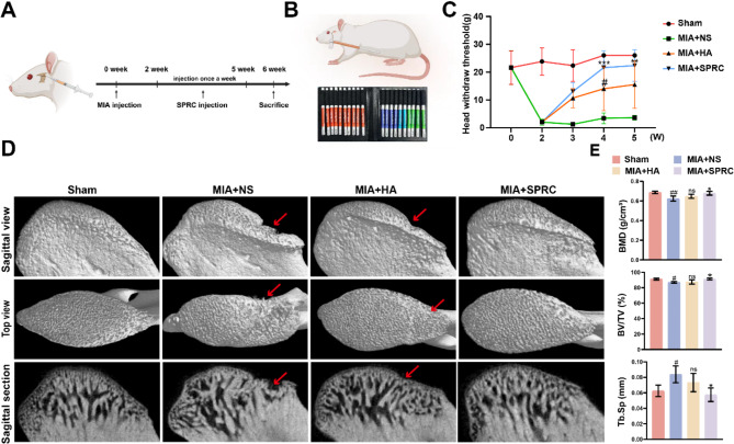
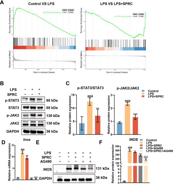
Background: Temporomandibular joint osteoarthritis (TMJ-OA) is a disease characterized by cartilage degradation and synovial inflammation, with limited effective treatment currently. Synovial macrophage polarization is pivotal in TMJ-OA progression, making it a promising therapeutic aspect. This study investigated the effects of S-propargyl-cysteine (SPRC), an endogenous H2S donor, on macrophage polarization and its therapeutic potential in alleviating TMJ-OA.

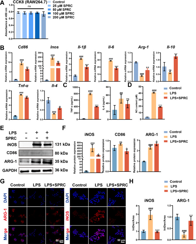
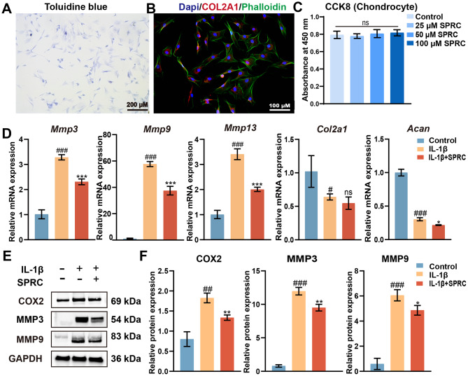
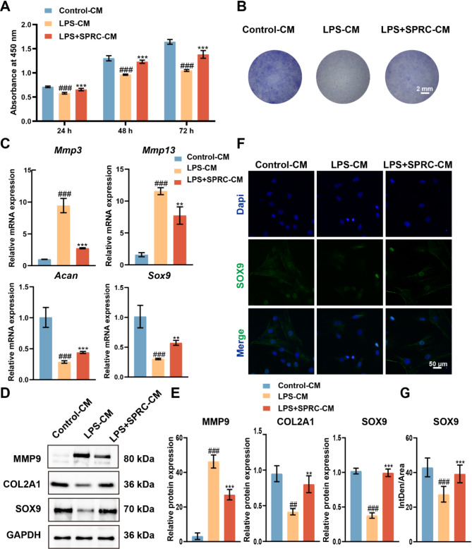
Methods: A MIA-induced TMJ-OA rat model and LPS-stimulated RAW264.7 macrophages were employed to evaluate the effects of SPRC in vivo and in vitro. TMJ bone and cartilage were analyzed via micro-CT and histological methods, while macrophage polarization markers expression were assessed via RT-qPCR, western blot, and immunofluorescence. RNA sequencing was performed on macrophages, and the JAK2/STAT3 signaling pathway was validated using the JAK2-specific inhibitor AG490. The direct effects of SPRC on rat primary condylar chondrocytes were examined by evaluating ECM synthesis and degradation. Co-culture experiments further assessed macrophage-chondrocyte interactions.

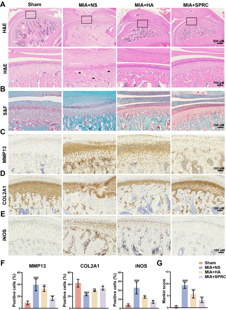
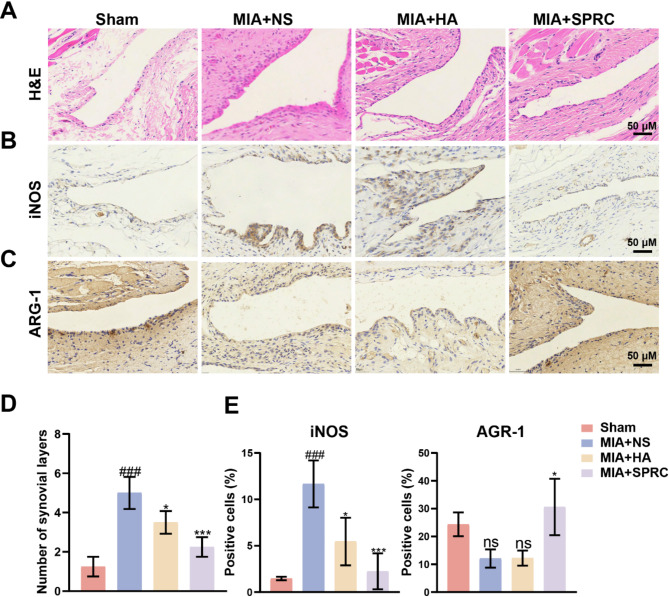
Results: SPRC significantly alleviated cartilage and bone damage in the TMJ-OA rat model, as demonstrated by improved bone volume and cartilage structure. SPRC reduced pro-inflammatory M1 macrophage infiltration and enhanced anti-inflammatory M2 macrophage polarization. SPRC effectively inhibited the JAK2/STAT3, leading to reduction of inflammatory markers, including TNF-α, IL-6, and iNOS. Co-culture experiments revealed that SPRC-treated macrophage-conditioned medium improved chondrocyte metabolic activity and restored ECM integrity.

Conclusions: SPRC-modulated macrophage polarization alleviates TMJ-OA via JAK/STAT downregulation, thereby reducing synovial inflammation and cartilage degradation. These findings position SPRC as a promising therapeutic candidate for TMJ-OA and provide insights into novel strategies targeting macrophage polarization and synovium-cartilage crosstalk.

Keywords: Hydrogen sulfide; JAK/STAT; Macrophage; S-propargyl-cysteine; Synovial; Temporomandibular joint osteoarthritis.


Conflict of interest statement

Declarations. Ethics approval and consent to participate: This study was conducted in accordance with the Declaration of Helsinki and approved by the Guangdong HUA WEI Testing Co., Ltd. Ethics Committee (No. 202310002). Consent for publication: Not applicable. Competing interests: The authors declare no competing interests.

Figures

Fig. 1
Fig. 1
SPRC promoted subchondral bone remodeling and reduced pain in TMJ-OA rats. (A) Experimental timeline outlining TMJ-OA induction and SPRC treatment in vivo. (B) Schematic representation of pain behavior assessment and the time-dependent changes in pain behavior observed during the experiment, including baseline measurements before MIA induction and post-treatment evaluation with SPRC. (C) Representative sagittal views, top views, and sagittal sections of the condyles. (D) Quantitative analysis of subchondral bone parameters, including BMD, BV/TV, and Tb.Sp. The data were analyzed via one-way ANOVA (n = 3). Statistical significance is denoted as follows: #p < 0.05 and ##p < 0.01 compared with the sham group; *p < 0.05, **p < 0.01, and ***p < 0.001 compared with the MIA + NS group. “ns” indicates no significant difference. Red arrows indicate bone defects
Fig. 2
Fig. 2
SPRC reduced condylar cartilage damage in TMJ-OA rats. (A-B) Representative images of condylar cartilage stained with H&E and S&F. (C-E) Representative images of immunohistochemical staining showing the expression of COL2A1, MMP13, and iNOS. (F) Quantitative analysis of COL2A1, MMP13, and iNOS expression levels. (G) Mankin score assessment of cartilage integrity. The data were analyzed via one-way ANOVA (n = 3). Statistical significance is denoted as follows: ###p < 0.001 compared with the sham group; *p < 0.05, **p < 0.01, and ***p < 0.001 compared with the MIA + NS group. “ns” indicates no significant difference. The black triangle indicates a decrease in the number of chondrocytes
Fig. 3
Fig. 3
SPRC reduced synovial inflammation and M1-phenotype macrophages in TMJ-OA rats. (A) Representative images of synovial tissue stained with H&E. (B-C) Representative images of immunohistochemical staining showing the expression of iNOS and ARG-1. (D) Quantitative analysis of the number of synovial lining layers. (E) Quantitative analysis of iNOS and ARG-1 expression levels. The data were analyzed via one-way ANOVA (n = 3). Statistical significance is denoted as follows: ###p < 0.001 compared with the sham group; *p < 0.05 and ***p < 0.001 compared with the MIA + NS group. “ns” indicates no significant difference
Fig. 4
Fig. 4
SPRC mediated the M1 to M2 macrophage phenotype polarization transition in vitro. (A) RAW264.7 macrophages were treated with various concentrations of SPRC (25, 50, 100, or 200 µM) for 24 h, and cell viability was evaluated via the CCK8 assay. (B) RT-qPCR analysis was conducted to measure the expression levels of M1-related genes (Cd86, Inos, Il-6, Il-1β, and Tnf-α) and M2-related genes (Arg-1, Il-10, and Il-4). (C) The levels of IL-6 and TNF-α in the supernatant of the culture medium were quantified via ELISA. (D) NO content in the culture medium supernatant was also assessed. (E-F) The protein levels of M1 macrophage markers (CD86 and iNOS) and the M2 macrophage marker ARG-1 were evaluated via western blot analysis. (G-H) Immunofluorescence staining for iNOS and ARG-1 was performed to assess macrophage polarization further. Statistical analysis was conducted via one-way ANOVA (n ≥ 3). Statistical significance is denoted as follows: #p < 0.05 and ###p < 0.001 compared with the control group; *p < 0.05, **p < 0.01, and ***p < 0.001 compared with the LPS group. “ns” indicates no significant difference
Fig. 5
Fig. 5
SPRC inhibited the JAK/STAT signaling pathway in vitro. (A) GSEA revealed significant enrichment of the JAK/STAT signaling pathway. (B-C) Western blot analysis of phosphorylated STAT3 (p-STAT3), total STAT3, phosphorylated JAK2 (p-JAK2), and total JAK2. (D) RT-qPCR analysis of Inos expression. (E-F) Western blot analysis of iNOS protein levels. The data were analyzed via one-way ANOVA (n ≥ 3). Statistical significance is denoted as follows: ###p < 0.001 compared with the control group; *p < 0.05, **p < 0.01, ***p < 0.001 compared with the LPS group; &&&p < 0.001 compared with the LPS + AG490 group
Fig. 6
Fig. 6
SPRC reduced condylar chondrocytes ECM catabolism in vitro. (A) Representative images of condylar chondrocytes stained with toluidine blue to assess extracellular matrix (ECM) integrity. (B) Immunofluorescence staining of chondrocytes showing DAPI-stained nuclei (blue), phalloidin-stained F-actin (green), and COL2A1 (red). (C) Cell viability of rat primary condylar chondrocytes (rPCCs) treated with SPRC (25, 50, or 100 µM) for 24 h, as evaluated by the CCK8 assay. (D) RT-qPCR analysis of matrix degradation-related genes (Mmp3, Mmp9, and Mmp13) and matrix synthesis-related genes (Col2a1 and Acan). (E-F) Western blot analysis of COX2, MMP3, and MMP9 protein expression levels. The data were analyzed via one-way ANOVA (n ≥ 3). Statistical significance is denoted as follows: #p < 0.05, ##p < 0.01, and ###p < 0.001 compared with the control group; *p < 0.05, **p < 0.01, and ***p < 0.001 compared with the IL-1β group. “ns” indicates no significance
Fig. 7
Fig. 7
CM from SPRC-treated macrophages restored metabolic activity, chondrogenic differentiation, and matrix destruction in rPCCs. (A) The metabolic activity of rPCCs cultured with CM from macrophages was evaluated at 24, 48, and 72 h via the CCK8 assay. (B) Representative images of toluidine blue-stained rPCCs cultured with macrophage CM for 7 days to assess ECM integrity. (C) RT-qPCR analysis of matrix degradation-related genes (Mmp3, Mmp13) and matrix synthesis-related genes (Acan, Sox9) in rPCCs. (D-E) Western blot analysis of MMP9, COL2A1, and SOX9 protein expression levels in rPCCs cultured with macrophage CM. (F-G) Immunofluorescence staining of SOX9 in rPCCs to assess chondrogenic differentiation. The data were analyzed via one-way ANOVA (n ≥ 3). Statistical significance is denoted as follows: ##p < 0.01, ###p < 0.001 compared with the control-CM group; **p < 0.01, ***p < 0.001 compared with the LPS-CM group

Similar articles

Cited by

References

    1. Bechelli C, Macabrey D, Deglise S, Allagnat F. Clinical potential of hydrogen sulfide in peripheral arterial disease. Int J Mol Sci 2023, 24(12). - PMC - PubMed
    1. Burguera EF, Meijide-Failde R, Blanco FJ. Hydrogen sulfide and inflammatory joint diseases. Curr Drug Targets. 2017;18(14):1641–52. - PubMed
    1. Cardoneanu A, Macovei LA, Burlui AM, Mihai IR, Bratoiu I, Rezus II, Richter P, Tamba BI, Rezus E. Temporomandibular joint osteoarthritis: pathogenic mechanisms involving the cartilage and subchondral bone, and potential therapeutic strategies for joint regeneration. Int J Mol Sci 2022, 24(1). - PMC - PubMed
    1. Chen BY, Pathak JL, Lin HY, Guo WQ, Chen WJ, Luo G, Wang LJ, Sun XF, Ding Y, Li J, et al. Inflammation triggers chondrocyte ferroptosis in TMJOA via HIF-1alpha/TFRC. J Dent Res. 2024;103(7):712–22. - PubMed
    1. Cirino G, Szabo C, Papapetropoulos A. Physiological roles of hydrogen sulfide in mammalian cells, tissues, and organs. Physiol Rev. 2023;103(1):31–276. - PubMed

MeSH terms

LinkOut - more resources