. 2025 Jul;9(7):e2400583.
doi: 10.1002/adbi.202400583. Epub 2025 Apr 8.

Activation of SIRT1 Reduces Renal Tubular Epithelial Cells Fibrosis in Hypoxia Through SIRT1-FoxO1-FoxO3-Autophagy Pathway

Affiliations

Activation of SIRT1 Reduces Renal Tubular Epithelial Cells Fibrosis in Hypoxia Through SIRT1-FoxO1-FoxO3-Autophagy Pathway

Guangyu Wang et al. Adv Biol (Weinh). 2025 Jul.

Abstract

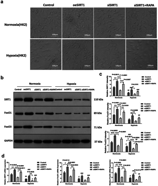
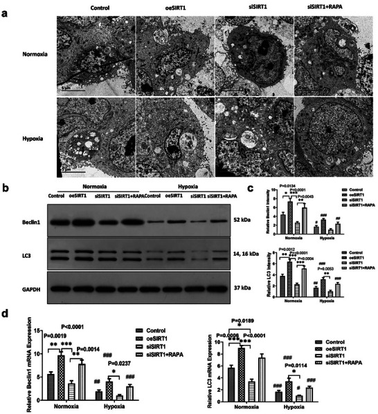
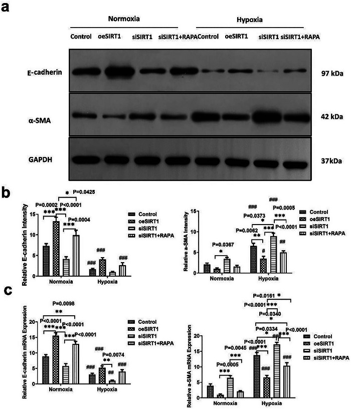
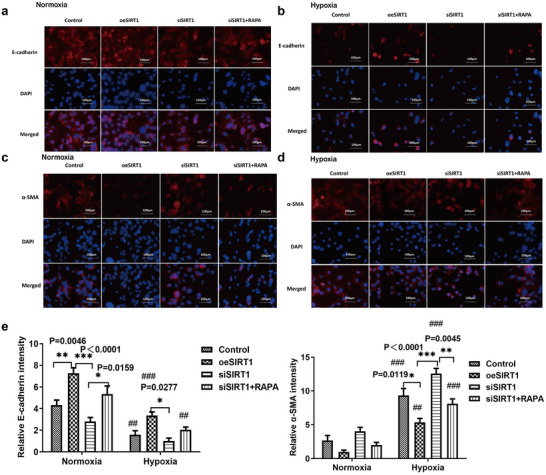
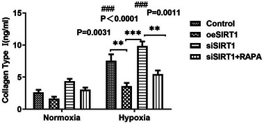
Heart failure-induced renal tubular epithelial cell fibrosis is an important pathological process that leads to chronic kidney disease. This study is to investigate the regulatory mechanism of over-expression or knock-down SIRT1 gene, alleviating hypoxia-induced HK2 cell fibrosis in heart failure. The focus is on the SIRT1-FoxO1-FoxO3-Autophagy pathway. In vitro experiments are performed by HK2cell line to simulate the normal oxygen state (Normoxia) and the hypoxia state (Hypoxia) caused by heart failure, SIRT1 gene over-expression by transfected vectors, knock-down and Rapamycin (RAPA)-induced cellular autophagy, and the cell models are divided into four subgroups, named control group, oeSIRT1, siSIRT1 and siSIRT1+RAPA. Western blotting (WB), real-time qPCR, immunofluorescence (IF), ELISA, and transmission electron microscopy are used to quantitatively or semi-quantitatively analyze the expression of FoxO1, FoxO3, SIRT1, Beclin1, LC-3, α-SMA, E- Cadherin, and collagen-I in cells or supernatants. It is demonstrated that activation of SIRT1 regulates the expression and activity of FoxO1 and FoxO3, thereby affecting autophagy. This modulation leads to a reduction in HK2 fibrosis markers (α-SMA and E-cadherin) and extracellular matrix deposition (collagen I), which ultimately attenuates renal tubular epithelial cell fibrosis. These findings provide new insights into potential therapeutic strategies for treating heart failure-induced renal tubular epithelial cell fibrosis by targeting the SIRT1-FoxO1-FoxO3-Autophagy pathway.

Keywords: SIRT1; autophagy; fibrosis; foxO1; heart failure; hypoxia.


Conflict of interest statement

The authors declare no conflict of interest.

Figures

Figure 1
Figure 1
Effects of over‐expression, knock‐down of SIRT1 and induction of autophagy on HK2 cell growth. a) Morphology of HK2 cells in the Control, oeSIRT1, siSIRT1, and siSIRT1+RAPA groups was observed. In the Control group, cells displayed typical morphology with uniform size and shape. In the oeSIRT1 group, cells showed increased density and altered shape. In the siSIRT1 group, cells appeared smaller and less organized. In the siSIRT1+RAPA group, cells presented a distinct morphology compared to the siSIRT1 group. (n = 3 samples per group). b) Western blot analysis was performed to detect the expression levels of SIRT1, FoxO1, FoxO3, and GAPDH (used as a normalization gene) under normoxia and hypoxia conditions. (n = 3 samples per group, Control versus oeSIRT1 versus siSIRT1 versus SIRT1+RAPA: * p < 0.05, ** p < 0.01, *** p < 0.001. Group under normoxia versus hypoxia: # p < 0.05, ## p < 0.01, ### p < 0.001). c) The relative intensity of SIRT1, FoxO1, and FoxO3 compared to the Control group was determined by western blot analysis (The Discovery Series Quantity One 1‐D Analysis Software Version 4.6.8, Bio‐Rad, USA). (n = 3 samples per group, Control versus oeSIRT1 versus siSIRT1 versus SIRT1+RAPA: * p < 0.05, ** p < 0.01, *** p < 0.001. Group under normoxia versus hypoxia: # p < 0.05, ## p < 0.01, and ### p < 0.001). d) The relative mRNA expression levels of SIRT1, FoxO1, and FoxO3 were evaluated. (n = 3 samples per group, Control versus oeSIRT1 versus siSIRT1 versus SIRT1+RAPA: * p < 0.05, ** p < 0.01, and *** p < 0.001. Group under normoxia versus hypoxia: # p <0.05, ## p < 0.01, and ### p < 0.001). All experiments were repeated three times independently of each other. Note: Control versus oeSIRT1 versus siSIRT1 versus SIRT1+RAPA: * p < 0.05, ** p < 0.01, and *** p < 0.001. Group under normoxia versus hypoxia: # p < 0.05, ## p < 0.01, and ### p < 0.001. oeSIRT: overexpression of SIRT; siSIRTI: SIRTI knockdown using siRNA; siSIRTI+RAPA: knockdown of SIRT1 using siRNA with rapamycin‐induced autophagy.
Figure 2
Figure 2
The effect of knock‐down, over‐expression of SIRT1, and autophagy inducing by RAPA on HK2 cell autophagy (n = 3), All experiments were repeated three times independently of each other. a) Autophagosome of HK2 cell in the Control, oeSIRT1, siSIRT1, and siSIRT1+RAPA groups was observed using transmission electron microscope(TEM) Images. b) Western blot analysis was performed to detect the expression levels of Beclin1, LC3, and GAPDH (used as a normalization gene) under normoxia and hypoxia conditions. c) The relative intensity of Beclin1 and LC3 compared to the Control group was determined by western blot analysis software(The Discovery Series Quantity One 1‐D Analysis Software Version 4.6.8, Bio‐Rad, USA). (n = 3 samples per group, Control versus oeSIRT1 versus siSIRT1 versus SIRT1+RAPA: * p < 0.05, ** p < 0.01, and *** p < 0.001.Group under normoxia versus hypoxia: # p < 0.05, ## p < 0.01, and ### p < 0.001). d) The relative mRNA expression levels of Beclin1 and LC3 were evaluated using real‐time‐q‐ PCR. (n = 3 samples per group, Control versus oeSIRT1 versus siSIRT1 versus SIRT1+RAPA: * p < 0.05, ** p < 0.01, and *** p < 0.001.Group under normoxia versus hypoxia: # p < 0.05, ## p < 0.01, and ### p < 0.001). Note: Control versus oeSIRT1 versus siSIRT1 versus SIRT1+RAPA: * p < 0.05, ** p < 0.01, and *** p < 0.001. Group under normoxia versus hypoxia: # p < 0.05, ## p < 0.01, and ### p < 0.001. oeSIRT: overexpression of SIRT; siSIRTI: SIRTI knockdown using siRNA; siSIRTI+RAPA: knockdown of SIRT1 using siRNA with rapamycin‐induced autophagy.
Figure 3
Figure 3
Characterization of the epithelial‐mesenchymal transition (EMT) in HK2 cells under normoxic and hypoxic conditions (n = 3). a) Western blot analysis was performed to detect the expression levels of E‐cadherin, α‐SMA, and GAPDH (used as a normalization gene) under normoxia and hypoxia conditions. b) The relative intensity of E‐cadherin and α‐SMA compared to the Control group was determined by western blot analysis software(The Discovery Series Quantity One 1‐D Analysis Software Version 4.6.8, Bio‐Rad, USA). (n = 3 samples per group, Control versus oeSIRT1 versus siSIRT1 versus SIRT1+RAPA: * p < 0.05, ** p < 0.01, and *** p < 0.001.Group under normoxia versus hypoxia: # p < 0.05, ## p < 0.01, and ### p < 0.001.) c) The relative mRNA expression levels of E‐cadherin and α‐SMA were evaluated using real‐time q‐PCR. (n = 3 samples per group, Control versus oeSIRT1 versus siSIRT1 versus SIRT1+RAPA: * p < 0.05, ** p < 0.01, and *** p < 0.001.Group under normoxia versus hypoxia: # p < 0.05, ## p < 0.01, and ### p < 0.001.) All experiments were repeated three times independently of each other. Note: Control versus oeSIRT1 versus siSIRT1 versus SIRT1+RAPA: * p < 0.05, ** p < 0.01, and *** p < 0.001. Group under normoxia versus hypoxia: # p < 0.05, ## p < 0.01, and ### p < 0.001. oeSIRT: overexpression of SIRT; siSIRTI: SIRTI knockdown using siRNA; siSIRTI+RAPA: knockdown of SIRT1 using siRNA with rapamycin‐induced autophagy.
Figure 4
Figure 4
Semi‐quantification of E‐cadherin and α‐SMA in HK2 cells under normoxia and hypoxia conditions (n = 3). a) The analysis of E‐cadherin using Immunofluorescence (IF) assay in normoxia condition. b) The analysis of E‐cadherin using Immunofluorescence (IF) assay in hypoxia condition. c) The analysis of a‐SMA using Immunofluorescence (IF) assay in normoxia condition. d) The analysis of a‐SMA using Immunofluorescence (IF) assay in hypoxia condition. e) The relative fluorescence levels of E‐cadherin and α‐SMA were evaluated by Image J/fiji software. All experiments were repeated three times independently of each other. (n = 3 samples per group, Control versus oeSIRT1 versus siSIRT1 versus SIRT1+RAPA: * p < 0.05, ** p < 0.01, and *** p < 0.001.Group under normoxia versus hypoxia: # p < 0.05, ## p < 0.01, and ### p < 0.001.) Note: Control versus oeSIRT1 versus siSIRT1 versus SIRT1+RAPA: * p < 0.05, ** p < 0.01, and *** p < 0.001. Group under normoxia versus hypoxia: # p < 0.05, ## p < 0.01, and ### p < 0.001. oeSIRT: overexpression of SIRT; siSIRTI: SIRTI knockdown using siRNA; siSIRTI+RAPA: knockdown of SIRT1 using siRNA with rapamycin‐induced autophagy.
Figure 5
Figure 5
Quantification of collagen I concentration in HK2 cell culture supernatants. The concentration of collagen I in 8 groups(normaxia and hypoxic) was detected using ELISA (n = 3). All experiments were independently repeated thrice. (n = 3 samples per group, Control versus oeSIRT1 versus siSIRT1 versus SIRT1+RAPA: * p < 0.05, ** p < 0.01, and *** p < 0.001.Group under normoxia versus hypoxia: # p < 0.05, ## p < 0.01, and ### p < 0.001.) Note: Control versus oeSIRT1 versus siSIRT1 versus SIRT1+RAPA: * p < 0.05, ** p < 0.01, and *** p < 0.001. Group under normoxia versus hypoxia: # p < 0.05, ## p < 0.01, and ### p < 0.001. oeSIRT: overexpression of SIRT; siSIRTI: SIRTI knockdown using siRNA; siSIRTI+RAPA: knockdown of SIRT1 using siRNA with rapamycin‐induced autophagy.

Similar articles

References

    1. Minhas A. M. K., Ijaz S. H., Jamal S., Dani S. S., Khan M. S., Greene S. J., Fudim M., Warraich H. J., Shapiro M. D., Virani S. S., Nasir K., Khan S. U., Circ. Heart Fail 2022, 15, 008943. - PubMed
    1. Tolwani A., N. Engl. J. Med. 2012, 367, 2505. - PubMed
    1. Johansson I., Joseph P., Balasubramanian K., McMurray J. J. V., Lund L. H., Ezekowitz J. A., Kamath D., Alhabib K., Bayes‐Genis A., Budaj A., Dans A. L. L., Dzudie A., Probstfield J. L., Fox K. A. A., Karaye K. M., Makubi A., Fukakusa B., Teo K., Temizhan A., Wittlinger T., Maggioni A. P., Lanas F., Lopez‐Jaramillo P., Silva‐Cardoso J., Sliwa K., Dokainish H., Grinvalds A., McCready T., Yusuf S., Circulation 2021, 143, 2129. - PubMed
    1. Free D., https://www.mdpi.com/2075‐4418/14/15/1635, (accessed: July 31, 2024).
    1. Shanah L., Mir T., Uddin M. M., Hussain T., Parajuli T., Bhat Z., Int. J. Cardiol. 2023, 370, 244. - PubMed

LinkOut - more resources