Review
. 2025 Jul 1;74(7):1037-1046.
doi: 10.2337/dbi24-0041.

What Is Gestational Diabetes-Really?

Affiliations
Review

What Is Gestational Diabetes-Really?

Thomas A Buchanan et al. Diabetes. .

Abstract

Gestational diabetes mellitus (GDM) is one of the most common medical complications of pregnancy. It is generally defined as glucose intolerance with onset or first recognition during pregnancy. The pathogenesis of GDM has long been attributed to inadequate pancreatic β-cell compensation for the physiological insulin resistance of pregnancy. This defect is thought to resolve after pregnancy but become manifest in later life as an increased risk of diabetes. Examination of mechanisms underlying GDM does not support this commonly held picture. In this Perspective, we present evidence that, like diabetes outside of pregnancy, GDM has no single etiology. It results from multiple causes of a common physiological manifestation, inadequate β-cell function, which leads to a common clinical manifestation, elevated glucose levels. We provide evidence that GDM often represents detection of chronic and progressive β-cell dysfunction that is temporally but not mechanistically related to pregnancy. We provide detailed characterization of the β-cell defect in one high-risk group, Hispanic Americans. Finally, we address some of the clinical and research implications of these findings.

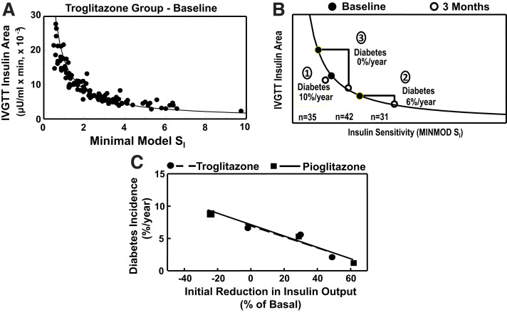
Article highlights: Gestational diabetes mellitus (GDM) is not one disease but many that share inadequate β-cell function as a common cause for elevated glucose levels. Inadequate β-cell function may result from factors that occur outside of pregnancy, such as autoimmunity, monogenic disorders, obesity, and insulin resistance. Pregnancy-specific causes may exist as well but remain to be defined. Detailed physiological studies in women with obesity reveal that inadequate β-cell function is likely a chronic condition that is detected by routine glucose screening in pregnancy and that worsens over time, leading to diabetes in later life. The authors' studies in Hispanic patients identify obesity and insulin resistance as important causes of β-cell dysfunction, providing a rationale for treating both to prevent diabetes after GDM. Additional work is needed to define the full breadth of underlying causes of GDM as the basis for precision management during and, especially, after pregnancy.


Conflict of interest statement

Duality of Interest. No potential conflicts of interest relevant to this article were reported.

Figures

Figure 1
Figure 1
A: Theoretical diagram of conventional wisdom that GDM develops during pregnancy when β-cells fail to compensate for increasing insulin resistance. This pathogenesis predicts that women would move from a normal to an abnormal sensitivity-secretion relationship during pregnancy. The upper arrow represents reaching a maximum capacity for β-cell compensation. The middle arrow represents an inadequate rate of increasing compensation. The lower arrow indicates inability to increase compensation. B–D: Actual results from three studies (9–11) in which women with normal glucose tolerance and women with GDM had insulin sensitivity and insulin secretion (B) or responses (C and D) measured in the third trimester and when they were not pregnant. Curved lines represent the product of mean insulin sensitivity (x-axis) and mean insulin response (y-axis) in nonpregnant women with normal glucose tolerance or participants with GDM. Percentages next to GDM symbols represent fractions of analogous (third trimester or not pregnant) disposition index in women with normal glucose tolerance. In all three studies, women with GDM had similar reductions in β-cell compensation for insulin resistance whether pregnant or not, in contrast to conventional wisdom for the pathogenesis of GDM depicted in A. MINMOD SI, minimal model assessment of insulin sensitivity. B and C are reproduced from Buchanan et al. (11). D was created using results published in Catalano et al. (10).
Figure 2
Figure 2
Plasma glucose levels during OGTTs conducted before pregnancy (75-g test) (A) and during the third trimester (100-g tests) (B) of pregnancies involving normal glucose tolerance (NGT) or pregnancies complicated by GDM. Differences in mean glucose levels between groups at each time point appear below curves. The sum of those differences is depicted in C, which demonstrates that most glucose differences in the third trimester were already present before pregnancy (77% for 3 h, 88% for the 2-h interval currently used to diagnose GDM). Figure was created using results published in Catalano et al. (10).
Figure 3
Figure 3
A and B: Insulin sensitivity and secretion from hyperglycemic clamps conducted in the third trimester or after pregnancy in women who had normal glucose tolerance (n = 8) or GDM (n = 7) during pregnancy. P values reflect differences between normal glucose-tolerant and GDM groups under each condition. C: Insulin sensitivity estimated from by the Matsuda index and β-cell response measured by the Stumvoll first-phase index from 75-g OGTTs administered to 809 women (8.3% with GDM) between 24 and 30 weeks’ gestation. The GDM group was divided into physiological subtypes who had insulin responses (Secretion), insulin sensitivity (Sensitivity), or both (Mixed) below the 25th percentile for the respective distributions in normal glucose-tolerant pregnant women. All three GDM subtypes have the same reduction in β-cell compensation for insulin resistance. They differ in insulin sensitivity as well as BMI. DI, disposition index. A and B were created using results published in Homko et al. (9). C was created using results published in Powe et al. (14).
Figure 4
Figure 4
A: Mean (95% CI) disposition index at baseline in physiological cohort of Hispanic women with prior GDM who remained diabetes free or developed diabetes during up to 135 months after the index pregnancy, grouped according to whether and when they developed diabetes. B: Disposition index over time in the same cohort. Results are plotted relative to final study visit because follow-up ended with development of diabetes. Numbers by symbols are sample sizes. Reproduced from Xiang et al. (19).
Figure 5
Figure 5
Relationship between disposition index and fasting glucose in women described in Fig. 4 during first 5 years after index pregnancies. Solid horizontal lines denote thresholds for impaired and diabetic glucose levels. Arrows denote direction of change over time. Round symbols represent means for women with prior GDM, and the star denotes the mean from 30 nonpregnant women of age, parity, and BMI similar to those of women in the GDM group but who had normal 1-h, 50-g glucose screen during pregnancy. Adapted from Xiang et al. (21).
Figure 6
Figure 6
A: Relationship between insulin sensitivity (minimal model SI [MINMOD SI], where SI is insulin sensitivity) and total area under the insulin curve during tolbutamide-modified IVGTTs at baseline in women in the intervention cohort (defined in the text) who were randomized to the troglitazone arm. Symbols represent individual participants; curved line is the mean disposition index (SI × insulin area) for all women. B: Changes in insulin sensitivity and insulin area between baseline and 3 months on treatment in the active treatment arm. The three subgroups are indicated by circled numbers: group 1, women who did not have an increase in insulin sensitivity; group 2, women who had an increase in insulin sensitivity and a change in insulin area below the median; and group 3, women who had an increase in insulin sensitivity and a change in insulin area above the median. “Diabetes” denotes annual average incidence rates. C: Relationship between early change from baseline in total IVGTT insulin area and average annual diabetes incidence in the three subgroups in the active troglitazone treatment arm depicted in B and analogous subgroups from the follow-on study of open-label pioglitazone treatment in participants who did not have diabetes after washout in the troglitazone study. Symbols represent tertiles of change from baseline in IVGTT area in each study, and lines represent linear fit of tertile values in each study. A and B were adapted from Buchanan et al. (24), and C was adapted from Xiang et al. (26).
Figure 7
Figure 7
Conceptual diagram of the interplay between β-cell secretory loading (insulin output) and susceptibility or resistance of β-cells to deteriorate under such loading, based on evidence from Hispanic women who develop GDM. Open arrows represent β-cells that are relatively susceptible to loading, and closed arrows represent β-cells that are relatively resistant to loading. In each case, increased loading accelerates the rate of deterioration; the effect is more prominent for susceptible β-cells. Women with resistant β-cells and low loading should have the slowest rate of deterioration and may not develop GDM during reproductive years. Women with susceptible β-cells and high loading should deteriorate most rapidly and may develop diabetes before they become pregnant. While the diagram depicts two states for each variable, they are likely continuously distributed, with many possible combinations.

References

    1. Petersen JS, Dyrberg T, Damm P, Kühl C, Mølsted-Pedersen L, Buschard K. GAD65 autoantibodies in women with gestational or insulin dependent diabetes mellitus diagnosed during pregnancy. Diabetologia 1996;39:1329–1333 - PubMed
    1. Weng J, Ekelund M, Lehto M, et al. Screening for MODY mutations, GAD antibodies, and type 1 diabetes–associated HLA genotypes in women with gestational diabetes mellitus. Diabetes Care 2002;25:68–71 - PubMed
    1. Löbner K, Knopff A, Baumgarten A, et al. Predictors of postpartum diabetes in women with gestational diabetes mellitus. Diabetes 2006;55:792–797 - PubMed
    1. Kousta E, Ellard S, Allen LI, et al. Glucokinase mutations in a phenotypically selected multiethnic group of women with a history of gestational diabetes. Diabet Med 2001;18:683–684 - PubMed
    1. Ellard S, Beards F, Allen LI, et al. A high prevalence of glucokinase mutations in gestational diabetic subjects selected by clinical criteria. Diabetologia 2000;43:250–253 - PubMed

MeSH terms