. 2025 Jun 20;6(2):103710.
doi: 10.1016/j.xpro.2025.103710. Epub 2025 Apr 8.

Protocol for the functional evaluation of genetic variants using saturation genome editing

Affiliations

Protocol for the functional evaluation of genetic variants using saturation genome editing

Sofia Obolenski et al. STAR Protoc. .

Abstract

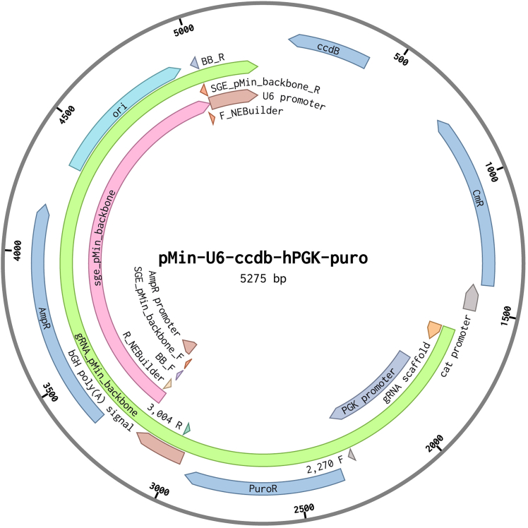
Saturation genome editing (SGE) employs CRISPR-Cas9 and homology-directed repair (HDR) to introduce exhaustive nucleotide modifications at specific genomic sites in multiplex, enabling the functional analysis of genetic variants while preserving their native genomic context. Here, we present a protocol for SGE-based variant evaluation in HAP1-A5 cells. We describe the steps for designing variant libraries, single-guide RNAs (sgRNAs), and oligonucleotide primers for PCR. We also detail the sample preparation before the SGE screen, the cellular screening process, and subsequent next-generation sequencing (NGS) library preparation. For complete details on the use and execution of this protocol, please refer to Radford et al.,1 Waters et al.,2 and Olvera-León et al.3.

Keywords: CRISPR; Genetics; Genomics; High-Throughput Screening.


Conflict of interest statement

Declaration of interests The authors declare no competing interests.

Figures

None
Graphical abstract
Figure 1
Figure 1
Overview of the complete SGE process workflow In a saturation genome editing (SGE) experiment, each region to be edited in a specific gene is targeted using a single guide RNA (sgRNA) in combination with an SGE homology-directed repair (HDR) template library in HAP1 cells deficient in DNA Ligase 4 (LIG4) and expressing endogenous Cas9 (HAP1-A5 cell line). Cas9 induces a double-strand break, which is repaired by the SGE HDR template libraries. Transfections are performed in triplicate, and genomic DNA (gDNA) samples are collected at varying time points depending on the screen length, typically at days 4, 7, 10, 14 and 21. This approach enables functional characterization through variant depletion kinetics, as deleterious alleles in essential loci are expected to decrease over time. The gDNA is then processed for edited gDNA library preparation and sequencing to generate functional scores. This figure is adapted from Olvera-León et al., with permission obtained.
Figure 2
Figure 2
Overview of SGE pre-screen process: Cloning SGE variant oligonucleotide libraries into linearized wild-type homology regions (A) The saturation genome editing (SGE) variant oligonucleotide library pool is PCR amplified using Primer Set 1 to increase the starting material of the SGE variant oligonucleotide library for subsequent cloning. Target region-specific SGE variant oligonucleotide libraries are generated by amplifying the SGE target region using ‘Primer Set 2’. (B) SGE_pMin_backbone is generated using Primer Set 3, which amplifies the promoter and coding sequence for ampicillin resistance as well as the origin of replication (ori) sequence from the pMin-U6-ccdb-hPGK-puro (pMin) plasmid. (C) Wild-type homology arms are amplified from HAP1-A5 genomic DNA (gDNA) using Primer Set 4, with one set designed for each exon. The resulting PCR amplicons are then ligated into the SGE_pMin_backbone, which contains homology regions. The wild-type homology region plasmids are then amplified via E. coli transformation and verified by Sanger sequencing. (D) Wild-type homology region plasmids are linearized using Primer Set 5, with specific primers for each target region. (E) The resulting linearized vectors are ligated with the individual target region-specific SGE library oligos to create SGE homology-directed repair (HDR) template libraries. These libraries are amplified via E. coli transformation and verified by next-generation sequencing.
Figure 3
Figure 3
Optimization of qPCR conditions to identify optimal amplification cycles for target-specific primers (A–D) qPCR amplification plots for samples 1–4, illustrating the exponential phase of amplification. The optimal cycle number is determined by identifying the point within the exponential phase prior to the onset of the non-exponential plateau phase. Based on the amplification curves, the optimal cycle numbers for maintaining saturation genome editing (SGE) variant oligonucleotide library complexity and avoiding over- or under-amplification are 19, 11, 13, and 15 cycles for samples 1, 2, 3, and 4, respectively, y-axis is normalized reporter (EvaGreen) fluorescence (Rn) and x-axis is cycle number, the theshold is also displayed by horizontal coloured line. (E) Agarose gel electrophoresis (2% agarose in TAE buffer) of qPCR products after 35 cycles. The expected band size (∼350 base pair (bp)) is observed with appropriate band intensity and no evidence of non-specific amplification.
Figure 4
Figure 4
Schematic representation of the pMin-U6-ccdb-hPGK-puro plasmid The pMin plasmid, with a total length of 5.275 kb, serves as the backbone for incorporating the wild-type homology arms (indicated by the pink annotation). Primer Set 3 is employed to amplify the ampicillin resistance (AmpR) gene and the origin of replication (ori) sequence. Additionally, the ‘pMin’ backbone is utilized to construct the ‘gRNA_pMin_backbone’ (green annotation). BbsI-HF restriction enzyme digestion facilitates the inclusion of the AmpR gene, ori sequence, and the puromycin resistance gene, and exclusion of the ccdB casette in downstream clones.
Figure 6
Figure 6
Imaging of HAP1-A5 cells under different tissue culture conditions for the SGE screen (A) HAP1-A5 cells transfected with single guide RNA (sgRNA) alone and selected using puromycin (P+) and blasticidin (B+) media. The efficacy of the sgRNA in inducing a cell fitness or cell death phenotype may result in fewer cells in control samples, where only sgRNAs are transfected. (B) HAP1-A5 cells transfected with both the saturation genome editing (SGE) homology-directed repair (HDR) template library and the corresponding sgRNA, and selected with puromycin (P+) and blasticidin (B+) media, exhibit 70–80% confluency. This is because the SGE HDR template library serves as an HDR template, and most variants in the library rescue the cell fitness phenotype. (C) Untransfected wild-type HAP1-A5 cells also display 70–80% confluency. All images were captured using the Evos XL microscope at 10x magnification (scale bar indicated).
Figure 5
Figure 5
Schematic representation of two SGE tissue culture screening strategies This figure illustrates the two potential saturation genome editing (SGE) screening approaches in tissue culture. For genes with strong essentiality, in the HAP1-A5 cell line, a 14-day SGE screen is appropriate. In contrast, for genes with slower depletion dynamics, a 21-day SGE screen is recommended.
Figure 7
Figure 7
Overview of SGE-edited genomic DNA library samples preparation for sequencing Once the saturation genome editing (SGE) tissue culture experiments are completed, cell pellets are collected, and genomic DNA is extracted to prepare the SGE edited genomic DNA (gDNA) library for next-generation sequencing (NGS). Library preparation includes three rounds of PCR. (A) Primary PCR: Amplification of the genomic region edited by SGE is performed using Primer Set 6, with a specific set of primers designed for each exon. (B) Secondary PCR: To amplify the specific target region from the sample and add Illumina adapters, Primer Set 7 is used. (C) Indexing PCR: To add unique barcodes to all samples for NGS (including replicates and time points), Primer Set 8 is used for each individual sample. After indexing, all samples are bead-purified, pooled and sequenced for subsequent analysis.
Figure 8
Figure 8
Control gel electrophoresis of bead-purified samples prior to sequencing submission 80 ng of DNA sample and an appropriate volume of loading dye were used for gel electrophoresis. Lane 1: 1 kb HyperLadder. Lane 2: Sample 1 following secondary PCR (non-indexed), with an expected fragment size between ∼245-300 base pair (bp). Lane 3: Sample 1 after indexing in the PCR round, showing an expected larger fragment size due to the addition of a unique barcode. Lanes 4–6: Three additional indexed sample examples, which show larger fragment sizes compared to the secondary PCR product. All band intensities are within an acceptable range, and no non-specific bands are observed.

References

    1. Radford E.J., Tan H.-K., Andersson M.H.L., Stephenson J.D., Gardner E.J., Ironfield H., Waters A.J., Gitterman D., Lindsay S., Abascal F., et al. Saturation genome editing of DDX3X clarifies pathogenicity of germline and somatic variation. Nat. Commun. 2023;14:7702. doi: 10.1038/s41467-023-43041-4. - DOI - PMC - PubMed
    1. Waters A.J., Brendler-Spaeth T., Smith D., Offord V., Tan H.K., Zhao Y., Obolenski S., Nielsen M., Van Doorn R., Murphy J.-E., et al. Saturation genome editing of BAP1 functionally classifies somatic and germline variants. Nat. Genet. 2024;56:1434–1445. doi: 10.1038/s41588-024-01799-3. - DOI - PMC - PubMed
    1. Olvera-León R., Zhang F., Offord V., Zhao Y., Tan H.K., Gupta P., Pal T., Robles-Espinoza C.D., Arriaga-González F.G., Matsuyama L.S.A.S., et al. High-resolution functional mapping of RAD51C by saturation genome editing. Cell. 2024;187:5719–5734.e19. doi: 10.1016/j.cell.2024.08.039. - DOI - PubMed
    1. Kotecki M., Reddy P.S., Cochran B.H. Isolation and characterization of a near-haploid human cell line. Exp. Cell Res. 1999;252:273–280. doi: 10.1006/excr.1999.4656. - DOI - PubMed
    1. Andersson B.S., Collins V.P., Kurzrock R., Larkin D.W., Childs C., Ost A., Cork A., Trujillo J.M., Freireich E.J., Siciliano M.J. KBM-7, a human myeloid leukemia cell line with double Philadelphia chromosomes lacking normal c-ABL and BCR transcripts. Leukemia. 1995;9:2100–2108. - PubMed

MeSH terms

Substances

LinkOut - more resources