. 2025 Aug;62(8):10333-10348.
doi: 10.1007/s12035-025-04885-7. Epub 2025 Apr 8.

Epigenetics and Mitochondrial Biogenesis: The Role of Sirtuins in HIV Neuropathogenesis

Affiliations

Epigenetics and Mitochondrial Biogenesis: The Role of Sirtuins in HIV Neuropathogenesis

James Haorah et al. Mol Neurobiol. 2025 Aug.

Abstract

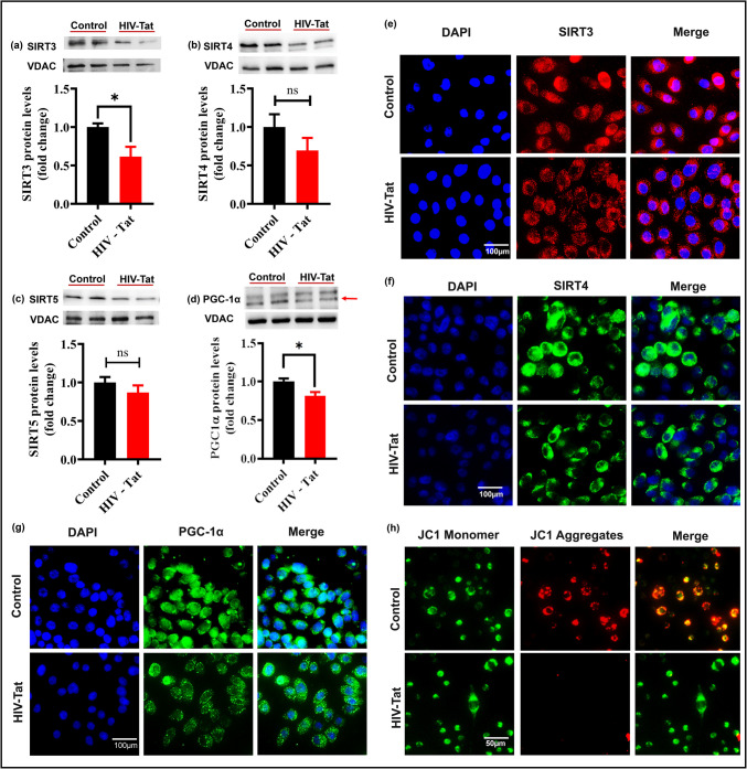
Mitochondrial energy deficits play a central role in HIV-associated neurocognitive disorder (HAND). HIV disrupts cellular functions, including epigenetic modifications such as class III histone deacetylation mediated by sirtuins (SIRTs). However, the role of SIRTs in HAND pathogenesis remains unclear. We hypothesize that HIV alters mitochondrial biogenesis and energy homeostasis by modifying SIRT family members 1-7, contributing to HAND progression. To test this hypothesis, we examined postmortem frontal lobe brain tissue from people with HIV (PWH) and HIV-negative controls, focusing on epigenetic alterations in SIRTs 1-7, the energy sensor adenosine monophosphate-activated protein kinase (AMPK), the mitochondrial master regulator peroxisome proliferator-activated receptor-γ coactivator-1α (PGC-1α), and transcription factors such as mitochondrial transcription factor A (TFAM), nuclear respiratory factors 1 and 2 (NRF-1/2), and factors associated with oxidative phosphorylation (OXPHOS). Our analysis revealed a significant increase in AMPK, OXPHOS, and PGC-1α levels, alongside a decrease in TFAM levels in PWH brains compared to uninfected controls. NRF-1 was upregulated in mitochondria but downregulated in the cytoplasm, while NRF-2 exhibited the opposite trend in PWH compared to HIV-negative controls. The epigenetic signatures of SIRTs 1, 2, 3, 4, 6, and 7 were upregulated in PWH, while SIRT5 was downregulated compared to uninfected brain tissues. We exposed primary human astrocyte and microglial cultures to the HIV-1 transactivator of transcription (Tat) protein to identify the cell types involved. These studies confirmed that HIV-induced epigenetic modifications of SIRTs and mitochondrial impairments occurred in both astrocytes and microglia, highlighting the crucial role of SIRTs in HAND pathogenesis.

Keywords: Epigenetic modification; HAND; HIV; Mitochondrial biogenesis; Sirtuins.


Conflict of interest statement

Declarations. Institutional Review Board Statement: Brain tissue samples from uninfected and HIV-positive frontal and temporal regions were obtained from the National NeuroAIDS Tissue Consortium (NNTC) and National Neurological AIDS Bank (NNAB) University of California (UCLA), California, USA. Informed Consent: Not applicable. Competing Interest: The authors declare no competing interests.

Figures

Fig. 1
Fig. 1
SIRTs levels in the frontal lobe of brain tissues from HIV-infected individuals. Alterations in sirtuin (SIRT) 1, 2, 6, and 7 levels in cytosolic fractions (ad) and nuclear fractions (eh). Changes in SIRT3, 4, and 5 and peroxisome proliferator-activated receptor-γ coactivator-1α (PGC-1α) levels in mitochondrial fractions (il). Representative bar graphs were derived from n = 5 independent experiments. Results were normalized by using beta-actin, Lamin B, and VDAC as loading control for cytosolic, nuclear, and mitochondrial fractions, respectively. The data are presented as mean ± SEM; unpaired t-test. * indicates p-value < 0.05, ** indicates p-value < 0.01. # indicates p-value = 0.05, and ns indicates non-significance
Fig. 2
Fig. 2
Effects of HIV on adenosine monophosphate-activated protein kinase (AMPK and phospho-AMPK), PGC-1α, nuclear respiratory factor-1/2 (NRF-1/2) levels in cytosol (ad) and TFAM, NRF-1, NRF-2, and oxidative phosphorylation (OXPHOS) levels in mitochondrial fractions (eh) from frontal lobe of brain tissues from HIV-infected individuals. Representative bar graphs were derived from n = 5 independent experiments. Results were normalized using beta-actin and VDAC and are presented as mean ± SEM; unpaired t-test. * indicates p-value < 0.05, and ns indicates non-significance. Western blot illustrating β-actin, COX-IV, and histone H3 from organelle-specific extractions of cell cultures (i). Schematic representation of separation of Cyto—cytosol; Mito—mitochondrial; Nuc—nuclear extracts from cell lysate (j)
Fig. 3
Fig. 3
HIV-TAT induces SIRTs alteration in human microglia cells: representative Western blot showing the protein levels of SIRTs 1, 2, 6, and 7 in cytosolic fractions (ad) and nuclear fractions (eh). Representative bar graphs were derived from n = 3 independent experiments consisting of two replicates each. Beta-actin and Histone H3 were probed as loading controls for cytosolic and nuclear fractions, respectively. The data are presented as mean ± SEM; unpaired t-test. * indicates p-value < 0.05, ** indicates p-value < 0.01, # indicates p-value = 0.05, and ns indicates non-significance. Qualitative imaging of SIRT1 in human microglia culture with/without human immunodeficiency virus-1 transactivator of transcription (HIV-1 Tat) (i)
Fig. 4
Fig. 4
HIV-TAT mediated downregulation of mitochondrial SIRTs in human microglia: Protein levels of SIRTs 3, 4, and 5 and PGC-1α in mitochondrial fractions (ad). Representative bar graphs were derived from n = 3 independent experiments consisting of two replicates each. Results were normalized using VDAC and are presented as mean ± SEM; unpaired t-test. * indicates p-value < 0.05, and ns indicates non-significance. Qualitative imaging of SIRT3, SIRT4, and PGC-1α (eg) in human microglia culture with/without HIV-1 Tat. Depolarization of mitochondrial membrane potential via JC-1 staining (h) following microglia exposure to HIV-1 Tat (50 ng/ml). Cell magnification is × 40
Fig. 5
Fig. 5
Alterations of cytosolic and nuclear SIRTs in HIV-Tat exposed human astrocytes: Western blot analysis of SIRTs 1, 2, 6, and 7 in cytosolic fractions (ad) and nuclear fractions (eh) following human astrocyte culture exposure to HIV-1 Tat. Representative bar graphs were derived from n = 3 independent experiments consisting of two replicates each. Results were normalized by using beta-actin and Histone H3 probed as loading controls for cytosolic and nuclear fractions, respectively. The data are presented as mean ± SEM; unpaired t-test. * indicates p-value < 0.05, ** indicates p-value < 0.01, and ns indicates non-significance. Qualitative imaging of SIRT1 (i) in primary human astrocyte culture with/without HIV-1 Tat. Cell magnification is × 40
Fig. 6
Fig. 6
Effect of HIV-Tat on mitochondrial biogenesis and epigenetics in astrocytes: Expression of SIRTs 3, 4, and 5 and PGC-1α in mitochondrial fractions (ad). Representative bar graphs were derived from n = 3 independent experiments consisting of two replicates each. Results were normalized using VDAC and are presented as mean ± SEM; unpaired t-test. * indicates p-value < 0.05, ** indicates p-value < 0.01, # indicates p-value = 0.05, and ns indicates non-significance. Qualitative imaging of SIRT3 and SIRT4 (e and f) and PGC-1α (g) in primary human astrocyte culture with/without HIV-1 Tat. Cell magnification is × 40. Activation of AMPK and phospho-AMPK levels in cytosol of HIV-Tat exposed astrocytes and microglia with corresponding bar graphs. Results were normalized with AMPK and represented as ratio
Fig. 7
Fig. 7
Differential regulation of SIRTs in brain cell types by HIV infection. Downregulation of SIRTs in HIV-infected microglia reduces mitochondrial biogenesis and OXPHOS by suppressing SIRT1/2-regulated PGC-1α and NRF-1 expression, leading to increased viral replication and neurodegeneration. SIRT upregulation in HIV-activated astrocytes increases mitochondrial biogenesis and OXPHOS by upregulating SIRT1/2-regulated PGC-1α and NRF1 levels

References

    1. Wang G et al (2019) Regulation of UCP1 and mitochondrial metabolism in brown adipose tissue by reversible succinylation. Mol Cell 74(4):844–857 e7. - PMC - PubMed
    1. Jurkowska K et al (2022) The effect of antiretroviral therapy on SIRT1, SIRT3 and SIRT6 expression in HIV-infected patients. Molecules 27(4) - PMC - PubMed
    1. Liu G et al (2017) Loss of NAD-dependent protein deacetylase sirtuin-2 alters mitochondrial protein acetylation and dysregulates mitophagy. Antioxid Redox Signal 26(15):849–863 - PMC - PubMed
    1. Pagans S et al (2005) SIRT1 regulates HIV transcription via Tat deacetylation. PLoS Biol 3(2):e41 - PMC - PubMed
    1. Pereira JM et al (2018) Infection reveals a modification of SIRT2 critical for chromatin association. Cell Rep 23(4):1124–1137 - PMC - PubMed

Substances

LinkOut - more resources