. 2025 Apr 8;21(4):e1013053.
doi: 10.1371/journal.ppat.1013053. eCollection 2025 Apr.

HOPS/CORVET tethering complexes are critical for endocytosis and protein trafficking to invasion related organelles in malaria parasites

Affiliations

HOPS/CORVET tethering complexes are critical for endocytosis and protein trafficking to invasion related organelles in malaria parasites

Joëlle Paolo Mesén-Ramírez et al. PLoS Pathog. .

Abstract

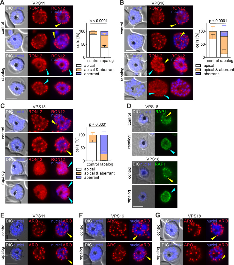
The tethering complexes HOPS/CORVET are central for vesicular fusion through the eukaryotic endolysosomal system, but the functions of these complexes in the intracellular development of malaria parasites are still unknown. Here we show that the HOPS/CORVET core subunits are critical for the intracellular proliferation of the malaria parasite Plasmodium falciparum. We demonstrate that HOPS/CORVET are required for parasite endocytosis and host cell cytosol uptake, as early functional depletion of the complex led to developmental arrest and accumulation of endosomes that failed to fuse to the digestive vacuole membrane. Late depletion of the core HOPS/CORVET subunits led to a severe defect in merozoite invasion as a result of the mistargeting of proteins destined to the apical secretory organelles, the rhoptries and micronemes. Ultrastructure-expansion microscopy revealed a reduced rhoptry volume and the accumulation of numerous vesicles in HOPS/CORVET deficient schizonts, further supporting a role of HOPS/CORVET in post-Golgi protein cargo trafficking to the invasion related organelles. Hence, malaria parasites have repurposed HOPS/CORVET to perform dual functions across the intraerythrocytic cycle, consistent with a canonical endocytic pathway for delivery of host cell material to the digestive vacuole in trophozoite stages and a parasite specific role in trafficking of protein cargo to the apical organelles required for invasion in schizont stages.


Conflict of interest statement

The authors have declared that no competing interests exist.

Figures

Fig 1
Fig 1. The HOPS/CORVET core subunits are important for intracellular replication.
(A) Left, schematic representation of the composition of the HOPS and CORVET complexes modified from the predicted architecture in yeast [41,42]. Right, model of HOPS/CORVET -mediated tethering of a vesicle and an acceptor compartment upon interaction with the corresponding Rab and SNARE proteins before fusion. Modified from [41]. (B) Live-cell fluorescence microscopy images of transgenic parasite lines expressing the indicated 2xFKBP-GFP-2xFKBP tagged endogenous VPS11, 16 and 18. (C) Live cell fluorescence microscopy images of transgenic parasite lines expressing endogenous VPS11/16/18-2xFKBP-GFP together with P40PX-mCherry as marker for endosomes and the digestive vacuole membrane. Nuclei were stained with DAPI. DIC, differential interference contrast. Scale bars: 5 μm. Enlarged micrographs (zoom: 650x) of the indicated white boxes are shown to visualise localization of the VPS subunit in relation to the P40PX (DV membrane). (D) Flow cytometry growth curves over four days of the indicated cell lines in absence (control) or presence of rapalog. Dots indicate mean parasitemia of n = 4 replicates and error bars indicate SD.
Fig 2
Fig 2. Stage specific functional depletion of HOPS/CORVET leads to developmental arrest in different stages.
(A) Giemsa-smears images of the indicated parasite cell lines at the indicated time points in absence (control) or presence of rapalog when KS was induced 0–4 h.p.i. (B) Cell size (μm2) of control and KS-induced (rapalog) late stages (36–48 h.p.i) of the indicated cell lines after early induction. Superplot from n = 3 independent experiments and a total of 88 (control) and 95 (rapalog) VPS11 parasites; 79 (control) and 87 (rapalog) VPS16 parasites; 74 (control) and 86 (rapalog) VPS18 parasites. Small dots represent parasites from n = 3 individual experiments defined by blue, purple and green. Large dots, average of each experiment. Mean, red line, error bars (SD), black lines. p values from a two-tailed unpaired t-test are indicated. (C) Number of nuclei in control and KS-induced parasites (rapalog) at 36-48 h.p.i when KS was induced early (0–4 h.p.i). Superplot from n = 3 independent experiments and a total of 66 (control) and 72 (rapalog) VPS11 parasites; 59 (control) and 60 (rapalog) VPS16 parasites; 60 (control) and 60 (rapalog) VPS18 parasites. Small dots represent parasites from n = 3 individual experiments defined by blue, purple and green. Large dots, average of each experiment. Mean, red line, error bars (SD), black lines. p values from a two-tailed unpaired t-test are indicated.
Fig 3
Fig 3. Functional depletion of VPS11 and VPS18 but not VPS16 causes an endocytosis defect.
(A) Bloated vacuole assay. Left: quantification of the number of cells with bloated and non-bloated DVs in control (-rapa) and KS-induced (+rapa) cultures. Results from n = 3 independent experiments with a total of 136 (control) and 123 (rapalog) VPS11 parasites; 136 (control) and 126 (rapalog) VPS16 parasites; 88 (control) and 88 (rapalog) VPS18 parasites. p values from a Fisher’s exact test are indicated. Right, representative live cell images of control and KS-induced parasites (rapalog) scored in a bloated vacuole assay. DIC, differential interference contrast. Scale bars: 5 μm. (B) Quantification of number of vesicles in synchronous trophozoites at the indicated time points after KS induction (+ rapalog) compared to controls (-). Superplots from n=3 independent experiments with a total of 78 (control) and 66 (rapalog) VPS11 parasites, 59 (control) and 66 (rapalog) VPS16 parasites, 65 (control) and 62 (rapalog) VPS18 parasites at 18-26 h.p.i; 73 (control) and 60 (rapalog) VPS11 parasites, 67 (control) and 60 (rapalog) VPS16 parasites, 70 (control) and 50 (rapalog) VPS18 parasites at 26-34 h.p.i. Small dots represent parasites from n = 3 experiments defined by blue, purple and green. Large dots, average of each experiment. Mean, red line, error bar (SD), black lines. p values from a two-tailed unpaired t-test are indicated. (C) Representative live cell fluorescence images of the indicated parasite cell lines expressing P40PX-mCherry after early KS induction (rapalog) compared to control parasites. Blue arrows indicate PI3P positive vesicles. (D, E) Live cell fluorescence images of the VPS18-2xFKBP-GFP parasites expressing an exported protein (GBP1-108-mScarlet) after early KS induction (rapalog) compared to control parasites. Enlarged micrographs (zoom: 600x) of the indicated white boxes are shown to visualise overlap of mScarlet signal with the vesicle-like structures visible in the DIC (blue arrows). In (E) parasites were incubated with E64. Bloated or non-bloated DV (pointed circle) is showed. (F) Live cell fluorescence images of the VPS11-2xFKBP-GFP parasites co-expressing Rab5b-mCherry, Rbns5-mCherry, mScarlet-Rab5a and Kelch13 mCherry. Enlarged micrographs (zoom: 650x) of the indicated white boxes are shown to visualise localization of the VPS11 in relation to the endolysosomal marker. DIC, differential interference contrast. Scale bars: 5 μm.
Fig 4
Fig 4. Knock sideways of HOPS/CORVET subunits leads to an invasion defect.
(A) Giemsa smear images of schizont stages of the indicated cell lines in absence (control) or presence of rapalog when KS was induced in late stages. Scale bars: 3 μm. (B) Number of nuclei in control and KS-induced parasites (rapalog) at 48-52 h.p.i when KS was induced late (32-36 h.p.i). Superplot from n = 3 independent experiments and a total of 58 (control) and 76 (rapalog) VPS11 parasites; 60 (control) and 61 (rapalog) VPS18 parasites. Small dots represent parasites from individual experiments defined by blue, purple and green. Large dots, average of each experiment. Mean, red line, error bar (SD), black lines. p values from a two-tailed unpaired t-test are indicated. (C-E) Live cell fluorescence microscopy images of C2- arrested control and late KS-induced (rapalog) schizont stages expressing IMC1c-mCherry. Nuclei were stained with DAPI. DIC, differential interference contrast. Scale bars: 5 μm (F) Egress rate of the indicated lines calculated from the number of control and KS-induced schizonts before and after removal of C2. Results from n=3 independent replicates defined by colours. Mean, red line; error bars, SD. p values from a two-tailed unpaired t-test are indicated. (G) Invasion rate of the indicated lines calculated from the number of rings per ruptured schizont upon removal of C2. Values from KS-induced parasites (rapalog) were plotted versus values of control parasites (set as 100%, green line).
Fig 5
Fig 5. Functional depletion of HOPS/CORVET subunits causes mislocalization of rhoptry resident proteins.
(A, B, C) Left, live-cell fluorescence microscopy images of C2-arrested control and KS-induced (rapalog) schizonts expressing RON12-mCherry (rhoptry neck luminal protein). Scale bars: 5 μm. Right, quantification of the number of cells with an apical, aberrant and apical & aberrant distribution of RON12. Results from n=3 independent experiments with a total of 75 (control) and 93 (rapalog) VPS11 parasites (A); 99 (control) and 113 (rapalog) VPS16 parasites (B); 126 (control) and 169 (rapalog) VPS18 parasites (C). Coloured error bars indicate SD of every phenotype. p values from a Chi-square test are indicated. (D) Immunofluorescence images of VPS16 and VPS18 C2-arrested control and KS-induced schizonts (rapalog) probed with anti-RAP1 (rhoptry luminal bulb protein). Nuclei stained with DAPI. DIC, differential interference contrast. Scale bars: 5 μm. (E, F, G) Live-cell fluorescence microscopy images of C2-arrested control and KS-induced (rapalog) schizonts expressing ARO-mCherry (rhoptry bulb cytosolic protein). Nuclei stained with DAPI. DIC, differential interference contrast. Scale bars: 5 μm. Yellow arrows show a typical rhoptry apical localization, light blue arrows show an aberrant localization: diffuse around or in the merozoites, PV or around the nucleus. Additional examples including magnifications are available in S5A.
Fig 6
Fig 6. Functional depletion of HOPS/CORVET subunits leads to aberrant localization of microneme proteins.
(A, B, C) Left, live-cell fluorescence microscopy images of C2-arrested control and KS-induced (rapalog) schizonts expressing EBA175-mCherry (transmembrane microneme protein) Scale bars: 5 μm. Right, quantification of the number of cells with an apical, aberrant and apical & aberrant distribution of EBA175. Results from n = 3 independent experiments with a total of 150 (control) and 161 (rapalog) VPS11 parasites (A); 97 (control) and 105 (rapalog) VPS16 parasites (B); 112 (control) and 86 (rapalog) VPS18 parasites (C). Coloured error bars indicate SD of every phenotype. p values from a Chi-square test are indicated. (D, E, F) Live-cell fluorescence microscopy images of C2-arrested control and KS-induced (rapalog) schizonts of the indicated cell lines expressing AMA1-mCherry (transmembrane microneme protein). Nuclei stained with DAPI. DIC, differential interference contrast. Scale bars: 5 μm. Yellow arrows show a typical microneme apical localization, light blue arrows show an aberrant localization: diffuse around and in the merozoites or around the nucleus. Additional examples including magnifications are available in S5B. (G) Confocal live-cell fluorescence microscopy images of C2-arrested control and KS-induced (rapalog) schizonts of VPS18 parasites expressing APH-mCherry (cytosolic surface microneme protein). Nuclei stained with DAPI. Scale bars: 2 μm.
Fig 7
Fig 7. Functional depletion of HOPS/CORVET subunits causes defect in vesicular trafficking to the rhoptries (A, B) Left panel, confocal images of control and KS-induced VPS16 (A) and VPS18 (B) schizonts prepared with U-ExM and stained with NHS-ester (rhoptries) and DAPI (nuclei).
Right panel, Imaris-generated 3D reconstructions shows nuclei (blue), rhoptries (magenta) and vesicles (green). Scale bars: 3 μm. Lower panels show enlargement of the region delimited by box in the upper panel. Zoom factor: 300%. Scale bars: 1 μm. Light blue arrows show apical ring (C) Number of rhoptries per nuclei in control and KS-induced schizonts (rapalog) analysed by U-ExM. Results from n = 2 independent experiments (distinguished by colours) with a total of 10 (control) and 8 (rapalog) VPS16 parasites; 9 (control) and 7 (rapalog) VPS18 parasites. Mean, red line; error bars, SD. p values from a two-tailed unpaired t-test are indicated. (D) Number of vesicles per nuclei in control and KS-induced schizonts (rapalog) analysed by U-ExM. Results from n = 2 independent experiments (distinguished by colours) with a total of 10 (control) and 8 (rapalog) VPS16 parasites; 9 (control) and 7 (rapalog) VPS18 parasites. Mean, red line; error bars, SD. p values from a two-tailed unpaired t-test are indicated. (E, F) Rhoptry volume of control and KS-induced schizonts (rapalog) of the indicated cell lines analysed in C and D. Results from n = 2 independent experiments with a total of 493 (control) and 424 (rapalog) rhoptries for VPS16 parasites (E); 480 (control) and 434 (rapalog) rhoptries for VPS18 parasites (F). Mean, red line; error bars, SD. p values from a two-tailed unpaired t-test are indicated.

References

    1. WHO. World malaria report 2023. 2023. Report No.: ISBN 978-92-4-008617-3.
    1. Poespoprodjo J, Douglas N, Ansong D, Kho S, Anstey N. Malaria. Lancet. 2023;402(10419):2328–45. - PubMed
    1. Venkatesan P. The 2023 WHO World malaria report. Lancet Microbe. 2024;5(3):e214. doi: 10.1016/S2666-5247(24)00016-8 - DOI - PubMed
    1. Spielmann T, Montagna GN, Hecht L, Matuschewski K. Molecular make-up of the Plasmodium parasitophorous vacuolar membrane. Int J Med Microbiol. 2012;302(4–5):179–86. doi: 10.1016/j.ijmm.2012.07.011 - DOI - PubMed
    1. Francis SE, Sullivan DJ Jr, Goldberg DE. Hemoglobin metabolism in the malaria parasite Plasmodium falciparum. Annu Rev Microbiol. 1997;51:97–123. doi: 10.1146/annurev.micro.51.1.97 - DOI - PubMed

MeSH terms

Substances

LinkOut - more resources