. 2025 Apr 8;18(881):eads6584.
doi: 10.1126/scisignal.ads6584. Epub 2025 Apr 8.

The antipsychotic drug thiothixene stimulates macrophages to clear pathogenic cells by inducing arginase 1 and continual efferocytosis

Affiliations

The antipsychotic drug thiothixene stimulates macrophages to clear pathogenic cells by inducing arginase 1 and continual efferocytosis

Yoko Kojima et al. Sci Signal. .

Abstract

Stimulating efferocytosis, the phagocytic removal of apoptotic cells by macrophages, has been proposed as a method to eliminate dying or dead cells that accumulate and contribute to diseases such as cancer, atherosclerosis, and infection. Toxicity related to the off-target clearance of healthy tissue has led to the premature termination of multiple clinical programs for proefferocytic therapies. To identify potential proefferocytic therapies with established risk profiles, we screened ~3000 US Food and Drug Administration (FDA)-approved drugs and other well-characterized compounds for their capacity to stimulate efferocytosis. We found that the antipsychotic drug thiothixene stimulated efferocytosis of apoptotic and lipid-laden cells by mouse and human macrophages and enhanced the continual efferocytosis of apoptotic cells. Consistent with thiothixene's suppressive effects on dopaminergic signaling, dopamine potently inhibited efferocytosis in a manner that was only partially reversed by thiothixene. The prophagocytic effects of thiothixene in mouse macrophages depended on increased expression of the gene encoding the retinol-binding protein receptor Stra6L, which, in turn, promoted the production of the continual efferocytosis stimulator arginase 1. Our findings demonstrate that dopamine inhibits efferocytosis in macrophages and identify thiothixene, a generic, FDA-approved antipsychotic drug that has been in use for more than 50 years, as a promising candidate for promoting continual efferocytosis and the removal of diseased tissue.


Conflict of interest statement

Completing Interests:

The authors declare that they have no competing interests.

Figures

Fig. 1.
Fig. 1.. Schematic of drug screen workflow.
High-throughput FACS-based screening of 2,996 compounds with cultured murine macrophages and apoptotic targets identified 16 hits that reproducibly increased efferocytosis of apoptotic RAW macrophages labelled with green dye by RAW macrophages labeled with red dye, as measured by an increase in the numbers of double-positive cells (arrow in the FACS plot). Of these, 8 compounds also promoted phagocytosis of oxLDL-laden target cells. In the second validation step, real-time Incucyte efferocytosis assays identified 5 compounds that were effective across species, increasing efferocytosis in both primary murine BMDMs and human PBMC-derived macrophages. Incucyte Annexin V assays were used as the final screen, which excluded 3 compounds that showed cellular toxicity. The Incucyte images show BMDM treated with drug 5 (Ceritinib) and stained with Incucyte Annexin V Green Dye. Scale bars, 100 μm. Following this screen, the remaining compounds were ranked according to potency, and thiothixene was prioritized for further investigation.
Fig. 2.
Fig. 2.. Dopamine inhibits efferocytosis in a concentration-dependent manner.
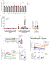
(A) Representative Incucyte-based kinetic curves of efferocytosis showing the effects of serotonin, histamine, or epinephrine treatment on the ability of unlabelled BMDM to clear pHrodo-labeled apoptotic BMDM. Engulfment of apoptotic cells was quantified as the total orange fluorescence integrated intensity per well captured by the Incucyte machine (n = 3 wells per condition in representative example; experiments repeated 3 times. (B) Representative kinetic curves for efferocytosis of pHrodo-labeled apoptotic BMDM by dopamine-treated BMDM (n= 3 wells per condition) with quantification of maximum total integrated orange intensity normalized to DMSO (n = 26 biologically independent samples differentiated from different animals, with 1–3 technical replicates per sample). (C) Quantification of efferocytosis-related transcripts measured by qPCR in BMDM treated with DMSO or different doses of dopamine (n = 5–8 biologically independent samples per condition). (D) Representative efferocytosis kinetic curves showing internalization of pHrodo-labeled apoptotic BMDM by nonlabelled BMDM stimulated with different concentrations of dopamine with or without 30 minutes of thiothixene pretreatment (n = 3 wells per condition in representative example) and quantification of maximum total integrated orange intensity normalized to DMSO (n = 26 biologically independent BMDM samples with 1–3 technical replicates per sample). Data are shown as mean ± SEM and analyzed by a Kruskal-Wallis followed by Dunn’s multiple comparisons test. *P < 0.05, ** P < 0.01, *** P < 0.005, **** P < 0.001. ns, not significant.
Fig. 3.
Fig. 3.. Thiothixene increases continual efferocytosis by inducing Arg1.
(A) qPCR of efferocytosis-related transcripts in BMDM stimulated with DMSO or thiothixene (n = 7 biologically independent BMDM samples per condition). (B) qPCR of cytokine and macrophage polarization–associated transcripts in BMDM pretreated with DMSO or thiothixene in monoculture (baseline) or in coculture with apoptotic Jurkat cells for 24 h (n = 6–22 biologically independent BMDM samples per condition ). Arg1 expression in thiothixene-pretreated BMDM cocultured with apoptotic Jurkat cells for 24 h, normalized to the DMSO-treated sample, is also shown (n = 22 biologically independent BMDM samples per condition). (C) Representative Western blotting image and quantification of Arg1 in BMDM treated with thiothixene or DMSO, with or without coculture with apoptotic Jurkat cells (AC) (n = 10 biologically independent samples; experiments repeated 3 times. ). GAPDH is a loading control. (D) Incucyte continual efferocytosis assay. BMDM were treated with thiothixene or DMSO and cocultured with unlabeled apoptotic EL4 cells (AC) for 45min or 2h. Then, pHrodo-labeled apoptotic EL4 cells were added, and orange fluorescent signals were measured by Incucyte. A representative efferocytosis kinetic curve (n= 3 wells) and quantification of maximum total integrated orange intensity normalized to DMSO (n = 14 biologically independent samples) are shown. (E) Representative kinetic curves of efferocytosis of pHrodo-labeled apoptotic EL4 cells by BMDM pretreated with 200 μM of arginase inhibitor (norNOHA), then stimulated with 2 μM of thiothixene. Quantification of the maximum total integrated orange intensity was normalized to the DMSO-treated sample (n = 37 biologically independent samples). Data are shown as mean ± SEM and analyzed by Mann-Whitney U test or a Kruskal-Wallis test followed by Dunn’s multiple comparisons test. *P < 0.05, ** P < 0.01, *** P < 0.005, **** P < 0.001. ns, not significant.
Fig. 4.
Fig. 4.. Thiothixene increases continual efferocytosis through increased expression of Stra6l and Dio2.
(A) Volcano plot of RNA-seq results from BMDM treated with thiothixene or vehicle for 24 hours (n = 3 biologically independent BMDM samples per group). Stra6l and Dio2 are highlighted. (B) Expression of Stra6l and Dio2 mRNAs as measured by qPCR in BMDM treated with thiothixene or DMSO (n = 13 biologically independent BMDM samples per group). (C) Representative kinetic curves of the efferocytosis of pHrodo-labeled apoptotic cells by BMDM transfected with siRNAs targeting Stra6l (siStra6l_1) or Dio2 (siDio2_1) and quantification of maximum total integrated orange intensity normalized to cells transfected with a scrambled control siRNA (siControl). n =18 biologically independent BMDM samples per group. Data are shown as mean ± SEM and analyzed by Mann-Whitney U test or a Kruskal-Wallis test followed by Dunn’s multiple comparisons test. ** P < 0.01, **** P < 0.001.
Fig. 5.
Fig. 5.. Vitamin A–dependent signaling mediates the proefferocytic effects of thiothixene.
(A) Representative kinetic curves of the efferocytosis of pHrodo-labeled apoptotic cells by ATRA-treated BMDM (n = 3 wells per treatment group) with quantification of maximum total integrated orange intensity normalized to DMSO (n = 14 biologically independent samples with 1–3 technical replicates per sample). (B) qPCR of transcripts related to efferocytosis and proresolving macrophages in BMDM stimulated with ATRA for 24 hours (n = 4 biological replicates per treatment group). (C) Representative kinetic curves of the efferocytosis of pHrodo-labeled apoptotic cells by retinol-treated BMDM (n = 3 wells per treatment group) with quantification of maximum total integrated orange intensity normalized to DMSO (n = 10 biologically independent samples). (D) Representative kinetic curves for efferocytosis of pHrodo-labeled apoptotic cells by thiothixene-treated BMDM following Stra6l knockdown with siStra6l_1 (n = 3 wells) and quantification of maximum total integrated orange intensity normalized to DMSO (n = 22 biologically independent samples). (E) Left, quantification of Arg1 transcripts in siStra6l_1-transfected BMDM pretreated with DMSO or thiothixene in monoculture (baseline) or in coculture with apoptotic Jurkat cells for 24h, normalized to the siControl, DMSO-treated samples (n = 10 biological replicates). Right, quantification of Arg1 transcripts in siStra6l_1-transfected, thiothixene-pretreated BMDM cocultured with apoptotic Jurkat cells for 24 h, normalized to the siStra6l_1-transfected DMSO-treated cocultured samples (n = 20 biologically independent samples). Data are shown as mean ± SEM and analyzed by Mann-Whitney U test or Kruskal-Wallis test followed by Dunn’s multiple comparisons test. *P < 0.05, ** P < 0.01, *** P < 0.005, **** P < 0.001. ns, not significant. (F) Summary model: Thiothixene increases macrophage efferocytosis by inhibiting dopaminergic signaling, which exerts a potent inhibitory effect on efferocytosis. In addition, thiothixene increases expression of the retinol-binding protein receptor Stra6, which in turn enhances vitamin A–dependent signaling and the expression of the continuous efferocytosis stimulator, Arg1. Model created with BioRender.com released under a Creative Commons Attribution-NonCommercial-NoDerivs 4.0 International license.

References

    1. Thorp EB, Mechanisms of failed apoptotic cell clearance by phagocyte subsets in cardiovascular disease. Apoptosis 15, 1124–1136 (2010); published online Epub2010/09// ( 10.1007/s10495-010-0516-6). - DOI - PMC - PubMed
    1. Green DR, Oguin TH, Martinez J, The clearance of dying cells: table for two. Cell Death Differ 23, 915–926 (2016); published online Epub2016/06// ( 10.1038/cdd.2015.172). - DOI - PMC - PubMed
    1. Kojima Y, Weissman IL, Leeper NJ, The Role of Efferocytosis in Atherosclerosis. Circulation 135, 476–489 (2017); published online EpubJan 31 ( 10.1161/CIRCULATIONAHA.116.025684). - DOI - PMC - PubMed
    1. Moon B, Yang S, Moon H, Lee J, Park D, After cell death: the molecular machinery of efferocytosis. Exp Mol Med 55, 1644–1651 (2023); published online Epub2023/08// ( 10.1038/s12276-023-01070-5). - DOI - PMC - PubMed
    1. Yurdagul A Jr., Metabolic Consequences of Efferocytosis and its Impact on Atherosclerosis. Immunometabolism 3, (2021) 10.20900/immunometab20210017). - DOI - PMC - PubMed

LinkOut - more resources