. 2025 Apr 23;147(16):13577-13591.
doi: 10.1021/jacs.5c00594. Epub 2025 Apr 8.

Membrane Charge Drives the Aggregation of TDP-43 Pathological Fragments

Affiliations

Membrane Charge Drives the Aggregation of TDP-43 Pathological Fragments

Giacomo Corucci et al. J Am Chem Soc. .

Abstract

TDP-43 protein is an RNA-binding protein linked to amyotrophic lateral sclerosis, frontotemporal dementia, and Alzheimer disease. While normally a protein that shuttles between the nucleus and cytoplasm, TDP-43 has recently been found also in extracellular vesicles. These are an important medium for cell-cell communication that allows the transfer of lipids, proteins, and genetic material among cells. An increasing concern in neurodegenerative diseases, however, is the possibility that extracellular vesicles can also provide an effective way to spread misfolded proteins that could "infect" other cells according to a "prion-like" mechanism. To characterize the interaction of TDP-43 with lipid membranes, we carried out a systematic biophysical study using a TDP-43 fragment lacking the first 84 N-terminal residues, called M85, and synthetic model phospholipid membranes. We utilized standard techniques, such as fluorescence and microscopy, complemented by neutron reflectivity measurements. Our results show that lipid charge affects the modality by which M85 interacts with membranes: a higher negative charge induces the protein to bind to the bilayer surface, promoting protein aggregation and decreasing lipid bilayer damage that this interaction causes. Thus, we speculate that the M85-lipid membrane interaction could play an important and previously undefined role in TDP-43-related neurodegenerative diseases.


Conflict of interest statement

The authors declare no competing financial interest.

Figures

Figure 1
Figure 1
Molecular structure of the synthetic lipids and model membranes used. (a) Common chains were made of palmitoyl and oleyl fatty acids (PO) account for the acyl chain composition, and they differ from the number of carbons in the acyl chain (16 and 18, respectively) and the number of double bonds (none and 1, respectively). This composition mimics the average composition of eukaryotic cells. The polar head region is different between the three lipids selected, where PC (phosphatidyl choline) is a neutral zwitterionic polar head, and PS (phosphatidyl serine) is negatively charged, as well as PA (phosphatidic acid) with a higher negative charge density due to its small volume and its negative charge of −2 (at pH 8). (b) Schematic representation of the three model membranes utilized in this work.
Figure 2
Figure 2
Purification and characterization of the M85 variant. (a) SDS-PAGE of the purification steps of the M85 variant, where lane 1 is the standard, lane 2, 3, and 4 are the sample immediately before addition of the TEV, after 3h, and after overnight incubation, respectively, and lane 5, 6, 7, and 8 are different aliquots during the reversed Ni affinity chromatography. The purified protein (M85 and its fragment) is in lane 5, and they were finally passed through an ion exchange chromatography column to remove contaminants. FT stands for flowthrough, whereas the percentages reported are the percentage of elution buffer utilized to elute that aliquot. (b) Electrospray mass spectrometry analysis of the two M85 fragments separated by using a size exclusion chromatography (SEC). (c) Schematic domain architecture of full-length TDP-43 (top) and the tagged M85 fragment (bottom) where it is highlighted the truncation point. (d) Sequence of the M85 fragment (residues 85–414). The first row contains the residues preceding RRM1 (85–104) expected to be unstructured. RRM1 (105–178) and RRM2 (191–262) are underlined in rows 2 and 4, separated by the linker between the two domains. The region 263–414 contains the unstructured gly-rich region and C-terminus. The RRM domains are defined according to the UniProt annotation. The positively and negatively charged residues are marked in blue and red, respectively. The DNA and amino acid sequences of full-length TDP-43 and of the construct used in this study are provided in the Supporting Information.
Figure 3
Figure 3
M85 aggregation assay in the presence of lipids. (a) ThT aggregation assay measurements of M85 aggregation kinetics with the presence of different composition LUVs (ThT intensity shown is subtracted by the buffer constituted of only LUVs samples), analyzed using a simple sigmoidal equation (provided in the Supporting Information) to obtain the t50 (displayed in Figure S5b). (b) Final ThT fluorescence intensity of each sample as well as a dot-blot of the supernatant after the aggregation, where duplicates are shown for each sample. (c) Plot of dot-blot intensity versus ThT final intensity. (d, e) Negative stain transmission electron microscopy of POPC:POPS ratio 8:2 (mol/mol) LUVs incubated in the absence (panel d) or in the presence (panel e) of M85 for 4 days. A TEM picture of M85 only is shown in the Supporting Information.
Figure 4
Figure 4
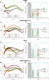
Fitting and analyses of neutron reflectivity data before (green colors) and after (red colors) the M85 addition under different SLB compositions. (a, b) POPC SLB, highlighted with an arrow is a broad Bragg peak most probably due to diluted multilamellar formations caused by issues in the vesicles fusion process, (c, d) POPC:POPS 8:2, (e, f) Dpol and (g, h) POPC:POPA 8:2 SLB. For the panels on the left-hand side (a, c, e, and g) in green color and in red color are data before and after ca. 20 h from the M85 injection, respectively. The solid lines represent the minimized theoretical curves. On the right-hand side (b, d, f, and h), the minimized models, where the dashed lines and solid lines represent the model before and after M85 injection, respectively. M85 protein is represented in red color.
Figure 5
Figure 5
Kinetics of interaction. (a) Trend of the neutron reflectivity-integrated kinetics measurements for the samples POPC, POPC:POPS 8:2, and POPC:POPA 8:2, highlighted with the arrows is the data set after the washing of the cell with buffer (analyzed and discussed in Figure 4). (b) Kinetics reflectivity profiles and analysis of the sample containing M85 and POPC:POPA 8:2 measured in H-buffer. (c) Minimized model from the analysis of the data in panel (b), where the fraction volume distribution of the deposition layer is shown with dashed lines. The “t0” is the data set collected just after the M85 injection into the cell (∼4 min from the injection). (d) Plot of the fraction volume of the deposition layer versus time.

References

    1. Prasad A.; Bharathi V.; Sivalingam V.; Girdhar A.; Patel B. K. Molecular Mechanisms of TDP-43 Misfolding and Pathology in Amyotrophic Lateral Sclerosis. Frontiers in Molecular Neuroscience 2019, 12, 25.10.3389/fnmol.2019.00025. - DOI - PMC - PubMed
    1. Neumann M.; Sampathu D. M.; Kwong L. K.; Truax A. C.; Micsenyi M. C.; Chou T. T.; Bruce J.; Schuck T.; Grossman M.; Clark C. M.; McCluskey L. F.; Miller B. L.; Masliah E.; Mackenzie I. R.; Feldman H.; Feiden W.; Kretzschmar H. A.; Trojanowski J. Q.; Lee V. M. Ubiquitinated TDP-43 in frontotemporal lobar degeneration and amyotrophic lateral sclerosis. Science 2006, 314 (5796), 130–133. 10.1126/science.1134108. - DOI - PubMed
    1. Cohen T. J.; Lee V. M.; Trojanowski J. Q. TDP-43 functions and pathogenic mechanisms implicated in TDP-43 proteinopathies. Trends Mol. Med. 2011, 17 (11), 659–67. 10.1016/j.molmed.2011.06.004. - DOI - PMC - PubMed
    1. Arai T.; Hasegawa M.; Akiyama H.; Ikeda K.; Nonaka T.; Mori H.; Mann D.; Tsuchiya K.; Yoshida M.; Hashizume Y.; Oda T. TDP-43 is a component of ubiquitin-positive tau-negative inclusions in frontotemporal lobar degeneration and amyotrophic lateral sclerosis. Biochem. Biophys. Res. Commun. 2006, 351 (3), 602–11. 10.1016/j.bbrc.2006.10.093. - DOI - PubMed
    1. Igaz L. M.; Kwong L. K.; Lee E. B.; Chen-Plotkin A.; Swanson E.; Unger T.; Malunda J.; Xu Y.; Winton M. J.; Trojanowski J. Q.; Lee V. M. Y. Dysregulation of the ALS-Associated Gene TDP-43 Leads to Neuronal Death and Degeneration in Mice. J. Clin. Invest. 2011, 121 (2), 726–738. 10.1172/JCI44867. - DOI - PMC - PubMed

LinkOut - more resources