

Comparison of TLR4, NF-κB and IRF3 expression in kidney tissue between lupus nephritis (LN) and systemic lupus erythematosus (SLE): a pristane-induced lupus mice model study
- PMID: 40199549
- PMCID: PMC11977469
- DOI: 10.1136/lupus-2024-001445
Comparison of TLR4, NF-κB and IRF3 expression in kidney tissue between lupus nephritis (LN) and systemic lupus erythematosus (SLE): a pristane-induced lupus mice model study
Abstract
Introduction and purpose: Lupus nephritis (LN) is a major cause of morbidity and mortality in patients with SLE, a complex autoimmune disease characterised by loss of tolerance to self-nuclear antigens. Toll-like receptor 4 (TLR4), the first line of defence in the innate immune system, has been linked to the pathogenesis of autoimmune diseases and LN by activating nuclear factor-κB (NF-κB) or interferon regulatory transcription factor 3 (IRF3). Local expression of those biomarkers in pristane-induced lupus mice is still unknown. Therefore, this study aimed to prove the role of TLR4, NF-κB and IRF3 in pristane-induced lupus mice.
Subjects and methods: The study subjects were female Balb/c pristane-induced lupus mice model, 8-12 weeks of age, n=30, divided into two groups, nephritis (LN group) and non-nephritis (SLE group). The control group were age-matched healthy female Balb/c mice, n=11. All mice were euthanised at weeks 16. Kidney tissue was taken for histopathology examination and TLR4, NF-κB, IRF3 immunofluorescence assay. The diagnosis of LN was based on proteinuria and histopathology examination according to the ISN/RPS 2004 classification of LN. Statistical analysis was performed using IBM SPSS Statistics V.25. P value <0.05 was considered statistically significant.
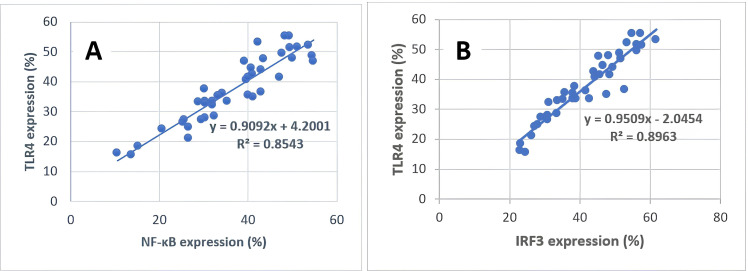
Results: There were significant differences in the expressions of TLR4, NF-κB and IRF3 among the LN, SLE and healthy control groups (p=0.000), with the highest expression found in the LN group for all markers. The linear regression between TLR4 and NF-κB resulted in p value=0.000; R2=0.817; β=0.904. Linear regression between TLR4 and IRF3 showed p value=0.000; R2=0.896; β=0.947, which means TLR4 had an 81.7% effect on NF-κB and 89.6% on IRF3 expression.
Conclusion: TLR4, NF-κB and IRF3 expression were increased in lupus, with the highest expression found in the LN group, suggesting that these biomarkers may be responsible for the development of nephritis in SLE, with TLR4 likely playing a dominant role in this pathway. Increased expression of these biomarkers in lupus without nephritis may indicate progression towards nephritis, which still needs to be proven with further research.
Keywords: autoimmune diseases; inflammation; lupus erythematosus, systemic; lupus nephritis.
© Author(s) (or their employer(s)) 2025. Re-use permitted under CC BY-NC. No commercial re-use. See rights and permissions. Published by BMJ Group.
Conflict of interest statement
Competing interests: None declared.
Figures


References
-
- Wallace DJ, Hahn BH. Dubois’ lupus erythematosus and related syndromes. 9th. China: Elsevier Inc; 2019. edn.
-
- Sutrisno RN, Rahmadi AR, Novita N, et al. Most Frequent Musculoskeletal Manifestation of Systemic Lupus Erythematosus Patients in Dr. Hasan Sadikin General Hospital Bandung. Ina J Rheum . 2019;9:13–7. doi: 10.37275/ijr.v9i2.71. - DOI
Publication types
MeSH terms
Substances
LinkOut - more resources
Full Text Sources
Medical