. 2025 Apr 8;13(4):e011066.
doi: 10.1136/jitc-2024-011066.

CD40L stimulates tumor-infiltrating B-cells and improves ex vivo TIL expansion

Affiliations

CD40L stimulates tumor-infiltrating B-cells and improves ex vivo TIL expansion

Renata Ariza Marques Rossetti et al. J Immunother Cancer. .

Abstract

Background: Adoptive transfer of tumor-infiltrating lymphocytes (TIL) is now a Food and Drug Administration (FDA)-approved treatment for melanoma. While this is a major milestone, there is room for improvement to increase clinical response rates and to further optimize the manufacturing of TIL products. In this study, we characterized the association of tumor-infiltrating B-cells (TIL-B) and tertiary lymphoid structures (TLSs) with clinical response to TIL therapy and tested whether the presence of B-cells in the tumor can be leveraged to optimize TIL manufacture.

Methods: Tumor sections from TIL responders (R, n=9) and non-responders (NR, n=11) were analyzed by RNA sequencing, and immune cell content was estimated in silico. To study the association between B-cells and TIL expansion, we quantified B-cell subsets and TIL phenotype by flow cytometry. CD40L-induced effects on melanoma-infiltrating B-cells were analyzed by flow cytometry and scRNA-sequencing.

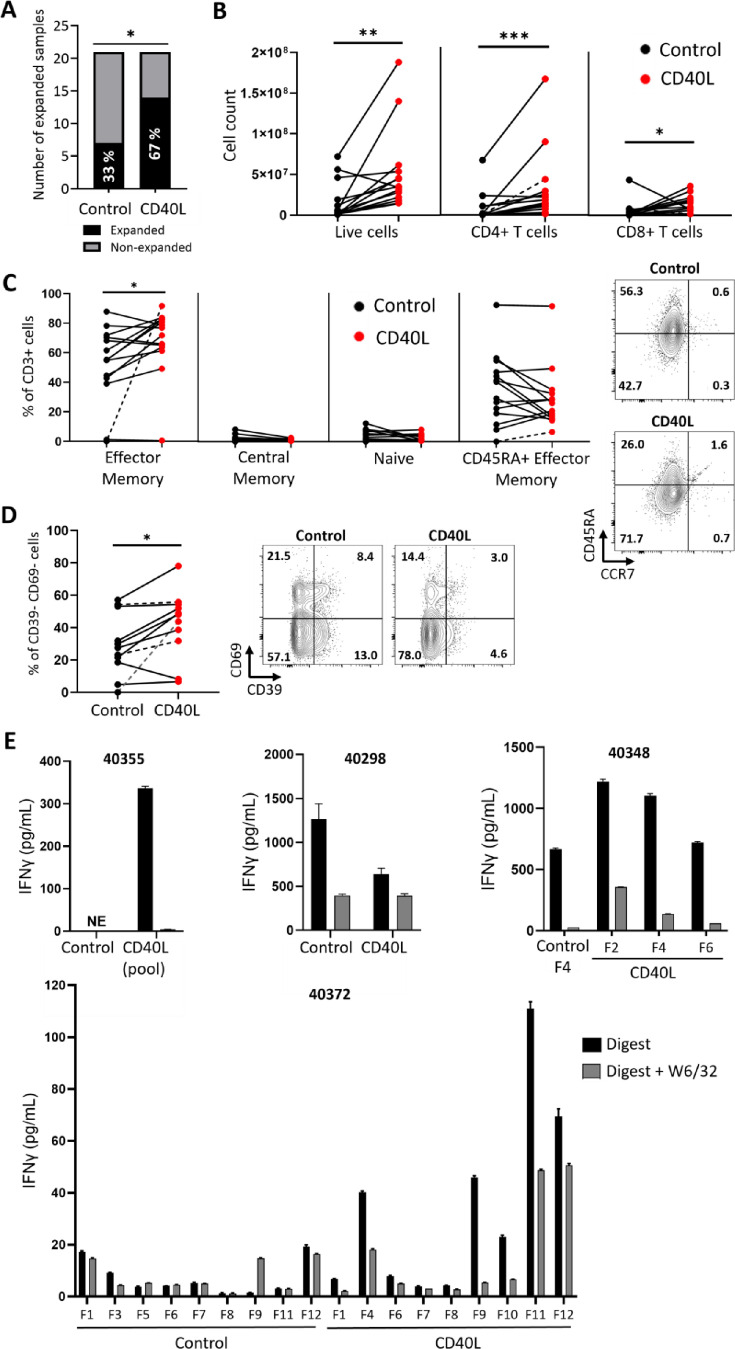
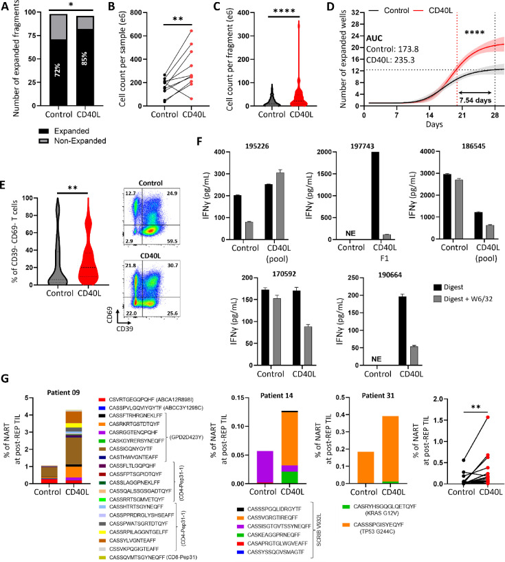
Results: Tumors from TIL clinical responders had greater abundance of class-switched B-cells (p=0.007) and a greater TLS score (p=0.03) than those of NRs. In addition, greater abundance of B-cells (p≤0.05) and switched memory B-cells (CD27+ IgD-, p≤0.05) in the tumors were associated with greater TIL expansion. Stimulation of TIL-B through addition of CD40L during TIL ex vivo culture improved their expansion success rate from 33% to 67% (p=0.03). Similarly, the addition of CD40L to non-small cell lung cancer (NSCLC) TIL cultures shortened the manufacturing period by 1 week. Moreover, CD40L-enhanced TIL showed more stem-like T-cells (CD39- CD69-, p≤0.05) and an enrichment of neoantigen-reactive T-cell clones in NSCLC TIL. Gene expression analysis showed that CD40L induced gene expression changes in TIL-B after 48 hours in culture (126 differentially expressed genes (DEGs)), with minimal to no changes observed in other immune cell types (including 12 DEG in macrophages, 10 DEG in dendritic cells, and none in monocytes). B-cell DEGs included upregulated co-stimulatory ligands (CD83, CD58), chemokines (CCL22, CCL17), among others. CD40L-induced upregulation of CD58 by melanoma infiltrating B-cells was associated with successful TIL expansion.

Conclusions: Our results show that CD40L-stimulated B-cells can be leveraged to enhance the quality and quantity of TIL. Clinical trial NCT05681780 is currently testing this concept applied to NSCLC TIL.

Keywords: Adoptive cell therapy - ACT; B cell; Melanoma; Non-Small Cell Lung Cancer; Tumor infiltrating lymphocyte - TIL.


Conflict of interest statement

Competing interests: RAMR, MB, SP-T and DA-D are inventors or coinventors in patent applications filed by H. Lee Moffitt Cancer Center and Research Center, related to technology described in the manuscript. Moffitt Cancer Center has licensed Intellectual Property (IP) related to the proliferation and expansion of tumor-infiltrating lymphocytes (TILs) to Iovance Biotherapeutics. SP-T and AS are coinventors on such Intellectual Property. AS and SP-T are coinventors on a patent application with Provectus Biopharmaceuticals. AS and SP-T participate in a sponsored research agreement with Turnstone Biologics. AS has received Ad hoc consulting fees from Iovance Biotherapeutics, Gerson Lehrman Group, Second City Science, Guidepoint and Blueprint Oncology Concepts. AS has received speaker fees from Clinical Education Alliance, MJN Holdings, International workshop CAR-T and Society for the ImmunoTherapy of Cancer. Moffitt has also licensed IP to Tuhura Biopharma. SP-T is an inventor of such Intellectual Property. SP-T participates in sponsored research agreements with Provectus Biopharmaceuticals, Intellia Therapeutics, Dyve Biosciences, Turnstone Biologics, and Iovance Biotherapeutics that are not related to this research. SP-T has received consulting fees from Seagen, Morphogenesis and KSQ Therapeutics. JJM has ownership interest in Aleta Biotherapeutics, CG Oncology, Turnstone Biologics, Ankyra Therapeutics, and AffyImmune Therapeutics, and is a paid consultant/paid scientific and/or clinical advisory board member for Turnstone Biologics, Vault Pharma, Ankyra Therapeutics, AffyImmune Therapeutics, UbiVac, Vycellix, and Aleta Biotherapeutics, as well as a Board of Directors member of CG Oncology. BP has received Ad hoc consulting fees from AstraZeneca, Bristol Myers Squibb, Daiichi Sankyo, G1 Therapeutics and Novocure. BP has received speaker fees from OncLive. BC has received Ad hoc consulting fees from Aptitude Health, AstraZeneca, Anchilles Therapeutics, Regeneron, Iovance Biotherapeutics, Johnson & Johnson, AbbVie, Boehringer, Bayer and G1 Therapeutics. BC has received speaker fees from AstraZeneca, Regeneron, Iovance Biotherapeutics, Johnson & Johnson, AbbVie, Boehringer, Bayer, G1 Therapeutics, American Association for Cancer Research, American Society of Clinical Oncology, European Society for Medical Oncology, Foundation of Pulmonary Cancer of Gran Canarias. BC participates in the Advisory Board of Achilles, and in the data safety monitoring board of Iovance Lung Steering Committee.

Figures

Figure 1
Figure 1. Immune cell content analysis in responders (R) and non-responders (NR) to melanoma TIL therapy. (A, B) Estimation of cell abundance in metastatic melanomas resected for TIL manufacture. Patients grouped based on clinical response status by RECIST criteria (total n=20; R=9 and NR=11) following TIL therapy. Boxplots are shown along with outliers’ individual data points. Gene expression was quantified by bulk RNA sequencing of FFPE sections. Cell content was estimated using the xCell deconvolution algorithm. Statistical analyses were performed by Wilcoxon signed-rank test. (C) The tertiary lymphoid structure (TLS) score was calculated based on expression of 12-chemokine genes combined, using principal component analysis for tumors resected. Boxplots are shown along with outliers’ individual data points. Statistical analyses were performed by Mann-Whitney U test. FFPE, formalin-fixed paraffin-embedded; TIL, tumor-infiltrating lymphocyte.
Figure 2
Figure 2. CD40L enhances the expansion of melanoma TIL. Melanoma single-cell suspensions were cultured in standard TIL expansion media (control) or media supplemented with an agonist of CD40 (CD40L) for 3–4 weeks. (A) TIL expansion success rate (n=21 patients) using control or CD40L-supplemented media. *p≤0.05 (χ2). (B) Cell counts of total viable cells, CD4+ and CD8+ T cells after 4 weeks of expansion. Individual values are plotted with lines connecting the paired samples, corresponding to individual patients. The dashed line represents one patient with no expansion in the control condition. This patient was not considered for the statistical analysis (n=14). *p≤0.05, **p≤0.01, ***p≤0.001 (Wilcoxon signed-rank test). (C) Differentiation phenotype analyzed by flow cytometry in pre-REP TIL (Effector Memory: CCR7 CD45RA; Central Memory: CCR7+ CD45RA; Naive: CCR7+ CD45RA+; CD45+Effector Memory: CCR7 CD45RA+). Individual values are plotted with lines connecting the paired samples, corresponding to individual patients. The dashed line represents one patient with no expansion in the control condition. This patient was not considered for the statistical analyses (n=14). *p≤0.05 (Wilcoxon signed-rank test). Scatter plot of a representative example is shown in the right panel. (D) Expression of CD39 and CD69 in TIL (gated on lymphoid, single, live, CD3+ cells) after 4 weeks of ex vivo expansion. Individual values are plotted with lines connecting the paired samples. The dashed lines represent patients with no expansion in the Control condition. For two of the patients a control sample expanded from fragments was used for comparison (black line). In the third patient, a value of zero was attributed to the control condition with no expansion (gray line). Those patients were not considered for the statistical analyses (n=11). *p≤0.05 (Wilcoxon signed-rank test). One outlier removed based on Grubb’s test. (E) Analysis of reactivity of melanoma TIL. Expanded T-cells were cultured with autologous tumor single-cell suspensions for 24 hours, at an E:T ratio of 1:1. Tumor reactivity was assessed by IFN-γ levels in the supernatant. Each graph represents an individual patient (n=4). TILs were expanded from tumor fragments (40355, 40348 and 40372) or tumor digest (40298). ‘Pool’ indicates TIL co-cultures generated from more than one fragment pooled together. ‘F’ indicates data from a single fragment. The black bar represents TIL co-cultured with autologous tumor cells (Digest). The gray bar represents TIL co-cultured with autologous tumor cells (Digest) preincubated with anti-HLA class I blocking antibody (W6/32). Reactivity was defined by secretion of >50 pg/mL of IFN-γ, inhibited by at least 20% when the HLA-I antibody was used. TILs, tumor-infiltrating lymphocytes.
Figure 3
Figure 3. Melanoma infiltrating B-cells upregulate costimulatory ligands in response to CD40L. (A–D) Percentage of CD45+cells (A), T-cells (CD45+CD3+CD19) (B), B-cells (CD45+CD3CD19+) (C), and non-B/non-T cells (CD45+CD3CD19) (D) present in melanoma single-cell suspensions, grouped based on their ability to yield TIL expansion. Individual values corresponding to independent patients are plotted with mean±SEM (n=20). *p≤0.05 (Mann-Whitney U test). (E) Relative abundance of different B-cell subsets (Switched memory: CD27+ IgD; Unswitched memory: CD27+ IgD+; Naive: CD27 IgD+; Double-Negative: CD27 IgD) present in melanoma tumor digests based on their ability to yield TIL expansion. Individual values are plotted with mean±SEM (n=24). *p≤0.05, ***p≤0.001 (Mann-Whitney U test). (F) Flow cytometry analysis of CD40 expression in myeloid cells (CD45+CD3CD19HLA-DR+) and B-cells (CD45+CD3CD19+) present in melanoma tumor digests. The individual values are plotted with lines connecting the paired samples corresponding to the same patient (n=11). ***p≤0.001 (paired Wilcoxon signed-rank test). A representative histogram of CD40 expression is also shown. (G, H) Flow cytometry analysis of HLA-DR, DQ, DP expression and percentage of CD80/CD86 double positive cells within myeloid cells (CD45+CD3CD19HLA-DR+) and B-cells (CD45+CD3CD19+) present in melanoma tumor digests. Tumor digests were cultured for 48 hours in standard TIL expansion media (control) or supplemented with a CD40 agonist (CD40L). Individual values corresponding to independent patients are plotted with mean±SEM (n=10). ***p≤0.001, ****p≤0.0001 (two-way ANOVA and Tukey’s multiple comparison test). ANOVA, analysis of variance.
Figure 4
Figure 4. Immune cell composition of melanoma tumors and response to CD40L. Melanoma digests were cultured in standard TIL expansion media (control) or supplemented with an agonist of CD40 (CD40L) for 48 hours (n=14 samples, 7 patients) and analyzed by single-cell RNA sequencing. (A) UMAP projection of different cell populations identified in the tumor digests. (B) UMAP projection of the tumor digests based on culture conditions (CD40L vs control). (C) UMAP projection of B-cells, showing expression of IgA, IgD, IgE, IgG1, IgG2, IgG3, IgG4 and IgM antibody/BCR isotypes. (D) UMAP projection of B-cells showing subclusters identified by the Seurat algorithm. Clusters corresponding to CD40L-treated samples are indicated with the red square. (E) Cross-referencing of B-cell clusters with previously described gene sets corresponding to naive, memory, germinal center (GC) and plasma cells. TIL, tumor-infiltrating lymphocyte.
Figure 5
Figure 5. CD40L induces gene expression changes in melanoma infiltrating B-cells. Melanoma digests were cultured in standard TIL (control) or CD40L-supplemented media for 48 hours (n=14 samples, 7 patients). (A) Volcano plot displaying differentially expressed genes in CD40L-treated versus control B-cells assessed by scRNA-seq (Log2FC=0.95; p≤0.05). Upregulated genes are shown in red. Downregulated genes are shown in blue. (B–D) UMAP projections of the B-cell compartment, showing expression of differentially expressed genes CD86, CCL22, and FcRL4. (E) Flow cytometry analysis of CCL22 expression in melanoma infiltrating B-cells. Individual values are plotted with lines connecting the paired samples corresponding to individual patients (n=22). ****p≤0.0001 (Wilcoxon signed-rank test). Flow cytometry scatter plot of a representative example shown in the right panel. (F) Flow cytometry analysis of FcRL4 expression in melanoma infiltrating B-cells. Individual values are plotted with lines connecting the paired samples (n=22). ***p≤0.001 (paired Wilcoxon signed-rank test). Representative example shown in the right panel. (G) UMAP projection of B-cells showing expression of CD58. (H) Flow cytometry analysis of CD58 expression in B-cells cultured in standard (control) or CD40L-supplemented media for 48 hours. Individual values are plotted with lines connecting the paired samples corresponding to individual patients (n=22). ****p≤0.0001 (paired Wilcoxon signed-rank test). (I) Percentage of CD58 positive B-cells in samples grouped based on their ability to yield TIL expansion (successful expansion, n=13; unsuccessful expansion, n=9). ns=not statistically significant, ***p≤0.01 (paired Wilcoxon signed-rank test). TIL, tumor-infiltrating lymphocyte.
Figure 6
Figure 6. CD40L enhances the expansion of NSCLC TIL and increases the frequency of neoantigen reactive T-cells. (A–E) Lung tumor fragments were cultured in standard TIL expansion media (control) or supplemented with an agonist of CD40 (CD40L) for 3–4 weeks. (A) TIL expansion success rate of individual tumor fragments, derived from 12 independent patients. *p≤0.05 (χ2). (B) Cell counts of total viable cells after 4 weeks of ex vivo expansion. Individual values are plotted with lines connecting the paired samples, corresponding to individual patients (n=12). **p<=0.01 (Mann-Whitney U test). (C) Cell counts of total viable cells per fragment (control: n=87; CD40L: n=92). ****p≤0.0001 (Mann-Whitney U test). (D) Number of TIL cultures arising from individual tumor fragments, over time (control: n=87; CD40L: n=92). ****p≤0.0001 (non-linear fit test). (E) Violin plots representing the percentage of CD39CD69 T-cells after 4 weeks of ex vivo expansion, as determined by flow cytometry (control: n=87; CD40L: n=92). **p≤0.01 (paired t-test). (F) Analysis of TIL reactivity against autologous tumor. NSCLC TILs were cultured with autologous tumor single-cell suspensions for 24 hours at an E:T ratio of 1:1. Reactivity was assessed by IFN-γ levels in the supernatant, quantified using the ELLA platform. Each graph represents an individual patient (n=5). TILs were expanded from fragments (samples 195226, 197743 and 186545) or tumor digest (170592 and 190664). ‘Pool’ indicates TIL cultures generated from more than one fragment pooled together. ‘F’ denotes TIL cultures derived from an individual fragment. Black bars represent TIL co-cultured with autologous tumor cells (Digest). Gray bar represents TIL co-cultured with autologous tumor cells (Digest) preincubated with anti-HLA class I blocking antibody (W6/32). (G) Lung tumor digests were cultured in standard TIL expansion media (control) or supplemented with an agonist of CD40 (CD40L) for 3–4 weeks, followed by a Rapid Expansion Protocol (REP) culture. The percentage of antigen-specific T-cell receptor clonotypes within post-REP TIL are represented for three patient samples. The neoantigen related to each clonotype is also described in the figure. NSCLC, non-small cell lung cancer; TIL, tumor-infiltrating lymphocyte.

References

    1. Julve M, Lythgoe MP, Larkin J, et al. Lifileucel: the first cellular therapy approved for solid tumours. Trends Cancer. 2024;10:475–7. doi: 10.1016/j.trecan.2024.04.003. - DOI - PubMed
    1. Rosenberg SA, Yang JC, Sherry RM, et al. Durable complete responses in heavily pretreated patients with metastatic melanoma using T-cell transfer immunotherapy. Clin Cancer Res. 2011;17:4550–7. doi: 10.1158/1078-0432.CCR-11-0116. - DOI - PMC - PubMed
    1. Lee S, Margolin K. Tumor-infiltrating lymphocytes in melanoma. Curr Oncol Rep. 2012;14:468–74. doi: 10.1007/s11912-012-0257-5. - DOI - PMC - PubMed
    1. Pilon-Thomas S, Kuhn L, Ellwanger S, et al. Efficacy of adoptive cell transfer of tumor-infiltrating lymphocytes after lymphopenia induction for metastatic melanoma. J Immunother. 2012;35:615–20. doi: 10.1097/CJI.0b013e31826e8f5f. - DOI - PMC - PubMed
    1. Chesney J, Lewis KD, Kluger H, et al. Efficacy and safety of lifileucel, a one-time autologous tumor-infiltrating lymphocyte (TIL) cell therapy, in patients with advanced melanoma after progression on immune checkpoint inhibitors and targeted therapies: pooled analysis of consecutive cohorts of the C-144-01 study. J Immunother Cancer. 2022;10:e005755. doi: 10.1136/jitc-2022-005755. - DOI - PMC - PubMed

LinkOut - more resources