. 2025 Jun;47(3):4187-4204.
doi: 10.1007/s11357-025-01653-2. Epub 2025 Apr 8.

Endothelial IGF- 1R deficiency disrupts microvascular homeostasis, impairing skeletal muscle perfusion and endurance: implications for age-related sarcopenia

Affiliations

Endothelial IGF- 1R deficiency disrupts microvascular homeostasis, impairing skeletal muscle perfusion and endurance: implications for age-related sarcopenia

Adam Nyul-Toth et al. Geroscience. 2025 Jun.

Abstract

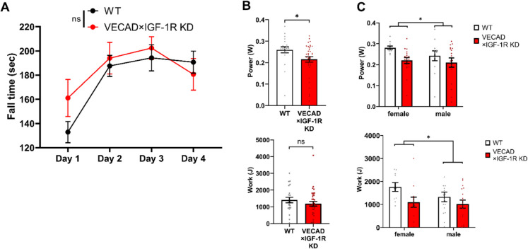
Aging is associated with a progressive decline in circulating insulin-like growth factor- 1 (IGF- 1) levels in humans, which has been implicated in the pathogenesis of sarcopenia. IGF- 1 is an anabolic hormone that plays a dual role in maintaining skeletal muscle health, acting both directly on muscle fibers to promote growth and indirectly by supporting the vascular network that sustains muscle perfusion. However, the microvascular consequences of IGF- 1 deficiency in aging muscle remain poorly understood. To elucidate how impaired IGF- 1 input affects skeletal muscle vasculature, we examined the effects of endothelial-specific IGF- 1 receptor (IGF- 1R) deficiency using a mouse model of endothelial IGF- 1R knockdown (VE-Cadherin-CreERT2/Igf1rf/f mice). These mice exhibited significantly reduced skeletal muscle endurance and attenuated hyperemic response to acetylcholine, an endothelium-dependent vasodilator. Additionally, they displayed microvascular rarefaction and impaired nitric oxide-dependent vasorelaxation, indicating a significant decline in microvascular health in skeletal muscle. These findings suggest that endothelial IGF- 1R signaling is critical for maintaining microvascular integrity, muscle perfusion, and function. Impaired IGF- 1 input to the microvascular endothelium may contribute to reduced muscle blood flow and exacerbate age-related sarcopenia. Enhancing vascular health by modulating IGF- 1 signaling could represent a potential therapeutic strategy to counteract age-related muscle decline.

Keywords: Aging; Claudication; Endothelial dysfunction; IGF- 1; IGF- 1R; Insulin-like growth factor- 1; Microvasculature; Sarcopenia; Skeletal muscle; Vascular function.


Conflict of interest statement

Declarations. Ethics approval and consent to participate: N/A. Consent for publication: N/A. Competing interest: Dr. Adam Nyul-Toth, Dr. Roland Patai, Dr. Tamas Csipo, Dr. Anna Csiszar, Dr. Holly Van Remmen, Dr. Andriy Yabluchanskiy, and Dr. Stefano Tarantini serve as Associate Editors for GeroScience. Dr. Zoltan Ungvari serves as Editor-in-Chief for GeroScience and has personal relationships with individuals involved in the submission of this paper.

Figures

Fig. 1
Fig. 1
Breeding strategy and experimental methods for assessing the role of endothelial IGF- 1R in skeletal muscle function. Schematic representation of the breeding strategy used to generate endothelial-specific IGF- 1R knockout (VE-Cadherin-CreERT2/Igf1rf/f) mice. VE-Cadherin-CreERT2 mice were crossed with Igf1rf/f mice to obtain experimental cohorts for functional analysis. The inset (left) illustrates vascular endothelial IGF- 1R signaling, highlighting IGF- 1 and IGF- 1R localization in endothelial cells. Shown is also the experimental workflow for assessing the impact of endothelial IGF- 1R deficiency on skeletal muscle function. Motor coordination and fatigue resistance were evaluated using the rotarod performance test and treadmill graded exercise test. Skeletal muscle blood flow was assessed via hind limb laser speckle imaging, while quadriceps vascularization and muscle fiber structure were analyzed by immunofluorescent staining and confocal microscopy. Endothelial function was further investigated by measuring aortic vasorelaxation in isolated vessel preparations. In vitro skeletal muscle strength measurements were also performed
Fig. 2
Fig. 2
Endothelial IGF- 1R deficiency impairs muscle endurance and fatigue resistance without affecting motor coordination. A Motor coordination and balance were assessed using a rotarod test over four consecutive days. No significant differences in fall time were observed between WT (black) and VECAD × IGF- 1R KD (red) mice, indicating that endothelial IGF- 1R deficiency does not impair motor coordination or balance (n = 22 WT, n = 35 KD; two-way ANOVA). B Treadmill fatigue resistance test revealed a significant reduction in muscle power in VECAD × IGF- 1R KD mice compared to WT controls (p < 0.05, upper panel). A trend toward reduced total work performed was observed in KD mice but did not reach statistical significance (p = ns, lower panel; unpaired Student’s t-test). C Sex-specific analysis of treadmill performance. Two-way ANOVA revealed a significant effect of IGF- 1R KD on muscle power (p < 0.05), with no significant sex effect (upper panel). However, total work performed was significantly reduced in female KD mice compared to their WT counterparts (p < 0.05, lower panel), suggesting a greater susceptibility of female mice to IGF- 1R deficiency-induced fatigue resistance impairment. Data are presented as mean ± SEM. p < 0.05 was considered statistically significant
Fig. 3
Fig. 3
Endothelial-specific IGF- 1R knockdown impairs skeletal muscle strength without causing significant muscle atrophy. A Average body weight of WT (white) and VECAD × IGF- 1R KD (red) mice. No significant differences were observed between groups, indicating that endothelial IGF- 1R deficiency does not induce overt muscular atrophy (WT: n = 5, KD: n = 9; unpaired two-tailed t-test). B Grip strength test results show a significant reduction in the maximal forelimb strength of VECAD × IGF- 1R KD mice compared to WT controls (p < 0.05), suggesting that endothelial IGF- 1R signaling plays a role in maintaining muscle force generation (WT: n = 16, KD: n = 15; unpaired two-tailed t-test). Data are presented as mean ± SEM. p < 0.05 was considered statistically significant
Fig. 4
Fig. 4
Endothelial IGF- 1R knockdown does not affect intrinsic muscle contractile strength. A, B Ex vivo muscle contractility assessment in isolated extensor digitorum longus muscles from WT (white) and VECAD × IGF- 1R KD (red) mice. A Maximum tetanic force and B specific force were measured using a muscle contractility assay to evaluate intrinsic muscle strength. No significant differences were observed between groups, indicating that endothelial IGF- 1R deficiency does not impair the intrinsic contractile properties of the skeletal muscle. These findings suggest that the observed reductions in endurance and muscle function in IGF- 1R KD mice are primarily due to vascular impairments rather than direct muscle weakness. Data are presented as mean ± SD. Statistical significance was set at p < 0.05 (unpaired two-tailed t-test)
Fig. 5
Fig. 5
Endothelial IGF- 1R knockdown impairs acetylcholine-induced hyperemia in skeletal muscle. A Representative laser speckle contrast images of the gastrocnemius muscle before (baseline) and after acetylcholine (ACh) superfusion (10⁻5 M) in WT (top) and VECAD × IGF- 1R KD (bottom) mice. WT mice exhibited a strong vasodilatory response, whereas KD mice displayed a blunted hyperemic response, indicating impaired endothelial function. B Time-course of perfusion changes in response to ACh superfusion in WT (black) and KD (red) mice. VECAD × IGF- 1R KD mice showed attenuated blood flow increases compared to WT controls. C Bar graph quantifying the total vasodilatory response (area under the curve, AUC). VECAD × IGF- 1R KD mice exhibited a significantly lower AUC, suggesting an inadequate vascular response to ACh stimulation (p < 0.001). Data are presented as mean ± SEM. ***p < 0.001 (unpaired two-tailed t-test)
Fig. 6
Fig. 6
Endothelial IGF- 1R knockdown impairs endothelium-dependent vasorelaxation. A–C Vasorelaxation responses to acetylcholine (A), ATP (B), and sodium nitroprusside (SNP, C) were measured in isolated aortic ring preparations from WT (black) and VECAD × IGF- 1R KD (red) mice following phenylephrine precontraction (10⁻⁶ M). A, B ACh- and ATP-induced vasodilation were significantly reduced in VECAD × IGF- 1R KD mice compared to WT controls (p < 0.05), indicating endothelial dysfunction. C SNP-induced vasodilation was not significantly different between groups, suggesting that vascular smooth muscle responsiveness to NO remained intact. Data are presented as mean ± SD (WT: n = 4, KD: n = 4). p < 0.05 was considered statistically significant (paired t-test)
Fig. 7
Fig. 7
Endothelial IGF- 1R knockdown induces microvascular rarefaction in mouse quadriceps. A Representative immunofluorescence images of quadriceps muscle sections from WT (top) and VECAD × IGF- 1R KD (bottom) mice. Endothelial cells were labeled with endomucin (green), muscle fibers with laminin (red), and nuclei with DAPI (blue). VECAD × IGF- 1R KD mice exhibited a striking reduction in endomucin-positive vessels, indicating microvascular rarefaction. B Quantification of vascular and muscle fiber density in quadriceps muscle of WT and KD mice (n = 12 per group). Vascular density was significantly reduced in KD mice, whereas muscle fiber density remained unchanged. C Vessel-to-fiber ratio in quadriceps muscle. KD mice showed a robust decrease in vascularization relative to muscle fiber content, suggesting capillary rarefaction independent of muscle loss (p < 0.001). Data are presented as mean ± SEM. p < 0.05 was considered statistically significant
Fig. 8
Fig. 8
Schematic representation of how aging-related IGF- 1 deficiency contributes to sarcopenia, muscle weakness, and fatigue through direct muscle-intrinsic effects and vascular dysfunction. The direct pathway (left) illustrates that reduced IGF- 1 signaling in skeletal muscle leads to atrophy, which contributes to sarcopenia. The vascular pathway (right) demonstrates that impaired IGF- 1 input to endothelial cells promotes microvascular rarefaction and endothelial dysfunction, leading to impaired skeletal muscle blood flow, exacerbating muscle weakness and fatigue. The endothelial IGF- 1R knockout model (VECAD-IGF- 1R KO) mimics key aspects of age-related vascular decline. Figure was created with https://BioRender.com

References

    1. Wiedmer P, Jung T, Castro JP, Pomatto LCD, Sun PY, Davies KJA, Grune T. Sarcopenia - molecular mechanisms and open questions. Ageing Res Rev. 2021;65: 101200. 10.1016/j.arr.2020.101200. - PubMed
    1. Sayer AA, Cooper R, Arai H, Cawthon PM, Ntsama Essomba MJ, Fielding RA, Grounds MD, Witham MD, Cruz-Jentoft AJ. Sarcopenia Nat Rev Dis Primers. 2024;10:68. 10.1038/s41572-024-00550-w. - PubMed
    1. Damluji AA, Alfaraidhy M, AlHajri N, Rohant NN, Kumar M, Al Malouf C, Bahrainy S, Ji Kwak M, Batchelor WB, Forman DE, Rich MW, Kirkpatrick J, Krishnaswami A, Alexander KP, Gerstenblith G, Cawthon P, deFilippi CR, Goyal P. Sarcopenia and cardiovascular diseases. Circulation. 2023;147:1534–53. 10.1161/CIRCULATIONAHA.123.064071. - PMC - PubMed
    1. He N, Zhang Y, Zhang L, Zhang S, Ye H. Relationship between sarcopenia and cardiovascular diseases in the elderly: an overview. Front Cardiovasc Med. 2021;8: 743710. 10.3389/fcvm.2021.743710. - PMC - PubMed
    1. Rivera FB, Escolano BT, Nifas FM, Choi S, Carado GP, Lerma E, Vijayaraghavan K, Yu MG. Interrelationship of sarcopenia and cardiovascular diseases: a review of potential mechanisms and management. J ASEAN Fed Endocr Soc. 2024;39:69–78. 10.15605/jafes.039.01.03. - PMC - PubMed

Substances