. 2025 Apr 8;15(1):12061.
doi: 10.1038/s41598-025-96976-7.

Transcriptomics-driven exploration of genetic variation and peptide discovery in the sea anemones Anthopleura midori and Actinia equina

Affiliations

Transcriptomics-driven exploration of genetic variation and peptide discovery in the sea anemones Anthopleura midori and Actinia equina

Han Zhang et al. Sci Rep. .

Abstract

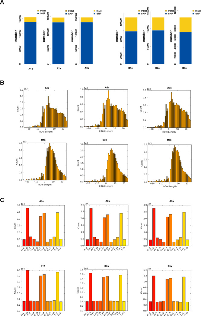
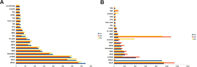
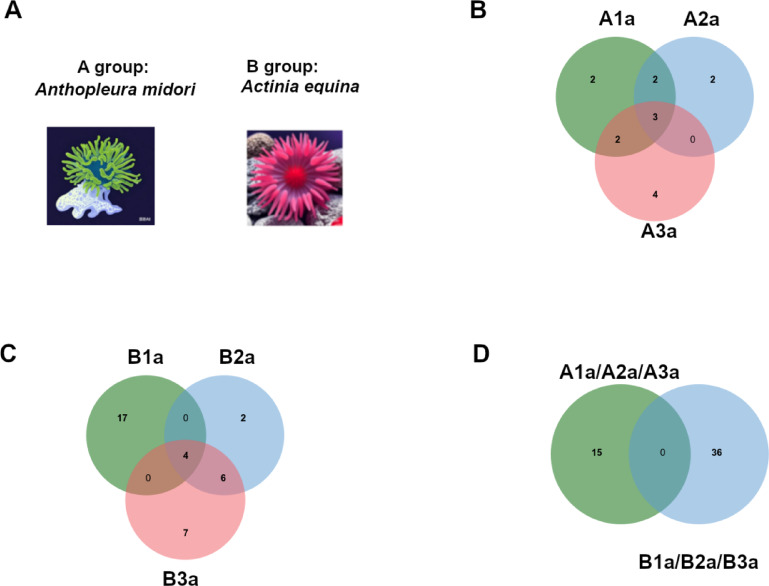
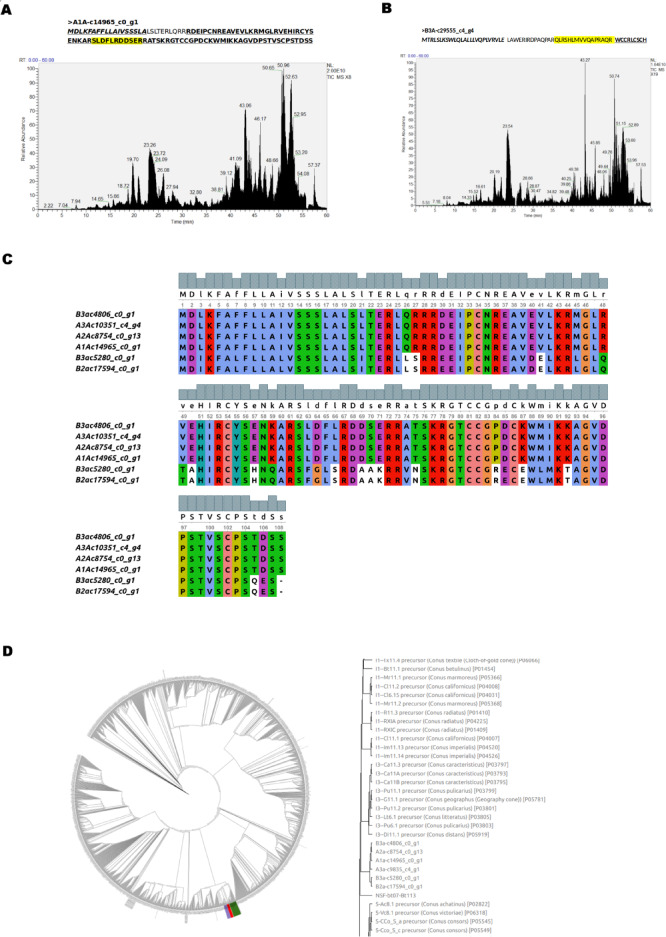
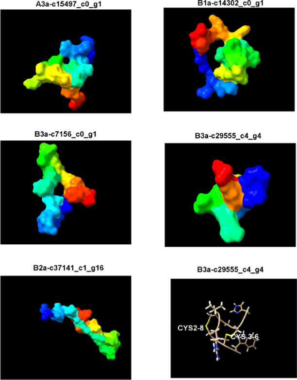
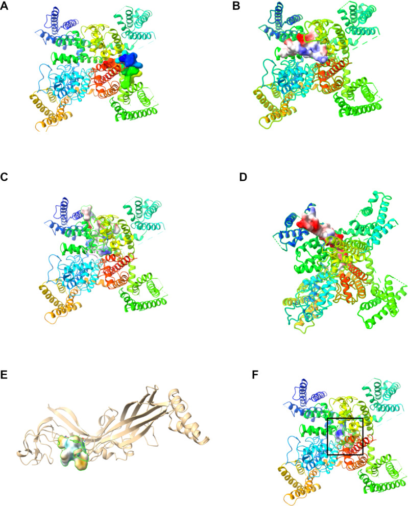
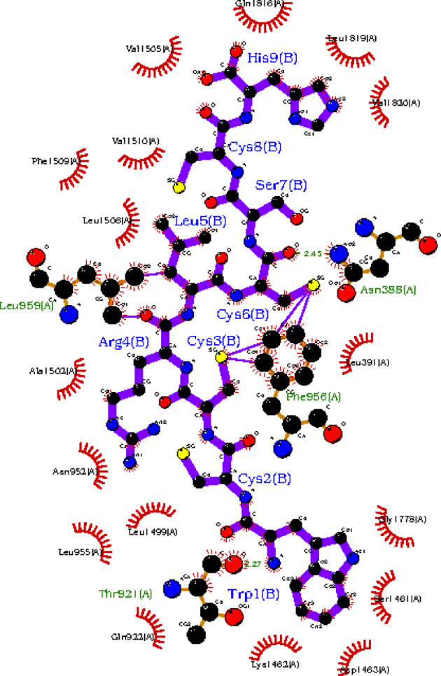
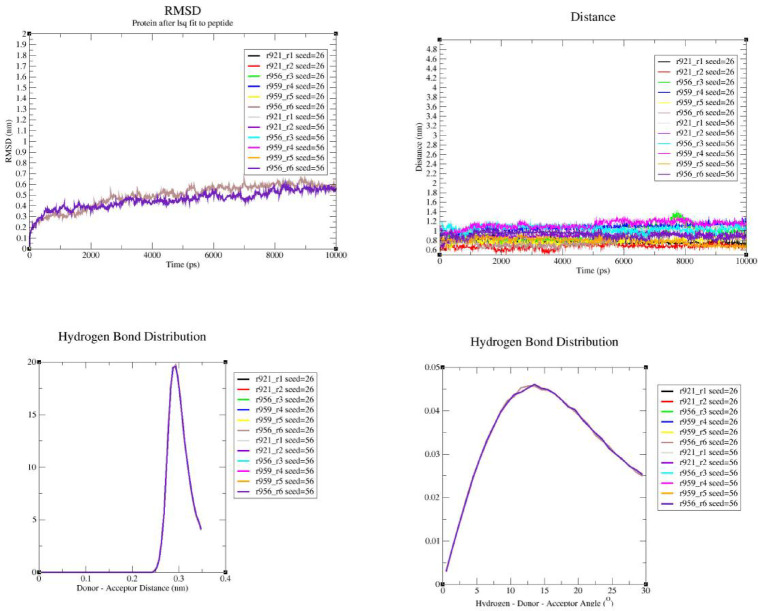
Exploring sea anemone polypeptides enables us to understand the evolutionary history and ecological adaptation strategies of species at the microscopic level. More importantly, it aims to provide a solid theoretical foundation for drug development and biodiversity conservation research. Through systematic research, we discovered a total of 51 toxin sequences in species Anthopleura midori and Acyinia equina. The toxin sequences between the two species exhibited significant differences, with notable diversity observed among individuals. In terms of genetic diversity, species Anthopleura midori primarily exhibits variations due to single nucleotide polymorphisms (SNPs), whereas species Actinia equina shows frequent insertion and deletion events. In transcription factor analysis, both species Anthopleura midori and Actinia equina share common transcription factors TEA (TEA Domain Transcription Factor), SPL(Squamosa Promoter Binding Protein-like), and bHLH (Basic Helix-Loop-Helix). Notably. Notably, bHLH is highly expressed in Actinia equina, which may give it advantages in muscle and nervous system development. On the other hand, Anthopleura midori may rely on other transcription factors. Furthermore, by employing transcriptomics and mass spectrometry techniques, two new gene families were successfully identified, and five structurally novel cyclic peptides were predicted. Kinetic simulations further confirmed that the peptide segment B3a-c29555_c4_g4 binds primarily through hydrogen bonds and hydrophobic interactions with the Cav3.1 (PDB ID:6 KZO) protein, and this peptide has the potential to act as a channel modulator for Cav3.1. Overall, this research not only deepens our understanding of the genetic basis of toxin diversity but also highlights the great potential of these toxins in the development of novel drugs.

Keywords: SNPs; Sea anemones; Toxins; Transcription factors.


Conflict of interest statement

Declarations. Competing interests: The authors declare no competing interests.

Figures

Fig. 1
Fig. 1
Genetic variation analysis of sea anemone. (A) Statistics of variation counts in each sample; (B) Distribution of indel lengths; (C) Distribution of SNP variation sites.
Fig. 2
Fig. 2
Transcription factor analysis. (A) Anthopleura midori; (B) Actinia equin. Note: (1) bHLH (Basic Helix - Loop - Helix); (2) CSL (CBF1, Su(H), and LAG − 1); (3) ETS (E twenty - six); (4) Fork_head (Forkhead Box); (5) HMG (High Mobility Group); (6) Homeobox (Homeodomain); (7) IRF (Interferon Regulatory Factor); (8) MBD (Methyl - CpG Binding Domain); (9) MYB (Myeloblastosis); (10) Others; 11. RHD (Rel Homology Domain); 12. TEA (TEA Domain Transcription Factor); 13. TF_bZIP (Transcription Factor Basic Leucine Zipper); 14. THAP (THAP Domain); 15. Tub (Tubby); 16. ZBTB (Zinc Finger and BTB Domain); 17. zf - C2H2 (Zinc Finger C2H2); 18. zf - GATA (GATA Zinc Finger); 19. zf - LITAF - like (Zinc Finger LITAF - like); 20. zf - MIZ (Zinc Finger MIZ - type).
Fig. 3
Fig. 3
Identification of sea anemone toxin peptides based on transcriptome. (A) Schematic diagram of Anthopleura midori and Actinia equin by Doubao AI (https://www.doubao.com/); (B) Venn diagram showing toxin peptide overlap among three individuals of Anthopleura midori; (C) Venn diagram showing toxin peptide overlap among three individuals of Actinia equin; (D) Venn diagram showing toxin peptide overlap between Anthopleura midori and Actinia equin.
Fig. 4
Fig. 4
Identification of peptides through proteomics and transcriptomics. (A) Protein ion spectrum of sample 3 from Anthopleura midori, with the yellow section representing identified sequences; (B) Protein ion spectrum of sample 3 from Actinia equin, with the yellow section representing identified sequences; (C) Alignment of new family toxin peptide sequences; (D) Phylogenetic tree analysis of toxin signal peptides.
Fig. 5
Fig. 5
Predicted structures of five candidate cyclic peptides.
Fig. 6
Fig. 6
Visualization of molecular docking of peptide B3a-c29555_c4_g4 with ion channels. (A) Molecular docking of B3a-c29555_c4_g4 with the ion channel Kv7.2 (KCNQ2, PDB ID: 6 V01); (B) Molecular docking of B3a-c29555_c4_g4 with the ion channel Kir2.1 (KCNA11, PDB ID: 3 JYC); (C) Molecular docking of B3a-c29555_c4_g4 with the ion channel Nav1.4 (SCN4 A, PDB ID: 6MBA); (D) Molecular docking of B3a-c29555_c4_g4 with the ion channel Nav1.7 (SCN9 A, PDB ID: 7LH2); (E) Molecular docking of B3a-c29555_c4_g4 with the P2Z Receptor(PDB ID:5U1U); (F) Molecular docking of B3a-c29555_c4_g4 with the ion channel Cav3.1 (PDB ID: 6 KZO).Note: The black region represents the binding pocket.
Fig. 7
Fig. 7
2D interaction map of peptide B3a-c29555_c4_g4 with membrane protein Cav3.1 (PDB ID:6 KZO).
Fig. 8
Fig. 8
Molecular dynamics simulation validation of peptide B3a-c29555_c4_g4 with membrane protein Cav3.1 (PDB ID:6 KZO). (A) Root Mean Square Deviation (RMSD) analysis of the protein residue 921 (Threonine) with peptide residues 1 (Tryptophan) and 2 (Cysteine), residue 956 (Phenylalanine) with residues 3 (Cysteine) and 6 (Cysteine), and residue 959 (Leucine) with residues 4 (Arginine) and 5 (Leucine), calculated randomly twice; (B) Distance calculations between protein residue 921 (Threonine) and peptide residues 1 (Tryptophan) and 2 (Cysteine), residue 956 (Phenylalanine) and residues 3 (Cysteine) and 6 (Cysteine), residue 959 (Leucine) and residues 4 (Arginine) and 5 (Leucine), calculated randomly twice; (C) Hydrogen bond distribution analysis: protein residue 921 (Threonine) with peptide residues 1 (Tryptophan) and 2 (Cysteine), residue 956 (Phenylalanine) with residues 3 (Cysteine) and 6 (Cysteine), residue 959 (Leucine) with residues 4 (Arginine) and 5 (Leucine), calculated randomly twice; (D) Hydrogen bond angle analysis: protein residue 921 (Threonine) with peptide residues 1 (Tryptophan) and 2 (Cysteine), residue 956 (Phenylalanine) with residues 3 (Cysteine) and 6 (Cysteine), residue 959 (Leucine) with residues 4 (Arginine) and 5 (Leucine), calculated randomly twice.

Similar articles

References

    1. Sigwart, J. D., Blasiak, R., Jaspars, M., Jouffray, J. B. & Tasdemir, D. Unlocking the potential of marine biodiscovery. Nat. Prod. Rep.38, 1235–1242. 10.1039/d0np00067a (2021). - PubMed
    1. Fu, X. Y. et al. Three rounds of Stability-Guided optimization and systematical evaluation of oncolytic peptide LTX-315. J. Med. Chem.67, 3885–3908. 10.1021/acs.jmedchem.3c02232 (2024). - PubMed
    1. Mackieh, R. et al. Voltage-Gated sodium channels: A prominent target of marine toxins. Mar. Drugs. 1910.3390/md19100562 (2021). - PMC - PubMed
    1. B, O. D. et al. AbeTx1 is a novel sea anemone toxin with a dual mechanism of action on Shaker-Type K⁺ channels activation. Mar. Drugs. 1610.3390/md16100360 (2018). - PMC - PubMed
    1. Norton, R. S. & Chandy, K. G. Venom-derived peptide inhibitors of voltage-gated potassium channels. Neuropharmacology127, 124–138. 10.1016/j.neuropharm.2017.07.002 (2017). - PubMed

LinkOut - more resources