. 2025 Apr 8;15(1):11935.
doi: 10.1038/s41598-025-95175-8.

The effective-compound compatibility of JHF inhibits fibroblast activation in pulmonary fibrosis by enhancing PINK1/PARK2-mediated mitophagy

Affiliations

The effective-compound compatibility of JHF inhibits fibroblast activation in pulmonary fibrosis by enhancing PINK1/PARK2-mediated mitophagy

Yunping Bai et al. Sci Rep. .

Abstract

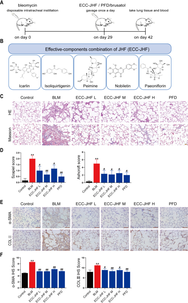
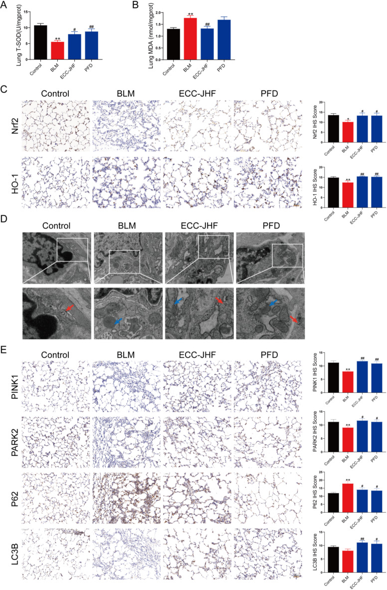
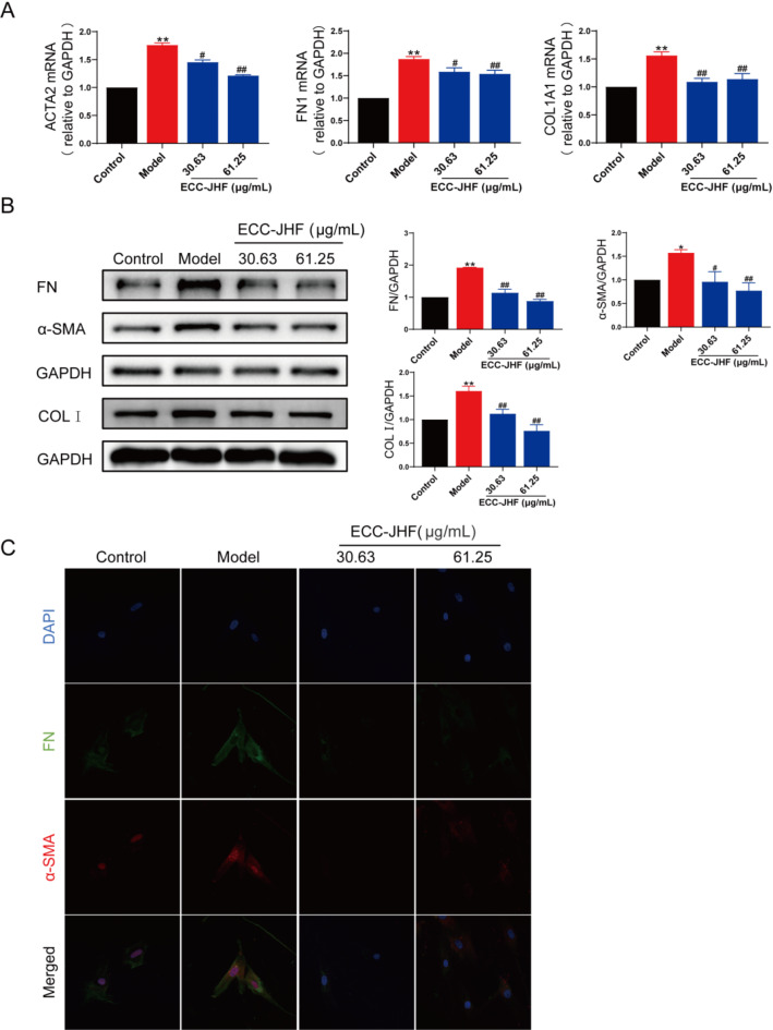
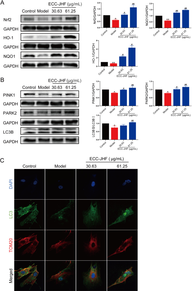
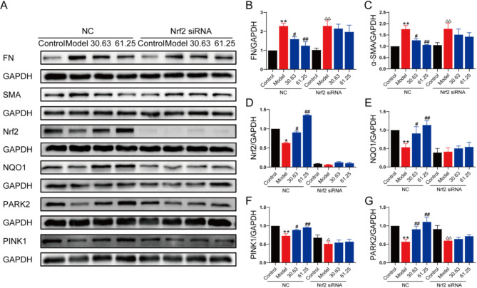
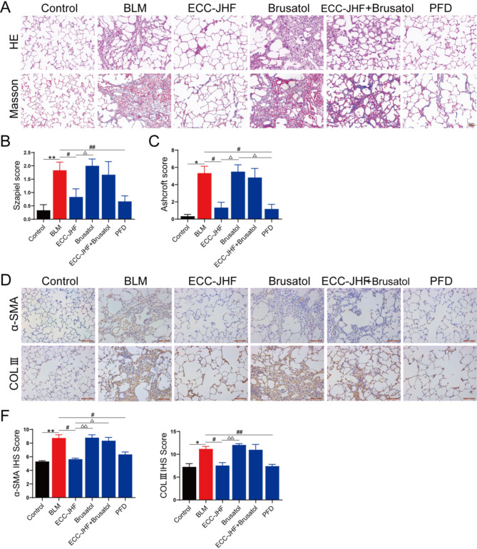
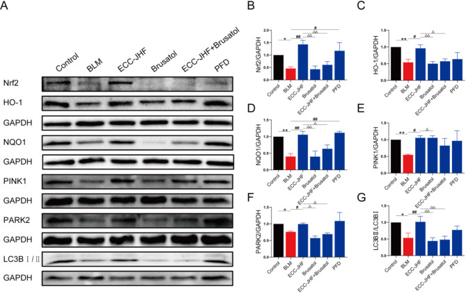
This work aimed to elucidate the anti-PF mechanism of ECC-JHF.The effects of ECC-JHF on lung fibrosis and fibroblast activation were investigated by establishing a BLM-induced PF rat model and a transforming growth factor-beta (TGF-β)-induced fibroblast activation model. Furthermore, the effects of ECC-JHF on Nrf2 signaling and mitophagy were explored both in vivo and in vitro. In the PF model rats, ECC-JHF mitigated pathological damage, reduced collagen deposition, decreased levels of malondialdehyde (MDA) and P62, and increased levels of total superoxide dismutase (T-SOD) as well as the expression of Nrf2, HO-1, PINK1, PARK2, and LC3B in lung tissues. These results suggest that the anti-PF mechanism of ECC-JHF may be associated with the inhibition of oxidative stress and the enhancement of mitophagy. The medium dose of ECC-JHF and pirfenidone were similar in improving pulmonary fibrosis in rats. In the TGF-β-induced lung fibroblast activation, ECC-JHF inhibited fibroblast activation by downregulating the levels of fibronectin, alpha-smooth muscle actin (α-SMA), and collagen I. Additionally, ECC-JHF upregulated the level of Nrf2 and its target proteins, including HO-1 and NQO1, as well as mitophagy-related proteins PINK1, PARK2, and LC3B. This led to an increase in the co-localization of TOM20 and LC3, thereby enhancing mitochondrial autophagy. The application of Nrf2 siRNA and Nrf2 inhibitors significantly diminished the effects of ECC-JHF on Nrf2 signaling, PINK1/PARK2-mediated mitophagy, and fibroblast activation. ECC-JHF exerts a protective effect against PF by suppressing fibroblast activation through the upregulation of Nrf2 and PINK1/PARK2-mediated mitophagy, it provides a new target and strategy for the treatment of pulmonary fibrosis.

Keywords: Effective-compound combination; Fibroblasts activation; Mitophagy; Nrf2; Pulmonary fibrosis.


Conflict of interest statement

Declarations. Competing interests: The authors declare no competing interests. Ethics approval and consent to participate: The Sprague-Dawley rats (license number: SCXK (Beijing) 2016-0006) were supplied by Beijing Vital River Laboratory Animal Technology Co. Ltd. (Beijing, China). All experimental procedures received approval from the First Affiliated Hospital of Henan University of Chinese Medicine, Zhengzhou, China (YFYDW2017013). Consent for publication: In this study, all participants provided written informed consent for their data to be used in publications.

Figures

Fig. 1
Fig. 1
The therapeutic effect of ECC-JHF on BLM-induced PF in rats. (A) The general process of animal experiments. (B) The compounds of ECC-JHF and their chemical structure. (C) HE and Masson staining (magnification, × 200). (D) The Szapiel and Ashcroft scores of lung tissue (Alveolitis and fibrosis scores). (E-F) Immunohistochemical analysis of α-SMA, Col III and IHS score assessment (magnification, × 200). All data were expressed as mean ± SE (n = 6)., **p < 0.01, vs. control group. #p < 0.05, ##p < 0.01, vs. BLM group.
Fig. 2
Fig. 2
ECC-JHF suppresses oxidative stress and enhances mitochondrial autophagy in lung tissue. (A) The activity of T-SOD. (B) The content of MDA. (C) Immunohistochemical analysis of Nrf2, HO-1, and IHS score assessment (magnification, × 200). (D) Transmission electron microscopy: the red arrows indicate autophagosomes and the blue indicate mitochondria (magnification, × 30000). (E) Immunohistochemical analysis of PINK1, Park2, p62 and LC3B and IHS score assessment (magnification, × 200). All data were expressed as mean ± SE (n = 6). *p < 0.05, **p < 0.01, vs. control group. #p < 0.05, ##p < 0.01, vs. BLM group.
Fig. 3
Fig. 3
Inhibitory effect of ECC-JHF on fibroblast activation. (A) Relative mRNA levels of ACTA2, FN1 and COL1A1. (B) The protein expression levels of α-SMA, FN and COL1 were detected by western blotting. (C) Immunofluorescent staining of the protein expression levels of α-SMA and FN (magnification, × 200). All data were expressed as mean ± SE. *p < 0.05, **p < 0.01, vs. control group. #p < 0.05, ##p < 0.01, vs. model group.
Fig. 4
Fig. 4
Effect of ECC-JHF on Nrf2 signaling and mitophagy in TGFβ1-induced HFL1 cells. (A) The protein expression levels of Nrf2, HO-1 and NQO1. (B) The protein expression levels of PINK1, PARK2 and LC3B. (C) Immunofluorescent staining of the co-localization of TOM20 and LC3 (magnification, × 400). All data were expressed as mean ± SE. *p < 0.05, **p < 0.01, vs. control group. #p < 0.05, ##p < 0.01, vs. model group.
Fig. 5
Fig. 5
ECC-JHF inhibit TGF-β1-induced fibroblast activation by activting Nrf2 singaling, strengthening mitophagy. Effect of Nrf2 in ECC -treated HFL1 cells. After transfection with siNrf2 for 24 h, HFL1 cells were treated with ECC-JHF (30.63 and 61.25 µg/ml) and TGF-β1 (5 ng/ml) for 24 h. Then, cells were collected for western blotting. (A-G)The protein expression levels of α-SMA, FN, Nrf2, HO-1, and PINK1, PARK2. All data were expressed as mean ± SE. *p < 0.05, **p < 0.01, vs. control group in normal cells. #p < 0.05, ##p < 0.01, vs. model group in normal cells. p < 0.05, △△p < 0.01, vs. control group in Nrf2 siRNA cells.
Fig. 6
Fig. 6
Nrf2-dependence for the anti-PF effect of ECC-JHF. (A) HE and Masson staining (magnification, × 200). (B-C) The Szapiel and Ashcroft scores of lung tissue (Alveolitis and fibrosis scores). (D-F) Immunohistochemical analysis of α-SMA, Col III and IHS score assessment (magnification, × 200). All data were expressed as mean ± SE (n = 6). *p < 0.05, **p < 0.01, vs. control group. #p < 0.05, ##p < 0.01, vs.BLM group. p < 0.05, △△p < 0.01, vs. ECC-JHF group.
Fig. 7
Fig. 7
ECC-JHF improves BLM-induced PF in rats by activating Nrf2, enhancing mitophagy. (A-G)The protein expression levels of Nrf2, HO-1, NQO1, PINK1, PARK2 and LC3B. All data were expressed as mean ± SE (n = 3). *p < 0.05, **p < 0.01, versus the control group. #p < 0.05, ##p < 0.01, vs. BLM group. p < 0.05, △△p < 0.01, vs. ECC-JHF group.

Similar articles

Cited by

References

    1. Raghu, G. et al. Idiopathic pulmonary fibrosis (an update) and progressive pulmonary fibrosis in adults: an official ATS/ERS/JRS/ALAT clinical practice guideline. Am. J. S Respir Crit. Care Med.205 (9), e18–e47 (2022). - PMC - PubMed
    1. Sgalla, G. et al. Idiopathic pulmonary fibrosis: pathogenesis and management. Respir Res.19 (1), 32 (2018). - PMC - PubMed
    1. Rossmann, M. P. et al. Mitochondrial function in development and disease. Dis. Model. Mech.14(6). (2021). - PMC - PubMed
    1. Chianese, M. et al. Pirfenidone and nintedanib in pulmonary fibrosis: lights and shadows. Pharmaceuticals (Basel). 17 (6), 709 (2024). - PMC - PubMed
    1. Liu, X. et al. HER2 drives lung fibrosis by activating a metastatic cancer signature in invasive lung fibroblasts. J. Exp. Med.219 (10), e20220126 (2022). - PMC - PubMed

MeSH terms