. 2025 Apr 8;8(1):555.
doi: 10.1038/s42003-025-07927-x.

Genetic and structural insights into the functional importance of the conserved gly-met-rich C-terminal tails in bacterial chaperonins

Affiliations

Genetic and structural insights into the functional importance of the conserved gly-met-rich C-terminal tails in bacterial chaperonins

C M Santosh Kumar et al. Commun Biol. .

Abstract

E. coli chaperonin GroEL forms nano-cages for protein folding. Although the chaperonin-mediated protein folding mechanism is well understood, the role of the conserved glycine and methionine-rich carboxy-terminal residues remains unclear. Bacteria with multiple chaperonins always retain at least one paralogue having the gly-met-rich C-terminus, indicating an essential conserved function. Here, we observed a stronger selection pressure on the paralogues with gly-met-rich C-termini, consistent with their ancestral functional importance. E. coli GroEL variants having mutations in their C-termini failed to functionally replace GroEL, suggesting the functional significance of the gly-met-rich C-termini. Further, our structural modelling and normal mode analysis showed that the C-terminal region shuttles between two cavity-specific conformations that correlate with the client-protein-binding apical domains, supporting C-termini's role in client protein encapsulation. Therefore, employing phylogenetic, genetic, and structural tools, we demonstrate that the gly-met-rich C-termini are functionally significant in chaperonin-mediated protein folding function. Owing to the pathogenic roles of the chaperonins having non-canonical C-termini, future investigations on the client protein selectivity will enable understanding the disease-specific client protein folding pathways and treatment options.


Conflict of interest statement

Competing interests: The authors declare no competing interests.

Figures

Fig. 1
Fig. 1. Phylogenetic analysis of actinobacterial chaperonins.
a Phylogenetic relationships between 325 actinobacterial chaperonin sequences were inferred using a neighbourhood-joining algorithm and presented as a phylogenetic tree. The phylogenetic tree shows three clades. The yellow-coloured branch represents the E. coli GroEL sequence, which was included in the alignment. b Site specific conservation rates were calculated using ConSurf’s Rate4Site algorithm from the multiple sequence analysis and mapped onto E. coli GroEL sequence. The graph represents the normalized conservation scores (lowest score represents conservation, with a standard deviation of one) as a function of GroEL’s primary sequence. The domain regions of the equatorial (E), intermediate (I) and apical (A) domains and the CTS region (C) are indicated. c Sequence Logo depicting the diversity in the CTS region of the actinobacterial chaperonins, starting with the conserved Proline. d Scatter plot showing hydrophobicity of the chaperonin CTSs (GRAVY Scores) as a function of their average charges (pI). Each dot represents one CTS and the spots are colour coded in the same way as the three branches in the phylogenetic tree. A detailed phylogenetic tree with taxon names and node ages is presented in the supplementary information.
Fig. 2
Fig. 2. The carboxy terminus is essential for full GroEL function.
The ability of groEL lacking the 13 c-terminal residues to functionally replace groEL was assessed in (a) the groEL deletion strain AI90; (b) the conditional expression strains LG6 and MGM100 and (c) temperature-sensitive strains groEL44 and groEL100. Serially diluted cultures of the indicated E. coli strains expressing either the wildtype (W) or the 13 residue carboxy termini lacking groEL variant GroELΔC13 (Δ) from a plasmid, or the vector only control (V), were spotted on LB agar plates supplemented as indicated. All the plates in a and b were incubated at 30 °C, while the plates in c were incubated at the indicated temperatures. The first and second plates in b and c represent permissive and restrictive growth conditions, respectively. Relevant genetic features of the strains in a and b are depicted schematically. In AI90, groEL has been replaced by the kanR cassette while a functional copy, regulated by lactose inducible Plac promoter, is provided on a shelter plasmid pTGroEL7 (p15A, camR). The ability of the incoming indicated groEL variants to allow loss of pTGroEL7 was assessed. In LG6 and MGM100, the chromosomal copies of the bicistronic groE operon are controlled by lactose-inducible Plac and arabinose-inducible PBAD promoters, respectively.
Fig. 3
Fig. 3. Flexible carboxy termini wobble between the cavity-specific tight (T) and relaxed (R”) conformations.
a Molecular model of asymmetric GroEL-GroES complexes showing filling of the void by CTSs (red space filled). GroEL and GroES are in pale-blue and pink, respectively. One GroEL subunit in each ring is colour-coded to make the change in domain architecture easier to visualise. A, I and E represent apical, intermediate, and equatorial domains, respectively. b Conformational snapshots showing the series of conformations visited by the GroEL heptamer during transition from the T to the R” state. Domains in all the subunits are colour-coded as in (a). Two subunits were removed in the display to reveal the dynamics of CTSs inside the cavity. c Bottom view of the cavity showing the gradual opening of the aperture during the transition, and the dynamic movement of the CTSs. d Cartoon representations of single subunits from the two heptameric rings of GroEL in T and R” conformational states that were subjected to NMA. The rotation and transition of the CTSs are indicated. The helices F, H, I and M are indicated as αF, αH, αI and αM, respectively. e Residue level fluctuations in the torsion angles and displacements of alpha carbon atoms. Fluctuations in the indicated angles and displacements with respect to the T state that were calculated for all seven subunits were averaged and plotted as a function of the primary structure of a subunit. E, I, A, and G represent the regions of GroEL primary structure spanning the corresponding domains as colour-coded in the molecular model. The bold lines mark the regions and are scaled according to the size of the indicated helices.
Fig. 4
Fig. 4. Variations in GroEL carboxy terminus affect chaperonin function.
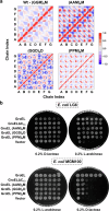
a Pairwise correlation matrices showing differential displacement of the indicated 13 residue carboxy-terminal peptides of the GroEL CTS variants that are shown and colour-coded as in Fig. S6. A to G indicate the subunit chains in the heptameric ring. b Serially diluted cultures of groEL conditional mutant strains, E. coli LG6 and E. coli MGM100 that are expressing the indicated groEL CTS variants, were spotted on LB agar plates. The plates were supplemented as indicated and incubated at 30 °C.
Fig. 5
Fig. 5. The possible mode of action of carboxy terminal segments.
Model depicting a hypothetical role for CTSs role in the chaperonin mechanism. Please refer to text for details.

Similar articles

References

    1. Horwich, A. L. & Fenton, W. A. Chaperonin-assisted protein folding: a chronologue. Q Rev. Biophys.53, e4 (2020). - PubMed
    1. Lund, P. A. Multiple chaperonins in bacteria- why so many? FEMS Microbiol Rev.33, 785–800 (2009). - PubMed
    1. Kumar, C. M., Mande, S. C. & Mahajan, G. Multiple chaperonins in bacteria-novel functions and non-canonical behaviors. Cell Stress Chaperones20, 555–574 (2015). - PMC - PubMed
    1. Hayer-Hartl, M., Bracher, A. & Hartl, F. U. The GroEL-GroES Chaperonin machine: a nano-cage for protein folding. Trends Biochem Sci.41, 62–76 (2016). - PubMed
    1. Braig, K. et al. The crystal structure of the bacterial chaperonin GroEL at 2.8 A. Nature371, 578–586 (1994). - PubMed

MeSH terms

LinkOut - more resources