

A three-step approach versus the inverted internal limiting membrane flap technique in large full thickness macular hole surgery: a comparative study
- PMID: 40200260
- PMCID: PMC11978172
- DOI: 10.1186/s12886-025-04003-0
A three-step approach versus the inverted internal limiting membrane flap technique in large full thickness macular hole surgery: a comparative study
Abstract
Objective: To evaluate the anatomical and functional outcomes of our novel surgery (a three-step approach) and the conventional inverted internal limiting membrane flap technique (IFT) in treating large full-thickness macular holes (FTMHs).
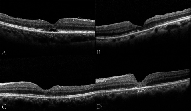
Methods: This was a retrospective, consecutive, nonrandomized comparative study of patients who underwent either the novel surgery (n = 27, Group A) or IFT (n = 27, Group B). The main outcomes of MH closure rates and the best corrected visual acuity (BCVA) at 1-, 3-, and 6-month follow-up were compared between the two groups.
Results: At 6 months postoperatively, MH closure was achieved in 24/27 patients in Group A and 22/27 patients in Group B (88.89% vs. 81.48%, P = 0.704) with U-shaped closure rates being significantly higher in Group A (P = 0.029). The average BCVA at month 6 was 0.69 ± 0.38 (LogMAR) in Group A and 0.91 ± 0.39 in Group B (P = 0.015) with the improvement in BCVA being significantly higher in Group A (0.50 ± 0.59 vs. 0.31 ± 0.59, P = 0.045). The recovery rates of ELM were significantly higher in Group A (P = 0.026).
Conclusions: Our three-step approach greatly improves anatomical and functional outcomes compared with IFT. This novel surgery has a dominant advantage in earlier and higher ultimate closure rate, U-type closure rate, and ELM recovery rate, and more importantly, better recovery of BCVA.
Keywords: A three-step approach; FTMH; Functional and anatomical outcome; IFT.
© 2025. The Author(s).
Conflict of interest statement
Declarations. Ethics approval and consent to participate: This study was approved by the Ethics Committee of Shanghai General Hospital, Shanghai Jiao Tong University School of Medicine and was implemented in accordance with the World Medical Association Declaration of Helsinki. The number of the ethical approval document is 2021SQ247, which is registered in Shanghai General Hospital. All patients were informed about the study and signed written informed consent forms before their enrollment. Consent for publication: Not applicable. Competing interests: The authors declare no competing interests.
Figures
Similar articles
-
EFFECT OF INVERTED INTERNAL LIMITING MEMBRANE FLAP ON CLOSURE RATE, POSTOPERATIVE VISUAL ACUITY, AND RESTORATION OF OUTER RETINAL LAYERS IN PRIMARY IDIOPATHIC MACULAR HOLE SURGERY.Retina. 2020 Oct;40(10):1955-1963. doi: 10.1097/IAE.0000000000002707. Retina. 2020. PMID: 31834129
-
Inverted internal limiting membrane flap technique versus complete internal limiting membrane peeling in large macular hole surgery: a comparative study.BMC Ophthalmol. 2020 Jan 6;20(1):11. doi: 10.1186/s12886-019-1294-8. BMC Ophthalmol. 2020. PMID: 31907015 Free PMC article.
-
INTERNAL LIMITING MEMBRANE PEELING VERSUS INVERTED FLAP TECHNIQUE FOR TREATMENT OF FULL-THICKNESS MACULAR HOLES: A COMPARATIVE STUDY IN A LARGE SERIES OF PATIENTS.Retina. 2018 Sep;38 Suppl 1:S73-S78. doi: 10.1097/IAE.0000000000001985. Retina. 2018. PMID: 29232338
-
Pars plana vitrectomy with internal limiting membrane flap versus pars plana vitrectomy with conventional internal limiting membrane peeling for large macular hole.Cochrane Database Syst Rev. 2023 Aug 7;8(8):CD015031. doi: 10.1002/14651858.CD015031.pub2. Cochrane Database Syst Rev. 2023. PMID: 37548231 Free PMC article. Review.
-
Comparative efficacy evaluation of inverted internal limiting membrane flap technique and internal limiting membrane peeling in large macular holes: a systematic review and meta-analysis.BMC Ophthalmol. 2020 Jan 8;20(1):14. doi: 10.1186/s12886-019-1271-2. BMC Ophthalmol. 2020. PMID: 31914954 Free PMC article.
References
-
- Bonińska K, Nawrocki J, Michalewska Z. Mechanism of flap closure after the inverted internal limiting membrane flap technique. Retina. 2018;38:2184–9. - PubMed
-
- Darian-Smith E, Howie AR, Allen PL, Vote BJ. Tasmanian macular hole study: whole population-based incidence of full thickness macular hole. Clin Exp Ophthalmol. 2016;44:812–6. - PubMed
-
- Duker JS, Kaiser PK, Binder S, De Smet MD, Gaudric A, Reichel E, Sadda SR, Sebag J, Spaide RF, Stalmans P. The international vitreomacular traction study group classification of vitreomacular adhesion, traction, and macular hole. Ophthalmology. 2013;120:2611–9. - PubMed
-
- Eckardt C, Eckardt U, Groos S, Luciano L, Reale E. [Removal of the internal limiting membrane in macular holes. Clinical and morphological findings]. Der Ophthalmologe: Z Der Deutschen Ophthalmologischen Gesellschaft. 1997;94:545–51. - PubMed
-
- Hayashi H, Kuriyama S. Foveal microstructure in macular holes surgically closed by inverted internal limiting membrane flap technique. Retina. 2014;34:2444–50. - PubMed
Publication types
MeSH terms
Grants and funding
LinkOut - more resources
Full Text Sources
