

Kaempferol inhibits cardiomyocyte pyroptosis via promoting O-GlcNAcylation of GSDME and improved acute myocardial infarction
- PMID: 40200275
- PMCID: PMC11980313
- DOI: 10.1186/s40360-025-00908-0
Kaempferol inhibits cardiomyocyte pyroptosis via promoting O-GlcNAcylation of GSDME and improved acute myocardial infarction
Abstract
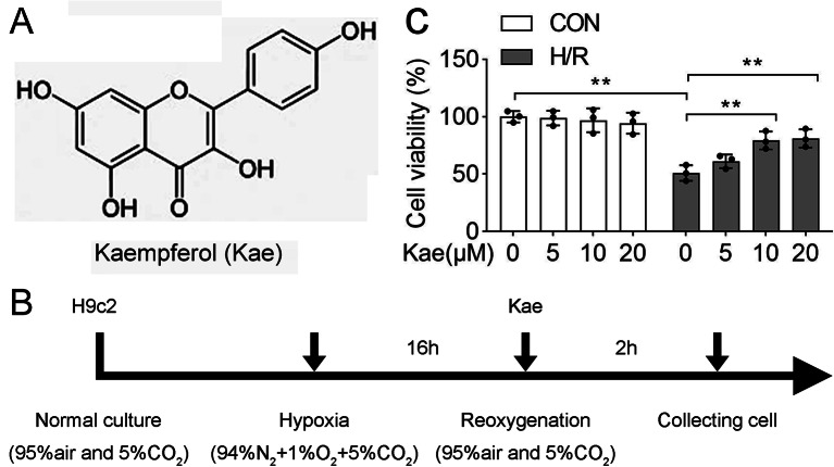
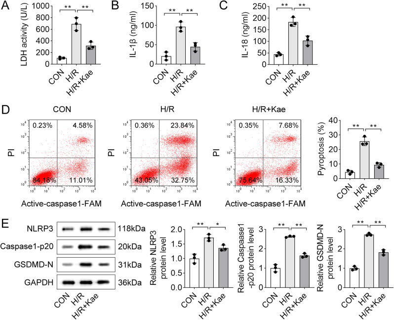
Acute myocardial infarction (AMI) is a leading fatal cardiovascular disease and poses a major threat to human health. Pyroptosis, an inflammation-related programmed cell death, plays a critical role in the progression of AMI. Kaempferol is a natural flavonoid compound with a variety of pharmacological effects, which exerts a significant cardioprotective function. The role of O-GlcNAcylation, a post-translation modification, has received attention in diseases including AMI. In this research, we explored the therapeutic potential of Kaempferol to AMI due to its well-known cardioprotective effect, including its antioxidant and anti-inflammatory properties. Hypoxia/reoxygenation (H/R) model was adopted to provoke myocardial injury and AMI mice model was established. Our findings indicated that H/R lessened cell viability and contributed to the release of LDH, IL-1β and IL-18, cell pyroptosis rate, and the expression of NLRP3, active caspase 1 and GSDMD-N-terminal domain (GSDMD-N). Kaempferol mitigated myocardial damage caused by H/R through repressing cell pyroptosis. Besides, we discovered that Kaempferol restored the levels of O-GlcNAcylation by regulating the activity of OGT (O-GlcNAc transferase) and OGA (O-GlcNAcase) in H/R-treated H9c2 cells. Notably, molecular docking revealed the binding relationship between Kaempferol and OGT. Further, we proved that knockdown of OGT abrogated the function of Kaempferol in H/R-induced pyroptosis. In AMI mice, Kaempferol relieved the myocardial tissue injury and decreased the NLRP3 and GSDME-N protein levels. More importantly, our results illustrated that OGT was responsible for the O-GlcNAcylation of GSDME at T94 site and acted as an inducing factor for GSDME phosphorylation. Namely, this study validated that Kaempferol facilitated GSDME O-GlcNAcylation to inhibit H/R-induced pyroptosis in an OGT-dependent manner.
Keywords: Acute myocardial infarction; GSDME; Kaempferol; O-GlcNAcylation; Pyroptosis.
© 2025. The Author(s).
Conflict of interest statement
Declarations. Ethics approval and consent to participate: This study was approved by the Ethics Committee of Huzhou Third Municipal Hospital. All animal experiments should comply with the ARRIVE guidelines. All methods were carried out in accordance with relevant guidelines and regulations. Consent for publication: Not applicable. Competing interests: The authors declare no competing interests.
Figures


Similar articles
-
Kaempferol-3-O-rutinoside exerts cardioprotective effects through NF-κB/NLRP3/Caspase-1 pathway in ventricular remodeling after acute myocardial infarction.J Food Biochem. 2022 Oct;46(10):e14305. doi: 10.1111/jfbc.14305. Epub 2022 Jun 27. J Food Biochem. 2022. PMID: 35758877
-
High glucose promotes lipopolysaccharide-induced macrophage pyroptosis through GSDME O-GlcNAcylation.J Periodontal Res. 2025 Jun;60(6):581-589. doi: 10.1111/jre.13349. Epub 2024 Sep 25. J Periodontal Res. 2025. PMID: 39319591
-
MiR-21 mediates the protection of kaempferol against hypoxia/reoxygenation-induced cardiomyocyte injury via promoting Notch1/PTEN/AKT signaling pathway.PLoS One. 2020 Nov 5;15(11):e0241007. doi: 10.1371/journal.pone.0241007. eCollection 2020. PLoS One. 2020. PMID: 33151961 Free PMC article.
-
O-GlcNAcylation dictates pyroptosis.Front Immunol. 2024 Dec 17;15:1513542. doi: 10.3389/fimmu.2024.1513542. eCollection 2024. Front Immunol. 2024. PMID: 39742284 Free PMC article. Review.
-
Decoding glycosylation in cardiovascular diseases: mechanisms, biomarkers, and therapeutic opportunities.Front Pharmacol. 2025 May 19;16:1570158. doi: 10.3389/fphar.2025.1570158. eCollection 2025. Front Pharmacol. 2025. PMID: 40458788 Free PMC article. Review.
References
-
- Reed GW, Rossi JE, Cannon CP. Acute myocardial infarction. Lancet. 2017;389(10065):197–210. - PubMed
-
- Caruntu F, Bordejevic DA, Buz B, Gheorghiu A, Tomescu MC. Independent predictors of in-hospital and 1-year mortality rates in octogenarians with acute myocardial infarction. Rev Cardiovasc Med. 2021;22(2):489–97. - PubMed
-
- Raskovalova T, Twerenbold R, Collinson PO, Keller T, Bouvaist H, Folli C, et al. Diagnostic accuracy of combined cardiac troponin and copeptin assessment for early rule-out of myocardial infarction: A systematic review and meta-analysis. Eur Heart J Acute Cardiovasc Care. 2014;3(1):18–27. - PMC - PubMed
MeSH terms
Substances
LinkOut - more resources
Full Text Sources
Medical
Miscellaneous
