. 2025 Apr 8;13(1):17.
doi: 10.1186/s40170-025-00386-2.

TRIM22 governs tumorigenesis and protects against endometrial cancer-associated cachexia by inhibiting inflammatory response and adipose thermogenic activity

Affiliations

TRIM22 governs tumorigenesis and protects against endometrial cancer-associated cachexia by inhibiting inflammatory response and adipose thermogenic activity

Liping Zhang et al. Cancer Metab. .

Abstract

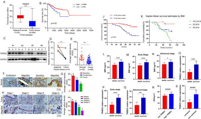
Background: Endometrial cancer (EC) is one of the most common cancers in women, with a short overall survival and poor prognosis. Besides the biologically aggressive EC properties, Cancer-associated cachexia is the main factor. However, the detailed mechanism underlying EC-related cachexia and its harmful effects on EC progression and patient prognosis remains unclear.

Methods: For clinical specimen and the vitro experiment, we detected TRIM22 expression level, EC patients' survival time, EC cell functional change, and adipose thermogenic changes to identify the function of TRIM22 in EC progression, EC-associated cachexia, and their molecular mechanisms. Then, for the vivo experiment, we exploited the xenografts in mice to identify the function of TRIM22 again, and to screen the drug therapeutic schedule.

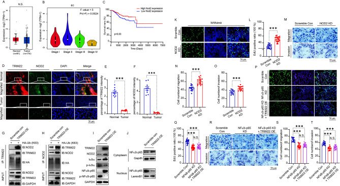
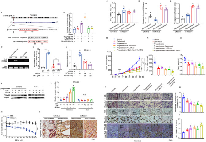
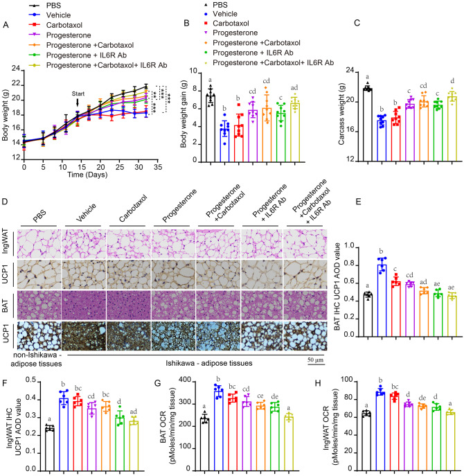
Results: Herein, we demonstrated that TRIM22 inhibited EC cell growth, invasion, and migration. Interleukin (IL)-6 mediated brown adipose tissue activation and white adipose tissue browning which induced EC-related cachexia. TRIM22 suppressed the EC cells' secretion of IL-6, and IL-6 mediated EC-related cachexia. Mechanistically, TRIM22 inhibited EC progression by suppressing the nucleotide-binding oligomerization domain 2(NOD2)/nuclear factor-kappaB (NF-κB) signaling pathway, with the purpose of impeding the production of IL-6. Moreover, we revealed that TRIM22 inhibited EC-associated cachexia by suppressing the IL-6/IL-6 receptor (IL-6R) signaling pathway. Therapeutically, we demonstrated that combination treatment with a TRIM22 inducer (progesterone) and a thermogenic inhibitor (IL-6R antibody) synergistically augmented the antitumor efficacy of carbotaxol (carboplatin and paclitaxel), in vivo.

Conclusion: Our data reveals that TRIM22-EC-IL-6-cachexia cross-communication has important clinical relevance and that the use of combined therapy holds great promise for enhancing the efficacy of anti-ECs. (Fig. graphical abstract).

Keywords: Antitumor therapy; Cancer-associated cachexia; Endometrial cancer; Tripartite motif-containing 22(TRIM22).


Conflict of interest statement

Declarations. Ethical approval and consent to participate: This study was approved by the research ethics committee of Fujian Medical University Union Hospital (Fuzhou, China), and carried out under the World Medical Association Declaration of Helsinki. Consent for publication: The authors confirm that they have obtained written consent from each patient to publish the manuscript. Competing interests: The authors declare no competing interests.

Figures

Fig. 1
Fig. 1
Lack of TRIM22 worsens overall survival (OS) in patients with EC accompanied by body mass index (BMI) reduction. (A) The TRIM22 expression level of EC tissue and its adjacent normal tissue in TCGA database. (B) The Kaplan-Meier analysis between TRIM22 and survival of EC patients in TCGA database. (C) Immunoblots of TRIM22 in 4 pairs of EC samples (N, normal endometrial tissue; A, adjacent normal tissue; T, tumor tissue). (D) Statistical pair analysis of TRIM22 expression derived from (C). (E) Statistical analysis of TRIM22 expression derived from (F and H). (F)Representative IHC staining of normal endometrial tissues from the proliferative and secretory during the menstrual cycle. (G) Statistical analysis of the expression of TRIM22 in EC tissues and normal endometrial tissues (proliferative and secretory) derived from (F and H). (H)Representative IHC staining of different stage EC tissues (stage I, II, III, IV). (I)Statistical analysis of TRIM22 expression in normal endometrial tissues and different stage EC tissues (stage I, II, III, IV) derived from (F and H). (J)The Kaplan-Meier analysis between TRIM22 and survival of EC patients according the TRIM22 expression derived from (H). (K)The Kaplan-Meier analysis between BMI and survival of EC patients (BMI: body mass index). (L)Statistical analysis of BMI of the death and survivor EC patients. (M) Statistical analysis of BMI of the death and survivor EC patients in the early stage (stage I and II). (N)Statistical analysis of BMI of the death and survivor EC patients in the advanced stage (stage III and IV). (O)Statistical analysis of TRIM22 expression of the death and survivor EC patients. (P)Statistical analysis of TRIM22 expression of the death and survivor EC patients in the early stage (stage I and II). (Q)Statistical analysis of TRIM22 expression of the death and survivor EC patients in the advanced stage (stage III and IV). (R) Statistical analysis of BMI between high-TRIM22 and low-TRIM22 expression groups based on mean TRIM22 expression levels. (S) Statistical analysis of TRIM22 expression between cachexia and non-cachexia group. Data were expressed as means ± SEM of three independent experiments. Scale bar, 50 μm. *p < 0.05, **p < 0.01, ***p < 0.001
Fig. 2
Fig. 2
TRIM22 OE suppressed EC progression and prevented body weight loss induced by cachexia. (A) Ratio of Edu-positive scramble control and TRIM22 OE Ishikawa cells in (A). (B) Representative images of scramble control and TRIM22 OE Ishikawa cells that cultured in trans-well plates. (C) The average number of migration Ishikawa cells in (C). (D) The average number of invasion Ishikawa cells in (C). (E) Representative images showing Edu incorporation in scrambling control and TRIM22 KD RL-952 cells. (F) Ratio of Edu-positive RL-952 cells in (F). (G) Representative images of scramble control and TRIM22 KD RL-952 cells that cultured in trans-well plates. (H) The average number of migration RL-952 cells in (H). (I) The average number of invasion RL-952 cells in (H). (J) A subcutaneous tumor growth curve from scramble control and TRIM22 OE Ishikawa cells at indicated time points (n = 9 mice per group). (K) Representative images of xenograft tumors in (K) at day 27 (n = 9 mice per group). (L) Weights of xenograft tumors in (L) at day 27 (n = 9 mice per group). (M)Representative H&E staining and IHC staining of Ki67 in xenograft tumors derived from (L). (N) Statistical analysis of Ki67 expression in xenograft tumors derived from (L) using IHC staining. (O) Statistical analysis of lipid aera in xenograft tumors derived from (L) using H&E staining. (P) A body weight curve from xenograft mice subcutaneously injected PBS, scramble control and TRIM22 OE Ishikawa cells. (Q) Statistical analysis of body weight gain of xenograft mice subcutaneously injected PBS, scramble control and TRIM22 OE Ishikawa cells. (R) Statistical analysis of carcass weight of xenograft mice subcutaneously injected PBS, scramble control and TRIM22 OE Ishikawa cells. (S) Statistical analysis of the mass of BAT from xenograft mice subcutaneously injected PBS, scramble control and TRIM22 OE Ishikawa cells. (T) Statistical analysis of the mass of IngWAT from xenograft mice subcutaneously injected PBS, scramble control and TRIM22 OE Ishikawa cells. (U) Statistical analysis of the mass of GonWAT from xenograft mice subcutaneously injected PBS, scramble control and TRIM22 OE Ishikawa cells. Data were expressed as means ± SEM of three independent experiments. Scale bar, 50 μm. *p < 0.05, **p < 0.01, ***p < 0.001
Fig. 3
Fig. 3
TRIM22 OE reversed EC-enhanced thermogenic activity in BAT. (A) Representative images of H&E and IHC-stained sections of BAT from xenograft mice subcutaneously injected PBS, scramble control and TRIM22 OE Ishikawa cells. (B) Statistical analysis of lipid aera in BAT from xenograft mice subcutaneously injected PBS, scramble control and TRIM22 OE Ishikawa cells in (A) using H&E staining. (C) Statistical analysis of UCP1 expression in BAT from xenograft mice subcutaneously injected PBS, scramble control and TRIM22 OE Ishikawa cells in (A) using IHC staining. (D) mRNA level of genes of BAT from xenograft mice subcutaneously injected PBS, scramble control and TRIM22 OE Ishikawa cells, including thermogenic genes, fatty acid oxidation related genes, lipolysis related genes, mitochondrial biogenic transcription factor. Data were expressed as means ± SEM of three independent experiments. Scale bar, 50 μm. *p < 0.05, **p < 0.01, ***p < 0.001
Fig. 4
Fig. 4
TRIM22 OE blocked EC-induced browning of WAT. (A) Representative images of H&E and IHC-stained sections of IngWAT from xenograft mice subcutaneously injected PBS, scramble control and TRIM22 OE Ishikawa cells. (B) Statistical analysis of lipid aera in IngWAT from xenograft mice subcutaneously injected PBS, scramble control and TRIM22 OE Ishikawa cells in (A) using H&E staining. (C) Statistical analysis of UCP1 expression in IngWAT from xenograft mice subcutaneously injected PBS, scramble control and TRIM22 OE Ishikawa cells in (A) using IHC staining. (D) mRNA level of genes of IngWAT from xenograft mice subcutaneously injected PBS, scramble control and TRIM22 OE Ishikawa cells, including thermogenic genes, fatty acid oxidation related genes, lipolysis related genes, mitochondrial biogenic transcription factor. (E) mRNA level of gene of beige cell marker (TMEM26) in IngWAT from xenograft mice subcutaneously injected PBS, scramble control and TRIM22 OE Ishikawa cells. (F) mRNA level of gene of beige cell marker (CD137) in IngWAT from xenograft mice subcutaneously injected PBS, scramble control and TRIM22 OE Ishikawa cells. (G) mRNA level of gene of beige cell marker (TBX1) in IngWAT from xenograft mice subcutaneously injected PBS, scramble control and TRIM22 OE Ishikawa cells. (H) Mean OCR from Seahorse in IngWAT from xenograft mice subcutaneously injected PBS, scramble control and TRIM22 OE Ishikawa cells. Data were expressed as means ± SEM of three independent experiments. Scale bar, 50 μm. *p < 0.05, **p < 0.01, ***p < 0.001
Fig. 5
Fig. 5
TRIM22 inhibited the enhancement of EC cells-mediated thermogenesis activity in adipocytes through the IL-6/IL-6R signaling pathway. (A) ELISA of IL-6 in blood from xenograft mice subcutaneously injected PBS, scramble control and TRIM22 OE Ishikawa cells. (B) mRNA level of IL-6R in BAT from xenograft mice subcutaneously injected PBS, scramble control and TRIM22 OE Ishikawa cells. (C) mRNA level of SOCS3 in BAT from xenograft mice subcutaneously injected PBS, scramble control and TRIM22 OE Ishikawa cells. (D) mRNA level of IL-6R in IngWAT from xenograft mice subcutaneously injected PBS, scramble control and TRIM22 OE Ishikawa cells. (E) mRNA level of SOCS3 in IngWAT from xenograft mice subcutaneously injected PBS, scramble control and TRIM22 OE Ishikawa cells. (F) mRNA level of TRIM22 in scramble control and TRIM22 OE Ishikawa cells. (G) ELISA of IL-6 in TRIM22 OE and scramble control Ishikawa cells condition medium (CM). (H) UCP1 mRNA in differentiated matured C3H10T1/2 cells after treated with the CM from scramble control and TRIM22 OE Ishikawa cells. (I) mRNA level of IL-6R in differentiated matured C3H10T1/2 cells after treated with the CM from scramble control and TRIM22 OE Ishikawa cells. (J) mRNA level of SOCS3 in differentiated matured C3H10T1/2 cells after treated with the CM from scramble control and TRIM22 OE Ishikawa cells. (K) OCR of differentiated matured C3H10T1/2 cells was evaluated using Seahorse after treated with CM from scramble control and TRIM22 OE Ishikawa cells. (L) Basal OCR from Seahorse in (K). (M) Uncoupled OCR from Seahorse in (K). (N) Maximal OCR from Seahorse in (K). (O) mRNA level of IL-6R in differentiated C3H10T1/2 cells when blocked the IL-6R (scramble control and IL-6R KD). (P) mRNA level of UCP1 in scramble control and IL-6R KD differentiated matured C3H10T1/2 cells after treated with the CM from Ishikawa cells. (Q)OCR of scramble control and IL-6R KD differentiated matured C3H10T1/2 cells were evaluated using Seahorse after treated with CM from Ishikawa cells. (R)Basal OCR from Seahorse in (Q). Data were expressed as means ± SEM of three independent experiments. *p < 0.05, **p < 0.01, ***p < 0.001
Fig. 6
Fig. 6
TRIM22 and NOD2 interaction restricted EC progression by inhibiting the NF‑κB pathway. (A) NOD2 expression level in the normal endometrial tissue and EC tissues in TCGA database. (B) NOD2 expression level in EC tissues from the different stage (I, II, III, IV). (C) The Kaplan-Meier analysis between NOD2 and survival of EC patients in TCGA database. (D) Immunofluorescent detection of TRIM22 and NOD2 in EC and normal endometrial tissue. Scale bar, 20 μm. (E) Statistical analysis of TRIM22 in (D). (F)Statistical analysis of NOD2 in (D). (G) Co-IP detection of TRIM22, NOD2 and HA-Ub in TRIM22 OE and scramble control Ishikawa cells. (H) Reciprocal Co-IP detection of TRIM22, NOD2 and HA-Ub in TRIM22 OE and scramble control Ishikawa cells. (I) Immunoblots of TRIM22, NOD2, NF‑κB-p65, IκB-α, p-NF‑κB-p65, p-IκB-α in TRIM22 OE and scramble control Ishikawa cells. (J) Immunoblots of nuclear NF‑κB-p65 and cytoplasmic NF‑κB-p65 in TRIM22 OE and scramble control Ishikawa cells. (K) Representative images showing Edu incorporation in the scramble control and NOD2 KD Ishikawa cells. (L) Ratio of Edu-positive scramble control and NOD2 KD Ishikawa cells in (K). (M) Representative images of scramble control and NOD2 KD Ishikawa cells that cultured in trans-well plates. (N)The average number of migration Ishikawa cells in (M). (O) The average number of invasion Ishikawa cells in (M). (P) Representative images showing Edu incorporation in the scramble control, NOD2 KD and NOD2 KD + TRIM22 OE Ishikawa cells. (Q) Ratio of Edu-positive scramble control, NOD2 KD and NOD2 KD + TRIM22 OE Ishikawa cells. (R) Representative images of scramble control, NOD2 KD and NOD2 KD + TRIM22 OE Ishikawa cells. that cultured in trans-well plates. (S) The average number of migration Ishikawa cells in (R). (T) The average number of invasion Ishikawa cells in (R). Data were expressed as means ± SEM of three independent experiments. Scale bar, 50 μm. *p < 0.05, **p < 0.01, ***p < 0.001
Fig. 7
Fig. 7
Progesterone enhanced the carbotaxol therapeutic effect on EC by inducing TRIM22 expression in vivo. (A) The TRIM22 putative progesterone responsive element (PRE) and the mutant site in the TRANSFAC database. (B) The Luciferase activity of TRIM22 and progesterone in Ishikawa cells. (C) ChIP detection of the association of PR with TRIM22 PRE in Ishikawa cells. (D) Statistical analysis of the association of PR with TRIM22 PRE in (C). (E) mRNA level of TRIM22 in Ishikawa cells treated with vehicle, MPA and MPA inhibitor RU486. (F) Immunoblots of TRIM22 in Ishikawa and KLE cells treated with vehicle and different concentration MPA. (G) Statistical analysis of TRIM22 in (F). (H) The CCK8 of Ishikawa and KLE cells treated with vehicle and different concentration MPA. (I) Representative images of pre-treatment and post-treatment IHC-stained (with an anti-TRIM22 antibody) sections of endometrial tissues from atypical endometrial hyperplasia (AEH) patients who accepted the high dose progesterone treatment for 3-6months. (J) Statistical analysis of TRIM22 in (I). (K) Statistical analysis of endometrial epithelium thickness in (I). (L) Statistical analysis of the ratio of endometrial glands to stroma in (I). (M) A subcutaneous tumor growth curve of xenograft mice treated with vehicle, carbotaxol, progesterone, combination progesterone and carbotaxol, combination progesterone, carbotaxol and IL-6R Ab at indicated time points (n = 5 mice per group). (N) Statistical analysis of last tumor volume in (M). (O) Statistical analysis of the last tumor weight of xenograft mice treated with vehicle, carbotaxol, progesterone, combination progesterone and carbotaxol, combination progesterone, carbotaxol and IL-6R Ab. (P) Representative H&E staining and IHC staining of Ki67 in xenograft tumors derived from (M). (Q) Statistical analysis of Ki67 expression in xenograft tumors derived from (M). (R)Statistical analysis of TRIM22 expression in xenograft tumors derived from (M). Data were expressed as means ± SEM of three independent experiments. Scale bar, 50 μm. *p < 0.05, **p < 0.01, ***p < 0.001
Fig. 8
Fig. 8
Progesterone-carbotaxol-IL-6R antibody combination therapy synergistically overcame the EC-associated cachexia in vivo. (A) A body weight curve from non-tumor bearing mice and tumor bearing mice treated with vehicle, carbotaxol, progesterone, combination progesterone and carbotaxol, combination progesterone, carbotaxol and IL-6R Ab at indicated time points (n = 5 mice per group). (B) Statistical analysis of body weight gain of non-tumor bearing mice and tumor bearing mice treated with vehicle, carbotaxol, progesterone, combination progesterone and carbotaxol, combination progesterone, carbotaxol and IL-6R Ab.(C) Statistical analysis of carcass weight of non-tumor bearing mice and tumor bearing mice treated with vehicle, carbotaxol, progesterone, combination progesterone and carbotaxol, combination progesterone, carbotaxol and IL-6R Ab.(D) Representative IHC staining of UCP1 in IngWAT and BAT from non-tumor bearing mice and tumor bearing mice treated with vehicle, carbotaxol, progesterone, combination progesterone and carbotaxol, combination progesterone, carbotaxol and IL-6R Ab.(E)Statistical analysis of UCP1 expression of BAT in (D).(F) Statistical analysis of UCP1 expression of IngWAT in (D). (G) OCR of BAT from non-tumor bearing mice and tumor bearing mice treated with vehicle, carbotaxol, progesterone, combination progesterone and carbotaxol, combination progesterone, carbotaxol and IL-6R Ab. (H) OCR of IngWAT from non-tumor bearing mice and tumor bearing mice treated with vehicle, carbotaxol, progesterone, combination progesterone and carbotaxol, combination progesterone, carbotaxol and IL-6R Ab. Data were expressed as means ± SEM of three independent experiments. Scale bar, 50 μm. *p < 0.05, **p < 0.01, ***p < 0.001

Similar articles

References

    1. Henley SJ, Ward EM, Scott S, Ma J, Anderson RN, Firth AU, et al. Annual report to the Nation on the status of cancer, part I: National cancer statistics. Cancer. 2020;126:2225–49. 10.1002/cncr.32802. - PMC - PubMed
    1. Sung H, Ferlay J, Siegel RL, Laversanne M, Soerjomataram I, Jemal A, Global Cancer. Statistics 2020: GLOBOCAN estimates of incidence and mortality worldwide for 36 cancers in 185 countries. CA Cancer J Clin. 2021;71:209–49. 10.3322/caac.21660. - PubMed
    1. Makker V, MacKay H, Ray-Coquard I, Levine DA, Westin SN, Aoki D. Endometrial cancer. Nat Rev Dis Primers. 2021;7(1):88. 10.1038/s41572-021-00324-8. - PMC - PubMed
    1. Crosbie EJ, Kitson SJ, McAlpine JN, Mukhopadhyay A, Powell ME, Singh N. Endometrial cancer. Lancet. 2022;399(10333):1412–28. 10.1016/S0140-6736(22)00323-3. - PubMed
    1. Tetsuji O, Hidemichi W, Masayoshi Ha, Takashi M, Yousuke K, Tatsuya K, et al. Multivariate survival analysis of the patients with recurrent endometrial cancer. J Gynecol Oncol. 2011;22(1):3–8. 10.3802/jgo.2011.22.1.3. - PMC - PubMed

LinkOut - more resources