

The potential of artificial intelligence reading label system on the training of ophthalmologists in retinal diseases, a multicenter bimodal multi-disease study
- PMID: 40200344
- PMCID: PMC11980151
- DOI: 10.1186/s12909-025-07066-1
The potential of artificial intelligence reading label system on the training of ophthalmologists in retinal diseases, a multicenter bimodal multi-disease study
Abstract
Objective: To assess the potential of artificial intelligence reading label system on the training of ophthalmologists in a multicenter bimodal multi-disease study.
Methods: The accuracy of 16 ophthalmologists with study duration ranging from one to nine years across multiple annotation rounds and its correlation with the number of rounds and ophthalmology study duration were analyzed. Additionally, this study evaluated the concordance between optical coherence tomography (OCT) or color fundus photography (CFP) and final case diagnosis.
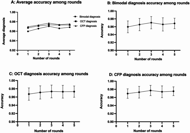
Results: The study involved 7777 pairs of OCT and CFP images, cases labeled with nine prevalent retinal diseases including diabetic retinopathy (DR, 2118 cases), retinal detachment (RD, 121 cases), retinal vein occlusion (RVO, 886 cases), dry age-related macular degeneration (dAMD, 549 cases), wet age-related macular degeneration (wAMD, 1023 cases), epiretinal membrane (ERM, 1061 cases), central serous retinopathy (CSC, 150 cases), macular schisis (MS, 128 cases), macular hole (MH, 86 cases) and normal fundus (1036 cases) were selected for further analysis. All images were assigned to 16 ophthalmologists over five rounds. The average diagnostic accuracy for the nine retinal diseases and normal fundus improved significantly across the five rounds (p = 0.013) and is closely correlated to the duration of ophthalmology study (p = 0.007). Furthermore, significant improvements were observed in the diagnostic accuracy of both OCT (p = 0.028) and CFP (p = 0.021) modalities as the number of rounds increased. Notably, OCT single modal diagnosis demonstrated higher consistency with the final diagnosis in cases of RD, ERM, MS, and MH compared to CFP, while CFP single modal diagnosis has higher consistency in DR, RVO and normal fundus.
Conclusion: The implementation of an artificial intelligence reading label system enhances the diagnostic accuracy of retinal diseases among ophthalmologists and holds potential for integration into future medical education.
Keywords: Artificial intelligence; Diagnostic accuracy; Medical education; Reading label system; Retinal diseases.
© 2025. The Author(s).
Conflict of interest statement
Declarations. Ethics approval and consent to participate: Our study agreed with the tenets of the Declaration of Helsinki. Ethics approval was approved from the PUMC Hospital Institutional Review Board (K3606). The PUMC Hospital Institutional Review Board has approved the waiver of informed consent for participants in this study. Consent for publication: Not applicable. Competing interests: The authors declare no competing interests.
Figures
References
-
- Litjens G, Kooi T, Bejnordi BE, Setio AAA, Ciompi F, Ghafoorian M, et al. A survey on deep learning in medical image analysis. Med Image Anal. 2017;42:60–88. - PubMed
-
- Cohen J. A coefficient of agreement for nominal scales. Educ Psychol Meas. 1960;20(1):37–46.
Publication types
MeSH terms
Grants and funding
LinkOut - more resources
Full Text Sources
Medical
Miscellaneous
