. 2025 Mar 25:16:1537484.
doi: 10.3389/fphys.2025.1537484. eCollection 2025.

Adipose tissue loss during neoadjuvant chemotherapy: a key prognostic factor in advanced epithelial ovarian cancer

Affiliations

Adipose tissue loss during neoadjuvant chemotherapy: a key prognostic factor in advanced epithelial ovarian cancer

Wassim Benouali et al. Front Physiol. .

Abstract

Background: Advanced epithelial ovarian cancer (EOC) patients often receive neoadjuvant platinum-based chemotherapy (NAC), with interval surgery (after three cycles of chemotherapy) considered as a major prognostic factors. We examined how changes in body composition (muscle and adipose tissue) during NAC influence prognosis.

Objective: Using CT images acquired before and during NAC in a cohort of women with advanced EOC, the aim of this study was to analyze body composition (muscle and fat mass) and see whether these parameters, at diagnosis or as they evolve during chemotherapy, can be linked to recurrence-free survival and overall survival (RFS and OS).

Material and methods: The study included 53 patients with FIGO stage III-IV epithelial ovarian cancer. CT images were analyzed to calculate skeletal muscle index (SMI), subcutaneous adipose tissue index visceral adipose tissue index estimated lean body mass (LBM) and estimated whole-body fat mass (WFM). Changes in tissue composition were normalized for 100 days and expressed as % change to account for intervals between scans at baseline and after three cycles of chemotherapy. The impact on survival was assessed by Log-rank test.

Results: At diagnosis, clinical criteria such as age or BMI did not correlate with RFS or OS. 60% of patients were considered sarcopenic (low SMI), including mainly underweight and normal-weight patients. Low SMI was not associated with RFS or OS. Twenty-six patients who underwent interval surgery demonstrated longer relapse-free intervals (p = 0.01). Notably, while muscle parameters showed minimal changes (-2%), parameters assessing adipose tissue showed significant decreases of 10, 12% and 7.6% per 100 days (VATI, SATI and estimated WFM, respectively). Obese patients were particularly affected by this loss of muscle and fat, compared with patients in other BMI categories. Rapid and severe loss of VATI (-28% per 100 days) and estimated WFM (-18% per 100 days) were significantly associated with shorter OS (p = 0.031 and p = 0.046 respectively).

Conclusion: Our findings suggests that early and substantial loss of visceral adipose tissue during NAC is a significant predictor of poor survival in advanced EOC. This highlights an urgent need for targeted nutritional or pharmaceutical strategies to mitigate fat loss and improve patients outcomes.

Keywords: body composition; epithelial ovarian cancer; neoadjuvant chemotherapy; ovarian cancer; sarcopenia; visceral adipose tissue.


Conflict of interest statement

The authors declare that the research was conducted in the absence of any commercial or financial relationships that could be construed as a potential conflict of interest.

Figures

FIGURE 1
FIGURE 1
Care pathway and CT scans analysis of ovarian cancer patients included in our retrospective clinical study. (A) During the diagnostic period, CT scan (defined as the reference CT scan) and laparoscopy were performed to allow staging of the cancer, definition of tumor histology and operability. When not eligible for primary surgery (unsatisfactory debulking), patients underwent neoadjuvant platinum-based chemotherapy. After three to four cycles of chemotherapy, further CT imaging (defined as C3-C4 CT scan) was performed to decide whether interval surgery was possible. Fifty-three women with available CT scans (at baseline and C3-C4) were included in this retrospective clinical study. Patients eligible for primary surgery were excluded. (B) Body composition analyzed on a patient’s CT scan using SliceOmatic software (Tomovision). Tissues are identified according to their contrast in Hounsfield Units (HU): skeletal muscle in red (−29 to +150 HU), subcutaneous adipose tissue in blue (−190 to −30 HU), intermuscular adipose tissue in green (−190 to −30 HU) and visceral adipose tissue in yellow (−150 to −50 HU).
FIGURE 2
FIGURE 2
BMI and body composition parameters at baseline do not influence the survival of patients with ovarian cancer. (A) Kaplan-Meier curves of recurrence-free survival (left) and overall survival (right). Patients were divided into BMI categories. P value were calculated by Log-rank tests. (B) Proportion of sarcopenic and non-sarcopenic patients by body mass index category (BMI - left) or age tertiles (right). Chi2 tests were used to generate p values. (C) Kaplan-Meier curves of recurrence-free survival (left) and overall survival (right). Patients were divided according to baseline body composition parameters: lean body mass (LBM) or whole-body fat mass (WFM). Black lines represent patients with low LBM (<34.3 kg, tertile 1) or low WFM (<17.6 kg, tertile 1). Red lines represent patients with high LBM (>38.6 kg, tertile 3) or high WFM (>26.4 kg, tertile 3). P value were calculated by Log-rank tests.
FIGURE 3
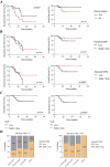
FIGURE 3
Loss of body fat, but not lean body mass, during neoadjuvant chemotherapy is associated with shorter survival for ovarian cancer patients. (A) Kaplan-Meier curves of recurrence-free survival (left) and overall survival (right) according to interval surgery eligibility. Black lines, patients with interval surgery. Red lines, patients without interval surgery. P value were calculated by Log-rank tests. (B) Kaplan-Meier curves of recurrence-free survival (left) and overall survival (right) according to changes in LBM or WFM during NAC. Black lines, patients who lost LBM or WFM. Red lines, patients who gained or had stable LBM or WFM. p value were calculated by Log-rank tests. (C) Kaplan-Meier curves of overall survival according to changes in subcutaneous (left) or visceral (right) adipose tissue index (SATI/VATI) during NAC. Black lines, patients who lost SATI or VATI. Red lines, patients who gained or had stable SATI or VATI. p value were calculated by Log-rank tests. (D) Proportion of patients with loss or gained/stable lean body mass (LBM - left) or whole-body fat mass (WFM - right) during NAC according to BMI category. Chi2 tests were used to generate p values.

References

    1. Armstrong D. K., Alvarez R. D., Bakkum-Gamez J. N., Barroilhet L., Behbakht K., Berchuck A., et al. (2021). Ovarian cancer, version 2.2020, NCCN clinical practice guidelines in oncology. J. Natl. Compr. Canc Netw. 19 (2), 191–226. 10.6004/jnccn.2021.0007 - DOI - PubMed
    1. Byerley L. O., Lee S. H., Redmann S., Culberson C., Clemens M., Lively M. O. (2010). Evidence for a novel serum factor distinct from zinc alpha-2 glycoprotein that promotes body fat loss early in the development of cachexia. Nutr. Cancer 62 (4), 484–494. 10.1080/01635580903441220 - DOI - PubMed
    1. Cai Y., Wang J., Zhang L., Wu D., Yu D., Tian X., et al. (2015). Expressions of fatty acid synthase and HER2 are correlated with poor prognosis of ovarian cancer. Med. Oncol. 32 (1), 391. 10.1007/s12032-014-0391-z - DOI - PMC - PubMed
    1. Chen R. R., Yung M. M. H., Xuan Y., Zhan S., Leung L. L., Liang R. R., et al. (2019). Targeting of lipid metabolism with a metabolic inhibitor cocktail eradicates peritoneal metastases in ovarian cancer cells. Commun. Biol. 2 (1), 281. 10.1038/s42003-019-0508-1 - DOI - PMC - PubMed
    1. Cotogni P., Bozzetti F., Goldwasser F., Jimenez-Fonseca P., Roelsgaard Obling S., Valle J. W. (2022). Supplemental parenteral nutrition in cancer care: why, who, when. Ther. Adv. Med. Oncol. 14, 17588359221113691. 10.1177/17588359221113691 - DOI - PMC - PubMed

LinkOut - more resources