. 2025 Apr 4:20:4123-4144.
doi: 10.2147/IJN.S500159. eCollection 2025.

Human Schwann Cell-Derived Extracellular Vesicle Isolation, Bioactivity Assessment, and Omics Characterization

Affiliations

Human Schwann Cell-Derived Extracellular Vesicle Isolation, Bioactivity Assessment, and Omics Characterization

Aisha Khan et al. Int J Nanomedicine. .

Abstract

Purpose: Schwann cell-derived extracellular vesicles (SCEVs) have demonstrated favorable effects in spinal cord, peripheral nerve, and brain injuries. Herein, a scalable, standardized, and efficient isolation methodology of SCEVs obtaining a high yield with a consistent composition as measured by proteomic, lipidomic, and miRNA analysis of their content is described for future clinical use.

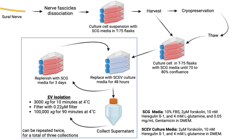
Methods: Human Schwann cells were obtained ethically from nine donors and cultured in a defined growth medium optimized for proliferation. At confluency, the culture was replenished with an isolation medium for 48 hours, then collected and centrifuged sequentially at low and ultra-high speeds to collect purified EVs. The EVs were characterized with mass spectrometry to identify and quantify proteins, lipidomic analysis to assess lipid composition, and next-generation sequencing to confirm miRNA profiles. Each batch of EVs was assessed to ensure their therapeutic potential in promoting neurite outgrowth and cell survival.

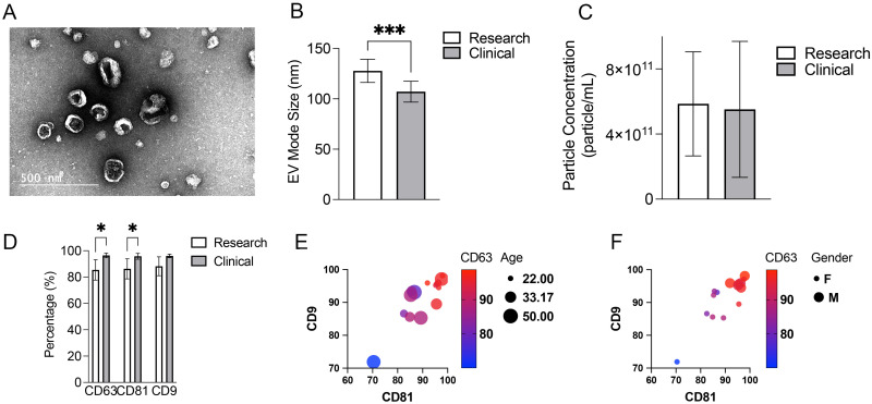
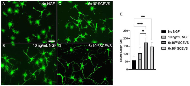
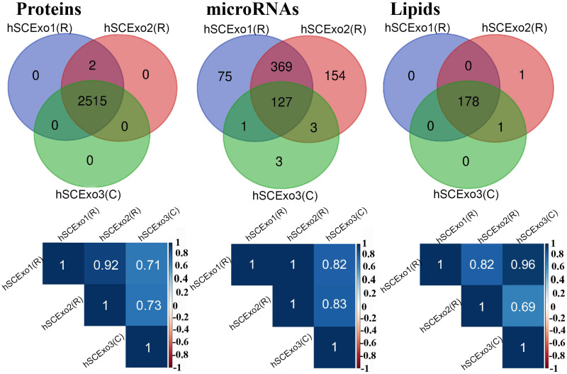
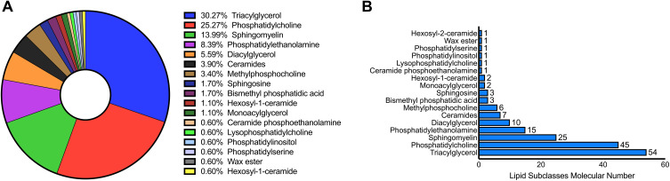
Results: High yields of SCEVs were consistently obtained with similar comprehensive molecular profiles across samples, indicating the reproducibility and reliability of the isolation method. Bioactivity to increase neurite process growth was confirmed in vitro. The predominance of triacylglycerol and phosphatidylcholine suggested its role in cellular membrane dynamics essential for axon regeneration and inflammation mitigation. Of the 2517 identified proteins, 136 were closely related to nervous system repair and regeneration. A total of 732 miRNAs were cataloged, with the top 30 miRNAs potentially contributing to axon growth, neuroprotection, myelination, angiogenesis, the attenuation of neuroinflammation, and key signaling pathways such as VEGFA-VEGFR2 and PI3K-Akt signaling, which are crucial for nervous system repair.

Conclusion: The study establishes a robust framework for SCEV isolation and their comprehensive characterization, which is consistent with their therapeutic potential in neurological applications. This work provides a valuable proteomic, lipidomic, and miRNA dataset to inform future advancements in applying SCEV to the experimental treatment of neurological injuries and diseases.

Keywords: axon growth; lipidomic; myelination; neuroprotection; regeneration.


Conflict of interest statement

The authors have no conflicts related to this paper.

Figures

Figure 1
Figure 1
Manufacture Process of SCs and SCEVs. Starting with sural nerve tissue, cells are dissociated, cultured in T-75 flasks to 70–80% confluence, and either cryopreserved or further cultured. Post-culture, the SCG medium is replaced with SCEV culture media to induce EV production. The supernatant is collected and processed through sequential centrifugation and filtration to isolate EVs. The procedure permits up to three EV harvests.
Figure 2
Figure 2
Phenotypical characterization of SCEVs from both research and clinical batches. (A) Electron micrographs of SCEVs revealed that the nanovesicles were round and elliptical. (B) Significant differences in EV Mode Size between research and clinical batches (***p<0.001). (C) Large variation in particle concentration of SCEVs between batches in both research and clinical production. Concentration can be affected by final product volume and donor variation. (D) Average expression of phenotypical markers of SCEVs, CD63, CD81, and CD9. Significant differences between research and clinical batches were observed for CD63 and CD81 expression (*p<0.05). One of the SCEVs research batches had lower expression of CD63 and CD81, which lowered the overall average of research batch expression. The rest of the SCEVs in the research batches were clustered with higher expressions. (E) No obvious clustering was observed with the age of donors to the phenotypical marker distribution of research batches of SCEVs. (F) Clustering of phenotypical marker distribution by gender observed in research batches of SCEVs.
Figure 3
Figure 3
Effects of SCEVs on Neurite Outgrowth. Fluorescent images (A–D) of rat hippocampal neurons stained with β-tubulin III. A notable increase in neurite length is observed in neurons treated with 6×10⁹ (C) or 6×10¹⁰ (D) particles/mL of SCEVs (n=7 donors) compared to the untreated group (A) and the group treated with 10 ng/mL of Nerve Growth Factor (NGF) (B). Quantitative analysis (E) confirms significantly longer neurites in the SCEVs treated group (*p<0.05, **p<0.01, ***p<0.001). Scale bar: 50 µM.
Figure 4
Figure 4
Comparative Analysis of Lipids, Proteins, and miRNAs in SCEVs. This figure illustrates the overlap and correlation in the contents of SCEVs (hSCExo1R, hSCExo2R, hSCExo3C) through Venn diagrams and Pearson correlation coefficient heat maps. The Venn diagrams reveal a substantial overlap in lipids and proteins across the samples, with a moderate overlap for miRNAs. A total of 180 lipids, 2517 proteins, and 732 miRNAs were identified. Meanwhile, the heat maps below each diagram demonstrate high to very high correlations in the intensity levels of these molecules among the three EVs samples, indicating consistency in exosomal content across different preparations.
Figure 5
Figure 5
Distribution of Lipid Subclasses in Human SCEVs. The composition and distribution of lipid molecules (total of 180) were within 17 distinct subclasses in SCEVs (A) emphasizing the dominance of triacylglycerol and phosphatidylcholine. Triacylglycerol and phosphatidylcholine are the most prevalent, constituting 55% of the total lipid content (B).
Figure 6
Figure 6
MicroRNA-target gene interaction networks. The interaction network map depicts the top 30 microRNAs and their potential target genes (FDR<0.05). MicroRNAs are illustrated in red as source nodes, and their respective target genes are in black as target nodes. Genes implicated in apoptosis, inflammation, and inhibition of regeneration, including caspase 3, caspase 9, IL-6, IL-1ß, and PTEN, are potential targets of hsa-let-7a-5p, hsa-let-7c-5p, hsa-let-7g-5p, hsa-98-5p, hsa-155-5p, hsa-23a-3p, hsa-21-5p, hsa-155-5p.

Similar articles

Cited by

References

    1. Illis LS. Central nervous system regeneration does not occur. Spinal Cord. 2012;50:259–263. [PMID: 22105462]. doi:10.1038/sc.2011.132 - DOI - PubMed
    1. Oliveira JT, Yanick C, Wein N, Gomez Limia CE. Neuron-Schwann cell interactions in peripheral nervous system homeostasis, disease, and preclinical treatment. Front Cell Neurosci. 2023;17:1248922. [PMID: 37900588]. doi:10.3389/fncel.2023.1248922 - DOI - PMC - PubMed
    1. Gomez-Sanchez JA, Carty L, Iruarrizaga-Lejarreta M, et al. Schwann cell autophagy, myelinophagy, initiates myelin clearance from injured nerves. J Cell Biol. 2015;210:153–168. [PMID: 26150392]. doi:10.1083/jcb.201503019 - DOI - PMC - PubMed
    1. Takenaka T, Ohnishi Y, Yamamoto M, Setoyama D, Kishima H. Glycolytic system in axons supplement decreased ATP levels after axotomy of the peripheral nerve. eNeuro. 2023:ENEURO.0353–22.2023. PMID: 36894321. doi:10.1523/ENEURO.0353-22.2023 - DOI - PMC - PubMed
    1. Mietto BS, Jhelum P, Schulz K, David S. Schwann cells provide iron to axonal mitochondria and its role in nerve regeneration. J Neurosci. 2021;41:7300–7313. [PMID: 34272312]. doi:10.1523/JNEUROSCI.0900-21.2021 - DOI - PMC - PubMed

LinkOut - more resources