

Artificial intelligence and consistency in patient care: a large-scale longitudinal study of mammographic density assessment
- PMID: 40201185
- PMCID: PMC11974406
- DOI: 10.1093/bjrai/ubaf004
Artificial intelligence and consistency in patient care: a large-scale longitudinal study of mammographic density assessment
Abstract
Objectives: To assess whether use of an artificial intelligence (AI) model for mammography could result in more longitudinally consistent breast density assessments compared with interpreting radiologists.
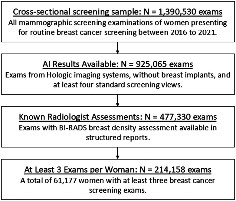
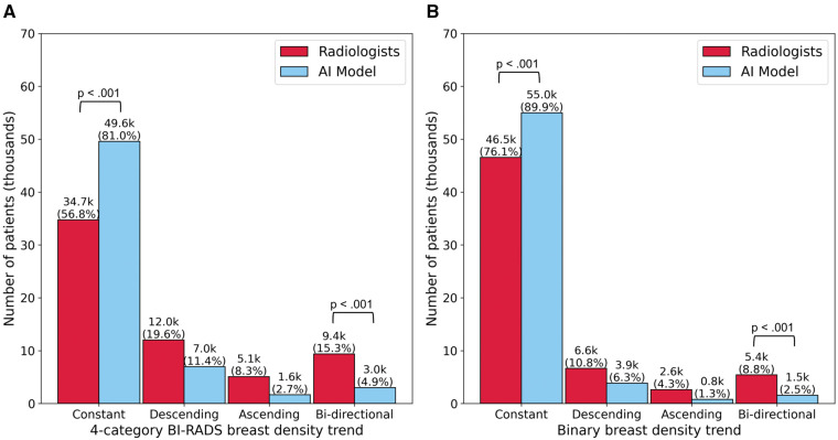
Methods: The AI model was evaluated retrospectively on a large mammography dataset including 50 sites across the United States from an outpatient radiology practice. Examinations were acquired on Hologic imaging systems between 2016 and 2021 and were interpreted by 39 radiologists (36% fellowship trained; years of experience: 2-37 years). Longitudinal patterns in 4-category breast density and binary breast density (non-dense vs. dense) were characterized for all women with at least 3 examinations (61 177 women; 214 158 examinations) as constant, descending, ascending, or bi-directional. Differences in longitudinal density patterns were assessed using paired proportion hypothesis testing.
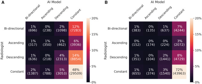
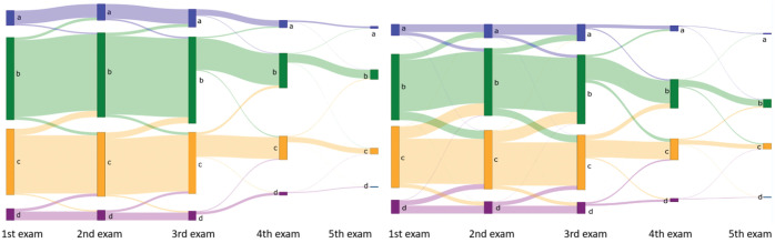
Results: The AI model produced more constant (P < .001) and fewer bi-directional (P < .001) longitudinal density patterns compared to radiologists (AI: constant 81.0%, bi-directional 4.9%; radiologists: constant 56.8%, bi-directional 15.3%). The AI density model also produced more constant (P < .001) and fewer bi-directional (P < .001) longitudinal patterns for binary breast density. These findings held in various subset analyses, which minimize (1) change in breast density (post-menopausal women, women with stable image-based BMI), (2) inter-observer variability (same radiologist), and (3) variability by radiologist's training level (fellowship-trained radiologists).
Conclusions: AI produces more longitudinally consistent breast density assessments compared with interpreting radiologists.
Advances in knowledge: Our results extend the advantages of AI in breast density evaluation beyond automation and reproducibility, showing a potential path to improved longitudinal consistency and more consistent downstream care for screened women.
Keywords: artificial intelligence; breast cancer screening; breast density; longitudinal analysis; mammography.
© The Author(s) 2025. Published by Oxford University Press on behalf of the British Institute of Radiology.
Conflict of interest statement
Washington University in St. Louis (WU) has equity interests in Whiterabbit.ai, Inc. and may receive royalty income and milestone payments from a “Collaboration and License Agreement” with Whiterabbit.ai, Inc. to develop a technology evaluated in this research. These agreements are managed by the WU Institutional COI Committee. The following authors analysed and controlled the data in this work: E.E.T., A.G., D.C., T.P.M., and R.M.H. In addition, the following authors are employed by and/or have equity interests in Whiterabbit.ai, Inc.: D.C., T.P.M., and R.M.H.
Figures
References
-
- Kelemen LE, Pankratz VS, Sellers TA, et al. Age-specific trends in mammographic density: the Minnesota Breast Cancer Family Study. Am J Epidemiol. 2008;167:1027-1036. - PubMed
-
- McCormack VA, dos Santos Silva I.. Breast density and parenchymal patterns as markers of breast cancer risk: a meta-analysis. Cancer Epidemiol Biomarkers Prev. 2006;15:1159-1169. - PubMed
Grants and funding
LinkOut - more resources
Full Text Sources