Case Reports
. 2024 Dec 16;11(2):107-112.
doi: 10.1016/j.aace.2024.12.003. eCollection 2025 Mar-Apr.

Bilateral Adrenal Nodules in the Setting of Disseminated Fungal Infection: An Important Consideration for Appropriate Management of Adrenal Pathology

Affiliations
Case Reports

Bilateral Adrenal Nodules in the Setting of Disseminated Fungal Infection: An Important Consideration for Appropriate Management of Adrenal Pathology

Bailey N Johnson et al. AACE Clin Case Rep. .

Abstract

Background/objective: Increased utilization of cross-sectional imaging has led to a rise in diagnosis of incidental adrenal lesions. Physicians in many clinical settings are increasingly faced with addressing these incidental lesions by initiating the correct workup, diagnosis, and long-term follow-up plan. Our objective was to demonstrate the importance of maintaining a broad differential and completing a thorough workup in determining the correct treatment plan for patients with bilateral adrenal lesions.

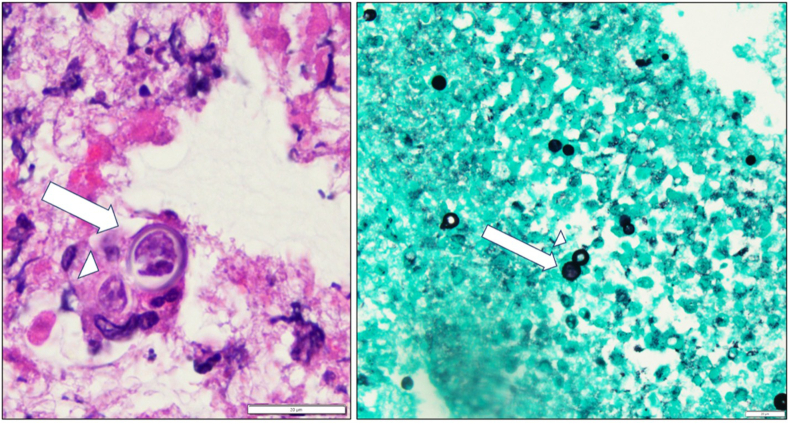
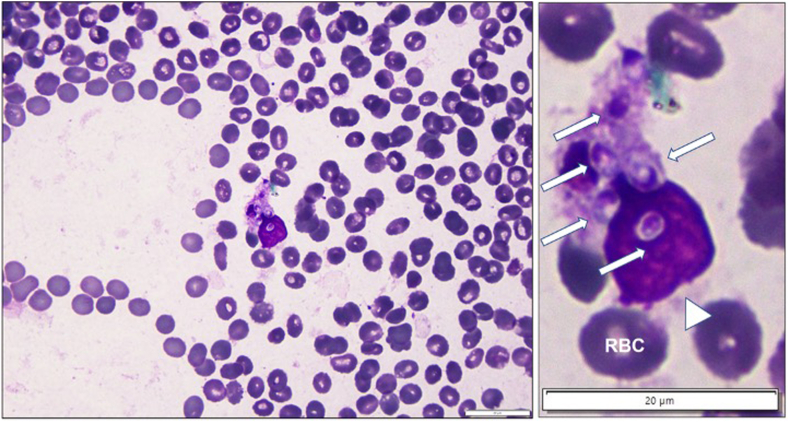
Case report: We present 2 patients who recently completed chemotherapy for lymphoma, found to have new bilateral adrenal lesions on postchemotherapy imaging. Urine antigen and/or adrenal biopsy was performed to confirm the diagnosis of disseminated fungal infection. This diagnosis has major implications on the treatment plan, which includes antifungal therapy instead of surgical management or additional chemotherapy. Cross-sectional imaging after initiation of antifungal treatment demonstrated decreasing size of nodules.

Discussion: A broad differential is critical when working up and developing treatment plans for adrenal nodules, specifically considering a fungal etiology in the setting of immunosuppression or primary extra-adrenal malignancy.

Conclusion: Incidentally found adrenal lesions are becoming more common, and in turn, the obligation for appropriate management of adrenal pathology not only falls to medical and surgical endocrinologists but also to general practitioners. It is prudent to consider atypical etiologies including disseminated fungal infection prior to surgical excision or initiation of chemotherapy as those treatment strategies would not benefit select patients.

Keywords: adrenal mycosis; bilateral adrenal nodules; blastomycosis; histoplasmosis; incidentaloma.


Conflict of interest statement

The authors have no conflicts of interest to disclose.

Figures

Fig. 1
Fig. 1
Postchemotherapy cross-sectional imaging for patient A demonstrating bilateral adrenal nodules. A, Right adrenal nodule measuring 2.6 cm. B, Left adrenal nodule measuring 3.7 x 2.2 cm. C, Positron emission tomography/computed tomography showing bilateral fluorodeoxyglucose-avid adrenal lesions.
Fig. 2
Fig. 2
Postchemotherapy cross-sectional imaging for patient B demonstrating bilateral adrenal nodules. A, Right adrenal nodule measuring 1.0 × 0.9 cm and left adrenal nodule measuring 1.9 × 1.1 cm. B, Positron emission tomography/computed tomography demonstrating fluorodeoxyglucose-avid bilateral adrenal nodules.
Fig. 3
Fig. 3
Fine needle aspiration of adrenal nodule demonstrating large, circular, broad-based budding yeasts. Material aspirated from the left adrenal nodule was initially stained via Diff-Quik and Papanicolaou (neither shown), revealing acute and granulomatous inflammation with scattered budding organisms morphologically consistent with Blastomyces species. Later, a cell black was generated and stained via hematoxylin and eosin (left) and Gomori methenamine silver (right) accentuating the morphologic features already described. Arrow, parent cell; Arrowhead, budding progeny cell. Bars, 20 μm.
Fig. 4
Fig. 4
Map of distribution of Blastomyces (blue) and Histoplasma (red).
Fig. 5
Fig. 5
Fine needle aspiration of adrenal mass demonstrating small, intraphagocytic yeasts. Material aspirated from the left adrenal mass was stained via a modified Wright-Giemsa method (Diff-Quik). Upon review, the specimen was hypocellular consisting primarily of contaminating blood, although adipose tissue and groups of benign adrenocortical cells were present. Small (3.2 × 2.5 μm), pyriform yeasts (arrows) were noted within phagocytic cells (arrowhead). These features were consistent with those expected for Histoplasma capsulatum yeasts. Bars, 20 μm. RBC = red blood cell.

Similar articles

References

    1. Vaidya A., Hamrahian A., Bancos I., Fleseriu M., Ghayee H.K. The evaluation of incidentally discovered adrenal masses. Endocr Pract. 2019;25(2):178–192. doi: 10.4158/DSCR-2018-0565. - DOI - PubMed
    1. Yip L., Duh Q.Y., Wachtel H., et al. American Association of Endocrine Surgeons guidelines for adrenalectomy: executive summary. JAMA Surg. 2022;157(10):870–877. doi: 10.1001/jamasurg.2022.3544. - DOI - PMC - PubMed
    1. Bancos I., Prete A. Approach to the patient with adrenal incidentaloma. J Clin Endocrinol Metab. 2021;106(11):3331–3353. doi: 10.1210/clinem/dgab512. - DOI - PMC - PubMed
    1. Williams A.R., Hammer G.D., Else T. Transcutaneous biopsy of adrenocortical carcinoma is rarely helpful in diagnosis, potentially harmful, but does not affect patient outcome. Eur J Endocrinol. 2014;170(6):829–835. doi: 10.1530/EJE-13-1033. - DOI - PMC - PubMed
    1. Vanderveen K.A., Thompson S.M., Callstrom M.R., et al. Biopsy of pheochromocytomas and paragangliomas: potential for disaster. Surgery. 2009;146(6):1158–1166. doi: 10.1016/j.surg.2009.09.013. - DOI - PubMed

Publication types

LinkOut - more resources