. 2025 Mar 25:19:1547165.
doi: 10.3389/fnana.2025.1547165. eCollection 2025.

Morphological analysis of the filum terminale and detailed description of the distal filum terminale externum: a cadaveric study

Affiliations

Morphological analysis of the filum terminale and detailed description of the distal filum terminale externum: a cadaveric study

Edgar Buloz-Osorio et al. Front Neuroanat. .

Abstract

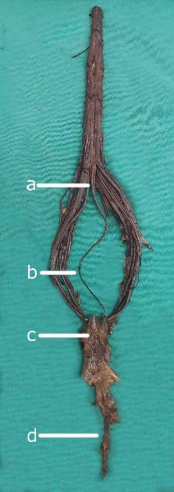
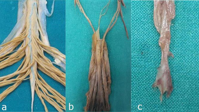
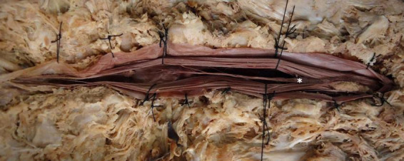
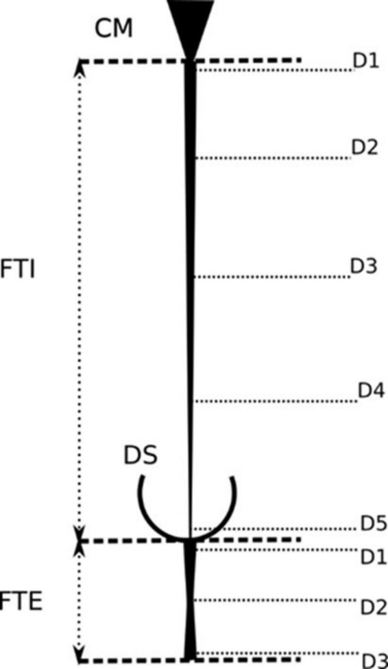
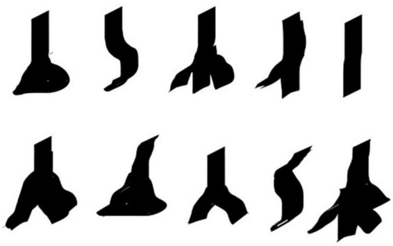
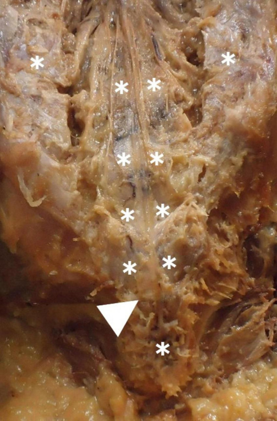
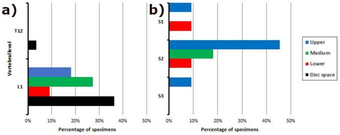
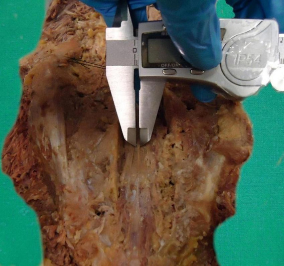
This observational, descriptive anatomical cadaveric study aimed to identify, characterize, and analyze the morphometric parameters of the filum terminale (FT) and macroscopically describe the distal insertion of the FTE. The FT is a complex, three-dimensional, fibro-cellular structure of connective tissue and glial cells, extending from the conus medullaris (CM) to the dural sac (DS) and coccyx. It is divided into two segments: an intradural filum terminale internum (FTI) and extradural filum terminale externum (FTE). Few studies have comprehensively addressed its morphometric characteristics in the last decades. Thirty-eight embalmed (M = 16, F = 22) human cadavers were examined to determine the CM-FTI and DS-FTE vertebral levels and FT, FTI, and FTE lengths and widths. FTI and FTE segmental diameters, correlations, gross characteristics, tension, and mobility in situ and ex vivo were assessed. FTE distal insertion is thoroughly described. FT, FTI, and FTE mean lengths were 254.32 mm (±26.46), 152.75 mm (±22.02), and 106.64 mm (±12.21), respectively. The CM-FTI junction was observed at the L1-L2 disk space (32.1%), DS-FTE fusion in the upper third of S2 (39.3%), and FTI-DS fusion in the DS midline (46.4%). FT length and FTI, FTE lengths were directly correlated, as were all FTI diameters. FT gross characteristics were an irregular surface (71.4%), bright hue (57.1%), macroscopic FTI-CM contrast (64.3%), filiform shape (60.7%), and movement-resistance (53.6%). The FTE exhibited a flattened shape (64.3%), immobility (60.7%), distal insertion at Cx1 (67.8%) and one distal strand (60.7%). FTI segments ≥ 2 mm were uncommon (21.4%). The FTE distal insertion is variable, inserting as strands, with vascular tissue surrounding it. A distal coccygeal venous plexus and single or multiple strand-like insertions of the distal FTE are for the first time described in detail. Discrepancies in the morphometric parameters of the FT between studies highlight the need for standardized protocols, especially given the FT's anatomic-clinical importance and potential role as a neural progenitor niche. We provide a comprehensive basis for future standardized morphometric analyses, acknowledging the limitations of embalmed cadaveric studies.

Keywords: cadaveric study; conus medullaris; dura mater; dural sac; filum terminale; neuroanatomy; spinal cord; tethered cord syndrome.


Conflict of interest statement

The authors declare that the research was conducted in the absence of any commercial or financial relationships that could be construed as a potential conflict of interest.

Figures

FIGURE 1
FIGURE 1
Neuroanatomical components of the filum terminale. (a) Conus medullaris: The tapered, distal portion of the spinal cord. (b) Filum terminale internum: The intradural continuation of the filum terminale, extending from the conus medullaris. (c) Dural sac: The membranous sheath formed by the dura mater surrounding the spinal cord and its roots. (d) Filum terminale externum: The portion of the filum terminale extending from the dural sac to its attachment at the coccyx.
FIGURE 2
FIGURE 2
Detailed anatomical relationships and segmental anatomy of the filum terminale. (a) Junction of the conus medullaris and filum terminale internum: The anatomical transition between the spinal cord and its terminal filum internum. (b) Dural sac: The continuation of the dura mater that encases the spinal cord and extends to the filum terminale internum. (c) Distal segment of the filum terminale externum: The extrameningeal portion of the filum terminale, passing beyond the dural sac to attach at the coccyx.
FIGURE 3
FIGURE 3
Dissection and exposure of the conus medullaris and filum terminale internum. Illustrative dissection highlighting the conus medullaris (*) and the closely associated filum terminale internum, demonstrating the spinal cord’s terminal portion and its intradural extension.
FIGURE 4
FIGURE 4
Morphometric parameters for the measurement of the filum terminale. Schematic representation of the anatomical landmarks and measurement protocols for assessing the length, width, and cross-sectional diameter of the filum terminale. These parameters are crucial for understanding its anatomical variation.
FIGURE 5
FIGURE 5
Macroscopical variability in the periosteal attachment of the filum terminale externum. Silhouettes illustrating the different forms of filum terminale externum insertion at the coccyx.
FIGURE 6
FIGURE 6
Periosteal insertion of the filum terminale externum at the dorsal coccyx and associated vascular structures. Shows the periosteal attachment of the filum terminale externum at the dorsal coccyx (arrow), highlighting the surrounding venous plexus (*). The vascular structures in this region may influence surgical approaches and pathology.
FIGURE 7
FIGURE 7
Frequency distribution of vertebral levels in relation to filum terminale anatomy. Bar graph illustrating the distribution of vertebral levels associated with: (a) The junction of the conus medullaris and filum terminale internum; (b) The fusion between the dural sac and the filum terminale externum. This data provides insight into the variability of these key anatomical landmarks along the spine.
FIGURE 8
FIGURE 8
Caliper-based measurement of the diameters of the filum terminale externum. Detailed illustration of the use of calipers to measure the diameters of the filum terminale externum.

Similar articles

Cited by

References

    1. Abdulrazeq H., Leary O., Tang O., Karimi H., McElroy A., Gokaslan Z., et al. (2023). The surgical histopathology of the Filum Terminale: Findings from a large series of patients with tethered cord syndrome. J. Clin. Med. 13:6. 10.3390/jcm13010006 - DOI - PMC - PubMed
    1. Arvidsson L., Fagerlund M., Jaff N., Ossoinak A., Jansson K., Hägerstrand A., et al. (2011). Distribution and characterization of progenitor cells within the human filum terminale. PLoS One 6:e27393. 10.1371/journal.pone.0027393 - DOI - PMC - PubMed
    1. Balodis A., Neverčika V., Zavertjajeva M., Mikijanskis R., Kalnina M., Breikša-Vaivode A., et al. (2024). Giant myxopapillary ependymoma with multi-site neural axis metastases: A rare case with suboptimal outcome. Am. J. Case Rep. 25:e942392. 10.12659/AJCR.942392 - DOI - PMC - PubMed
    1. Cai Y., Wang R., Wang J., Zhan Q., Wei M., Xiao B., et al. (2024). The discrepancy in triggered electromyography responses between fatty filum and normal filum terminale. BMC Surg. 24:60. 10.1186/s12893-024-02351-0 - DOI - PMC - PubMed
    1. Chern J., Dauser R., Whitehead W., Curry D., Luerssen T., Jea A. (2011). The effect of tethered cord release on coronal spinal balance in tight filum terminale. Spine (Phila Pa 1976) 36 E944–E949. 10.1097/BRS.0b013e3181fc2edd - DOI - PubMed

LinkOut - more resources