

Detailed morphological study of the tongue of forest dragon (Gonocephalus chamaeleontinus) by scanning electron and light microscopy
- PMID: 40201843
- PMCID: PMC11974288
- DOI: 10.5455/OVJ.2025.v15.i2.51
Detailed morphological study of the tongue of forest dragon (Gonocephalus chamaeleontinus) by scanning electron and light microscopy
Abstract
Background: The forest dragon (Gonocephalus chamaeleontinus) is a reptile of the Agamidae family, and its distribution includes Indonesia and Malaysia. The forest dragon uses its tongue to catch insects and invertebrates. In terms of morphology, the tongue of the Agamidae family is different from other reptiles. The study of morphology in the tongues of Agamidae is crucial for understanding their feeding behavior, prey capture mechanisms, and evolutionary relationships.
Aim: This research attempts to analyze the morphology of the dorsal surface of the tongue of G. chamaeleontinus by using the scanning electron microscope (SEM) and its histological structure by using hematoxylin-eosin (HE) staining.
Methods: This study achieves the SEM and light microscope images using hematoxylin eosin stains and employs six samples of G. chamaeleontinus.
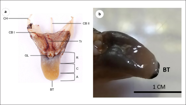
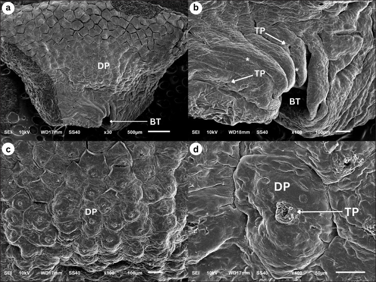
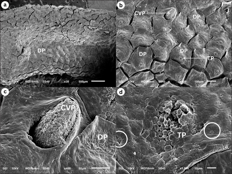
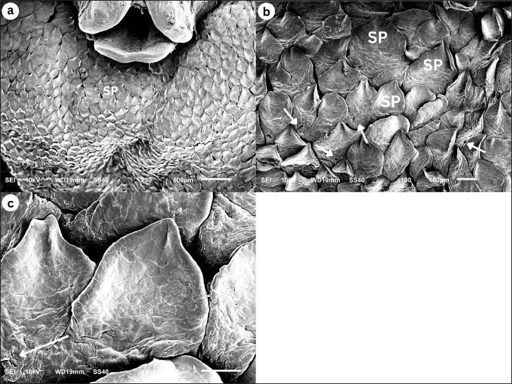
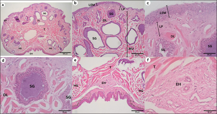
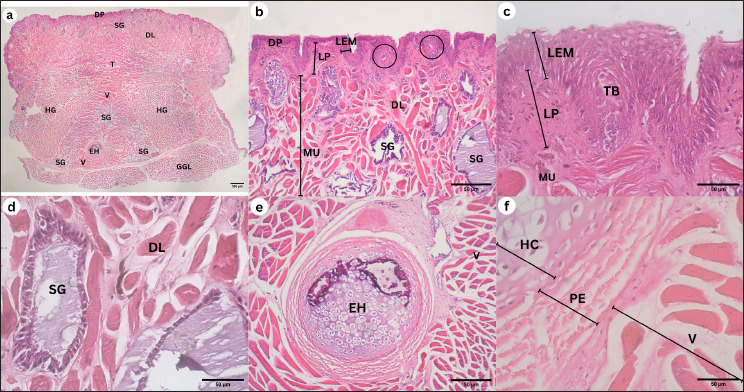
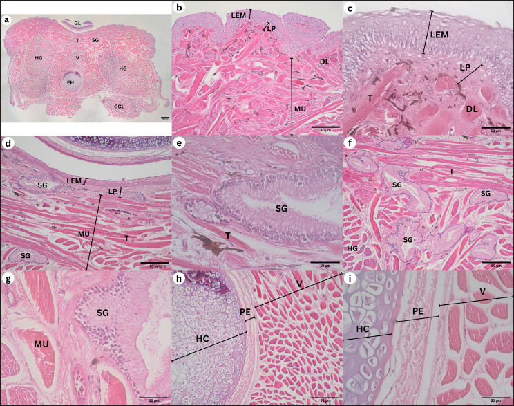
Results: The tongue of G. chamaeleontinus separated into three distinct parts: the apex (A), corpus (C), and radix ®. The structure's A contains dome-shaped papillae (DP). The C section contains DP and circumvallate papillae. The R contains scale-like papillae. Additionally, histological analysis using HE stains revealed the taste buds on the DP and circumvallate papillae and the presence of lingual salivary glands (SG) on the lamina propria mucosa.
Conclusion: The tongue's papillae of G. chamaeleontinus comprise sensory and mechanic papillae, which are also completed by lingual SG.
Keywords: Gonocephalus chamaeleontinus; Light microscopy; Scanning electron microscopy; Tongue.
Conflict of interest statement
The authors declare that there is no conflict of interest.
Figures
References
-
- Abbate F., Latella G., Montalbano G., Guerrera M.C., Germanà G., Levanti M. The lingual dorsal surface of the blue-tongue skink (Tiliqua scincoides) Anat. Histol. Embryol. 2009;38(5):348–350. - PubMed
-
- Abbate F., Latella G., Montalbano G., Guerrera M.C., Levanti M., Ciriaco E. Scanning electron microscopical study of the lingual epithelium of green iguana (Iguana iguana) Anat. Histol. Embryol. 2008;37(4):314–316. - PubMed
-
- Bayoumi S.S., Abd-Elhameed A.E., Mohamed E.S.M. Comparative studies on the dorsal lingual surface of two Egyptian squamate reptile with two different feeding habits. Egypt. J. Exp. Biol. (Zool.) 2011;7(2):203–211.
Publication types
MeSH terms
LinkOut - more resources
Full Text Sources
Miscellaneous