. 2025 May;21(20):e2410258.
doi: 10.1002/smll.202410258. Epub 2025 Apr 9.

Dynamics of Spatial Organization of Bacterial Communities in a Tunable Flow Gut Microbiome-on-a-Chip

Affiliations

Dynamics of Spatial Organization of Bacterial Communities in a Tunable Flow Gut Microbiome-on-a-Chip

Jeeyeon Lee et al. Small. 2025 May.

Abstract

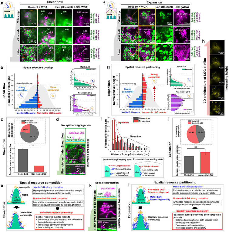
The human intestine, a biomechanically active organ, generates cyclic mechanical forces crucial for maintaining its health and functions. Yet, the physiological impact of these forces on gut microbiota dynamics remains largely unexplored. In this study, we investigate how cyclic intestinal motility influences the dynamics of gut microbial communities within a 3D gut-like structure (µGut). To enable the study, a tunable flow Gut Microbiome-on-a-Chip (tfGMoC) is developed that recapitulates the cyclic expansion and compression of intestinal motility while allowing high-magnification imaging of microbial communities within a 3D stratified, biomimetic gut epithelium. Using deep learning-based microbial analysis, it is found that hydrodynamic forces organize microbial communities by promoting distinct spatial exploration behaviors in microorganisms with varying motility characteristics. Empirical evidence demonstrates the impact of gut motility forces in maintaining a balanced gut microbial composition, enhancing both the diversity and stability of the community - key factors for a healthy microbiome. This study, leveraging the new tfGMoC platform, uncovers previously unknown effects of intestinal motility on modulating gut microbial behaviors and community organizations. This will be critical for a deeper understanding of host-microbiome interactions in the emerging field of microbiome therapeutics.


Conflict of interest statement

The authors declare no conflict of interest.

Figures

Figure 1
Figure 1
The tunable flow Gut Microbiome‐on‐a‐Chip (tfGMoC) recapitulates gut motility, 3D gut epithelium, and key functions. a) Cross‐sectional schematic of the tfGMoC model, illustrating the multilayered PDMS chip organization. Air is infused or withdrawn from the top‐air chamber, inducing deflection of the roof of the cell culture chamber, and resulting in cyclic compression and expansion. b) Schematic of the parallelized tfGMoC array chip, featuring the top layer with air chambers, the middle layer with cell culture chambers, and the glass cover slip. Inset: Magnified schematic of a section of the chip showcasing the bonding orientation of the PDMS layers. c) Image of the tfGMoC array chip with the inset highlighting the chip cross‐section under different conditions; expanded state, steady state, and compressed state, showcasing the elasticity of the cell culture channel roof. d) Fluid dynamic simulation illustrating velocity vectors within the tfGMoC model across one actuation cycle. The velocity vectors originate from two components: a constant volumetric flow rate pumping and the peristaltic motion of the air chambers. e) 3D self‐stratified biomimetic gut epithelium developed from Caco‐2 cells (µGut) within the tfGMoC. f) Magnified top‐view of the µGut cultured in the tfGMoC, signifying the convoluted villi‐like structures. g) Cross‐sectional view of the convoluted villi‐like projections of the µGut within the tfGMoC. The µGut exhibits h) micro‐villi structures and i) brush border. j) ZO‐1 expression indicates the formation of the µGut barrier. Computational Fluid dynamics (CFD) modeling shear stress (top) and pressure drop (bottom) in the tfGMoC model under k) shear flow and l) expansion mode. The top row highlights the schematic of the deflected cell channel at different time points. m) Height of the villi after 7 d of µGut culture under static, shear flow, and different modes of mechanical forces within the tfGMoC model. n = 4, **p < 0.01. n) Quantification of mucin secreted by µGut using Alcian blue staining, representing the fold change in mucin secretion relative to static Caco‐2 culture after 7 d. n = 5, **p < 0.01.
Figure 2
Figure 2
Motile bacteria utilize inherent motility for 3D spatial exploration in the µGut, with minimal influence from hydrodynamic forces. Top and cross‐sectional view of motile EcN attachment to the µGut surface under a) shear and b) expansion modes. c) Shear‐induced colonization of motile EcN on the 3D µGut, displaying the spatial presence throughout the 3D µGut. The top view of EcN colonization was captured at three spatial locations (villus base, side, and top) along the Z‐axis of the crypt‐villus axis. d) Deep learning‐based quantification of motile EcN distributed on the 3D µGut under the shear mode reveals 3D spatial presence but a gradient spatial distribution, predominantly localized near the villus base. e) Expansion‐induced spatial distribution of motile EcN on the 3D µGut. f) The 3D bacterial spatial presence and gradient spatial distribution of motile EcN on the µGut under expansion. g) Abundance of motile EcN colonizing 3D µGut under different mechanical forces. h) Schematics illustrating the 3D spatial exploration behavior of motile EcN and the resulting spatial distribution on the µGut under different mechanical modes.
Figure 3
Figure 3
Hydrodynamics generated by expansion facilitates efficient dispersal of nonmotile LGG, enabling 3D spatial exploration in the µGut. Top and cross‐sectional view of nonmotile LGG attachment to the µGut surface under a) shear and b) expansion modes. c) Shear‐induced nonmotile LGG colonizing in the 3D µGut, displaying localized spatial distribution, predominantly colonized near the base. The top view of LGG colonization was captured at five spatial locations along the Z‐axis of the crypt‐villus axis (base, side, and top of the villus), above the villus top and inter‐villus space. d) Deep learning‐based quantification of nonmotile LGG distributed on the 3D µGut under the shear mode. e) Expansion‐induced nonmotile LGG colonizing in the 3D µGut, displaying relatively even spatial distribution throughout the crypt‐villus axis. f) Quantification of nonmotile LGG under expansion, displaying even spatial distribution throughout the crypt‐villus axis. g) Abundance of nonmotile LGG under shear and expansion modes. h) Spatially varying shear stress along the Z‐axis of the crypt‐villus axis, influencing phenotype changes of nonmotile LGG in distinct spatial locations. i) Increased hydrostatic pressure under expansion, promoting cluster formation of nonmotile LGG. j) Schematics illustrating how hydrodynamic forces altered 3D spatial exploration behaviors of nonmotile LGG, resulting in distinct 3D spatial distribution and phenotypes in the 3D µGut.
Figure 4
Figure 4
Impact of hydrodynamic forces on spatial organization of bacterial communities with mixed motility, influencing compositions, diversity, and stability. a) Confocal imaging of the shear‐promoted spatially intermixed community, displaying motile EcN predominantly colonizing throughout the crypt‐villus axis while nonmotile LGG occupies the villus base in the 3D µGut. b) Deep learning‐based quantification of motile EcN and nonmotile LGG in the shear‐induced community reveals an overlapping spatial distribution of both bacterial species in the lower region of the villus. Because the motility of EcN enables efficient 3D spatial exploration, motile EcN becomes a strong competitor and dominates the shared spatial resource (µGut). c) Quantified species abundances and the resulting composition of the shear‐induced bacterial community exhibit imbalanced community composition. n = 5, ****p < 0.0001. d) Individual forms of nonmotile LGG surrounded by abundant EcN fail to provide spatial segregation. e) Schematics illustrating the intermixed spatial organization of the shear‐induced bacterial community. Motile EcN acquires spatial resources more efficiently than nonmotile LGG due to motility difference, dominating resources and resulting in a community with low diversity and stability. f) Confocal imaging of the expansion‐promoted spatially organized community reveals motile EcN predominantly localized in the lower region of the villus and nonmotile LGG localized in the upper region of the villus in the 3D µGut. g) Quantification of motile and nonmotile bacteria in the expansion‐induced community displays partitioning of spatial resources, with each species occupying a distinct spatial niche in the 3D µGut. h) Dense cluster formation of nonmotile LGG emanating from the top of the villus promoted by expansion. i) The distance distribution of motile EcN from the µGut surface under both mechanical modes. Broader distribution under shear mode suggests loosely bound bacteria with a high motility state, while a narrower distribution under expansion suggests tight bacterial adhesion inducing a low motility state. j) Quantified species abundance and the resulting composition of the expansion‐induced bacterial community, highlighting balanced composition. n = 5, ns > 0.05. k) The cluster phenotype adopted by nonmotile LGG under expansion force provides spatial segregation from motile EcN, contributing an additional strategy for the spatial organization of the bacterial community. l) Schematics illustrating the spatial organization of the expansion‐induced bacterial community. While expansion restricts rapid spatial resource acquisition of motile EcN by limiting its motility state, the same force enables resource acquisition of nonmotile LGG through dispersal, resulting in a diverse and stable community with a balanced composition.

Similar articles

References

    1. a) Bayliss W. M., Starling E. H., J. Physiol. 1899, 24, 99; - PMC - PubMed
    2. b) Gregersen H., Biomechanics of the Gastrointestinal Tract: New Perspectives in Motility Research and Diagnostics, Springer, London: 2003.
    1. Consortium H. M. P., Nature 2012, 486, 207. - PubMed
    1. a) Kashyap P. C., Marcobal A., Ursell L. K., Larauche M., Duboc H., Earle K. A., Sonnenburg E. D., Ferreyra J. A., Higginbottom S. K., Million M., Tache Y., Pasricha P. J., Knight R., Farrugia G., Sonnenburg J. L., Gastroenterology 2013, 144, 967; - PMC - PubMed
    2. b) Touw K., Ringus D. L., Hubert N., Wang Y., Leone V. A., Nadimpalli A., Theriault B. R., Huang Y. E., Tune J. D., Herring P. B., Farrugia G., Kashyap P. C., Antonopoulos D. A., Chang E. B., Physiol. Rep. 2017, 5, 13182; - PMC - PubMed
    3. c) Murakami T., Kamada K., Mizushima K., Higashimura Y., Katada K., Uchiyama K., Handa O., Takagi T., Naito Y., Itoh Y., Digestion 2017, 95, 55; - PubMed
    4. d) Kim H. J., Li H., Collins J. J., Ingber D. E., Proc. Natl. Acad. Sci. USA 2016, 113, E7. - PMC - PubMed
    1. a) Persat A., Nadell C. D., Kim M. K., Ingremeau F., Siryaporn A., Drescher K., Wingreen N. S., Bassler B. L., Gitai Z., Stone H. A., Cell 2015, 161, 988; - PMC - PubMed
    2. b) Dufrêne Y. F., Persat A., Nat. Rev. Microbiol. 2020, 18, 227; - PubMed
    3. c) Rusconi R., Garren M., Stocker R., Annu. Rev. Biophys. 2014, 43, 65; - PMC - PubMed
    4. d) Cremer J., Segota I., Yang C. Y., Arnoldini M., Sauls J. T., Zhang Z., Gutierrez E., Groisman A., Hwa T., Proc. Natl. Acad. Sci. USA 2016, 113, 11414. - PMC - PubMed
    1. a) Wadhwa N., Berg H. C., Nat. Rev. Microbiol. 2021, 20, 161; - PubMed
    2. b) Harshey R. M., Annu. Rev. Microbiol. 2003, 57, 249. - PubMed

LinkOut - more resources