. 2025 Apr;31(4):e70349.
doi: 10.1111/cns.70349.

Sanshen San Formula Hinders Cognitive Function and Pathology in Alzheimer's Disease Through Potentiating the Function of Synapse

Affiliations

Sanshen San Formula Hinders Cognitive Function and Pathology in Alzheimer's Disease Through Potentiating the Function of Synapse

Shiquan Chang et al. CNS Neurosci Ther. 2025 Apr.

Abstract

Background: Alzheimer's disease (AD) constitutes a devastating neurodegenerative disorder, manifested by amyloid-β aggregation, phosphorylated tau accumulation, and progressive cognitive deterioration. Current therapeutic interventions remain predominantly symptomatic, underscoring the urgency for more efficacious treatment strategies.

Purpose: This study elucidated the therapeutic potential of Sanshen San (SSS), a traditional Chinese herbal formula encompassing Polygala Radix, Pini Radix in Poria, and Acori Tatarinowii Rhizoma, on cognitive function and AD pathology.

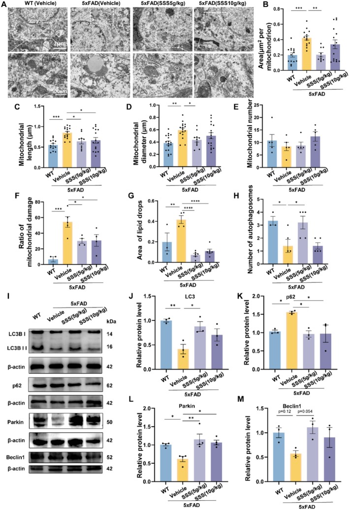
Methods: We implemented both acute Aβ1-42-injected mice and 5xFAD transgenic mouse models. The therapeutic efficacy of SSS was assessed through behavioral paradigms including Y-maze, Novel Object Recognition, and Morris Water Maze. Molecular mechanisms were delineated utilizing RNA sequencing, metabolomics analysis, immunofluorescence staining, Golgi-Cox staining, transmission electron microscopy, and Western blotting.

Results: Chemical analysis unveiled 10 principal bioactive compounds in SSS. The formula substantially ameliorated cognitive performance in both Aβ1-42-injected and 5xFAD mouse models, attenuated Aβ plaque burden, and augmented microglial phagocytosis. SSS safeguarded synaptic integrity, enhanced mitochondrial function, and facilitated autophagy. Transcriptomic and metabolomic analyses demonstrated that SSS predominantly operates by reinstating synaptic transmission and neurotransmitter function, particularly in the dopaminergic system.

Conclusion: SSS efficaciously mitigates AD pathology through potentiating synaptic function, optimizing mitochondrial homeostasis, and restoring neurotransmitter balance, exemplifying a promising multi-target therapeutic strategy for the treatment of AD.

Keywords: Alzheimer's disease; Sanshen san formula; Traditional Chinese medicine; autophagy; neuron.


Conflict of interest statement

The authors declare no conflicts of interest.

Figures

FIGURE 1
FIGURE 1
The components of Sanshensan and their interaction with Alzheimer's disease. (A) Composition of SSS, which is a mix of three traditional Chinese herbs, as well as a diagram of the extraction strategy; (B)Total Ion chromatograms and main components; (C) pC‐pT‐pP network of SSS components in the treatment of AD. The blue, orange, and red nodes represent active compounds, target genes, and pathways, respectively.
FIGURE 2
FIGURE 2
Sanshen San restrains learning and memory deficits in the AD‐like model of acute Aβ1‐42 injection. (A) The graph presents the experimental scheme. Aβ1‐42 or vehicle was injected into the hippocampus of C57BL/6J mice. (B)Y maze test. (C) Novel object recognition test. (D) Escape latency in the MWM test during the 5‐day training and learning regimen. Two‐way ANOVA followed by Student–Newman–Keuls post hoc analysis (time effect: F4,200 = 39.77, p < 0.001); group effect: F4,200 = 3.401, p = 0.0102); group × time effect: F16,200 = 0.6248, p = 0.8617). (E) Average swimming velocity. (F) Time in the target quadrant. (G) Frequency of crossing the platform. (H) Representative traces in MWM tests. The blue dots represent male mice, and the red dots represent female mice. The data were mean ± SEM (n = 8 per group). *p < 0.05; **p < 0.01; ***p < 0.001.
FIGURE 3
FIGURE 3
Sanshen San inhibited learning and memory deficits in 5×FAD mice. (A) The graph shows the experimental scheme. (B) Y maze test. (C) Novel object recognition test. (D) Escape latency in MWM test during the 5‐day training and learning regimen. Two‐way ANOVA followed by Student–Newman–Keuls post hoc analysis (group effect: F3,135 = 20.44, p < 0.001); time effect: F4,135 = 39.66, p < 0.001); group × time effect: F12,135 = 2.063, p = 0.023). (E) Swimming velocity. (F) Duration time in the target quadrant. (G) Frequency of platform crossing. (H) Representative trajectories in MWM tests. The data were mean ± SEM (n = 8 per group). *p < 0.05; **p < 0.01; ***p < 0.001.
FIGURE 4
FIGURE 4
Sanshen San impedes Aβ pathology and spine loss in 5xFAD mice. (A)Representative images of Aβ plaques (6E10) in DG areas of the hippocampus of 5xFAD and SSS‐treated 5×FAD mice. Scale bar =50 μm. (B, C) The bar graph represents the quantification of Aβ plaque area in the hippocampus. (D, E) Number of microglia. (F, G) Percentage of Aβ plaque area measured in the cortex. For (A) to (G), n = 3 mice per group, 3–5 slices each tissue). (H) Golgi‐Cox staining for the measurement of synapse density in the hippocampal neurons. High magnification imaging shows the spine density on basal dendrites per 10 μm. Scale bar = 10 μm. WT, 9.61 ± 0.25; AD, 4.67 ± 0.13; SSS‐M,8.42 ± 0.17; SSS‐H,7.56 ± 0.22. n = 4 mice per group; n = 47 to 50 dendrites per group were counted. (I) Representative Western blot data and (J) quantification of Synaptophysin and PSD95 proteins in the hippocampus. (K) Representative TEM images of synaptic structures. Scale bar = 2 μm/500 nm. (M, N) Quantification of postsynaptic density length and width. Data were presented as mean ± SEM. *p < 0.05; **p < 0.01; ***p < 0.001; ****p < 0.0001.
FIGURE 5
FIGURE 5
Sanshen San inhibits mitochondrial damage and enhances autophagy in 5×FAD mice. (A) Representative TEM images of mitochondrial structures. (B–H) Quantification of mitochondrial area, mitochondrial length, mitochondrial diameter, mitochondrial number, ratio of mitochondrial damage, lipid drop area, and autophagosomes. (I–M) Representative band diagram and quantification of LC3, p62, Parkin, and beclin1 proteins in the hippocampus. Data were presented as mean ± SEM. *p < 0.05; **p < 0.01; ***p < 0.001; ****p < 0.0001.
FIGURE 6
FIGURE 6
RNA‐seq and metabolomics analysis revealed that Sanshen San restored the neurotransmitter function between synapses in 5×FAD mice. (A) PCoA analysis; (B) Heatmap showing the differentially expressed genes in the cortex area of WT, AD, and SSS‐treated mice; (C, D) Number of differentially expressed genes; (E, F) Top 10 GO cellular component enrichment of differentially expressed genes from (C, D); (G) PCoA analysis; (H) Heatmap from mice serum metabolism; (I) KEGG classification of differential metabolites between SSS and AD groups; (J–M) Representative metabolites connecting to synapses differentially expressed.

Similar articles

References

    1. Spires‐Jones T. L. and Ritchie C. W., “A Brain Boost to Fight Alzheimer's Disease,” Science 361, no. 6406 (2018): 975–976, 10.1126/science.aau8060. - DOI - PubMed
    1. Mercerón‐Martínez D., Ibaceta‐González C., Salazar C., Almaguer‐Melian W., Bergado‐Rosado J. A., and Palacios A. G., “Alzheimer's Disease, Neural Plasticity, and Functional Recovery,” Journal of Alzheimer's Disease 82, no. s1 (2021): S37–S50, 10.3233/JAD-201178. - DOI - PubMed
    1. Association, A. S , “Alzheimer's Disease Facts and Figures,” Alzheimer's & Dementia 15, no. 3 (2019): 321–387.
    1. Hoy S. M., “Lecanemab: First Approval,” Drugs 83, no. 4 (2023): 359–365, 10.1007/s40265-023-01851-2. - DOI - PubMed
    1. Liu Q., Contreras A., Afaq M. S., et al., “Intensity‐Dependent Gamma Electrical Stimulation Regulates Microglial Activation, Reduces Beta‐Amyloid Load, and Facilitates Memory in a Mouse Model of Alzheimer's Disease,” Cell & Bioscience 13, no. 1 (2023): 138, 10.1186/s13578-023-01085-5. - DOI - PMC - PubMed