. 2025 Apr;31(4):e70368.
doi: 10.1111/cns.70368.

TRPC4 Mediates Trigeminal Neuropathic Pain via Ca2+-ERK/P38-ATF2 Pathway in the Trigeminal Ganglion of Mice

Affiliations

TRPC4 Mediates Trigeminal Neuropathic Pain via Ca2+-ERK/P38-ATF2 Pathway in the Trigeminal Ganglion of Mice

Xinlong Ke et al. CNS Neurosci Ther. 2025 Apr.

Abstract

Background: Trigeminal neuropathic pain (TNP) is a debilitating condition characterized by chronic facial pain, yet its underlying mechanisms remain incompletely understood. Transient Receptor Potential Canonical 4 (TRPC4) has been reported to promote the development of abnormal pain or pain hypersensitivity in neuropathic pain. However, the specific contribution of TRPC4 to TNP pathogenesis remains unclear.

Aim: This study aimed to investigate the role of TRPC4 in a mouse model of trigeminal neuropathic pain induced by chronic constriction of the unilateral infraorbital nerve (CION).

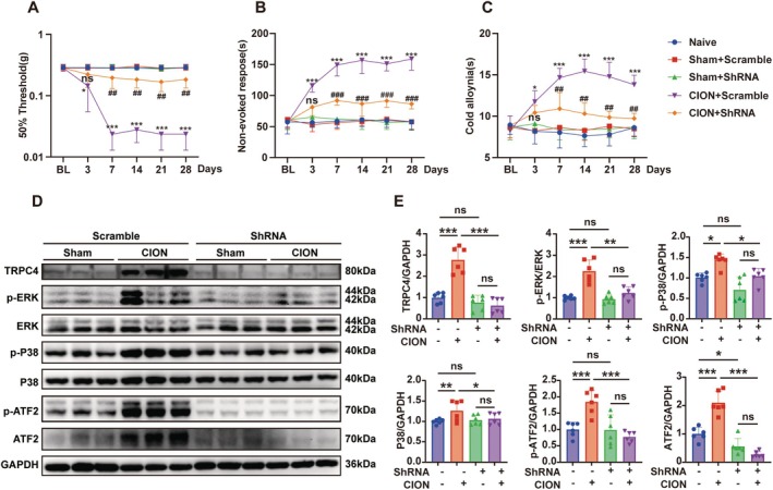
Methods: Adult male/female mice were subjected to either CION surgery or sham surgery. Behavioral assays were conducted to assess facial pain-like responses over a 28-day period. TRPC4 distribution in the trigeminal ganglion (TG) was evaluated using Immunofluorescence. TRPC4 inhibitor ML204 and agonist Englerin A were employed to evaluate the impact of TRPC4 on facial pain-like behaviors. A TRPC4-overexpressing HEK293 cell model was conducted via plasmid transfection. To assess the function of TRPC4, we employed cellular calcium imaging technology to investigate the effects of modulating TRPC4 function by analyzing dynamic changes in intracellular calcium ion concentrations in primary trigeminal ganglion neurons and HEK293 cells. Trpc4 shRNA was used to specifically knock down TRPC4 in the trigeminal ganglion. Western blot analysis was used to assess the activation of ERK, P38, and ATF2 signaling pathways.

Results: Mice subjected to CION exhibited persistent facial pain-like behaviors and a significant increase in TRPC4 expression in TG neurons. Trpc4 shRNA or pharmacological inhibition with ML204 attenuated CION-induced pain behaviors, while activation of TRPC4 with Englerin A induced pain-like responses in naive mice. Calcium imaging revealed that both Englerin A and TRPC4 overexpression elevated intracellular Ca²2+ levels in TG neurons and HEK293 cells. This Ca²2+ influx triggered the activation of ERK and P38, leading to enhanced ATF2 activation. Downregulation of TRPC4 in the TG reduced ERK/P38 phosphorylation and ATF2 expression and activation.

Conclusion: This study provides the first evidence that TRPC4 plays a critical role in CION-induced trigeminal neuropathic pain by promoting the activation of the downstream transcription factor ATF2 via the Ca²2+-ERK/P38 pathway.

Keywords: ATF2; CION; Ca2+; ERK; P38; TRPC4; trigeminal neuropathic pain.


Conflict of interest statement

The authors declare no conflicts of interest.

Figures

FIGURE 1
FIGURE 1
TRPC4 is associated with trigeminal neuropathic pain induced by the chronic constriction of the unilateral infraorbital nerve (CION). (A–C) In C57BL/6 male mice, constriction of the infraorbital nerve elicits mechanical allodynia (A), non‐evoked nociceptive behavior (B), and cold allodynia (C), persisting from day 3 until day 28 post‐surgery (*p < 0.05, **p < 0.01, and ***p < 0.001 versus sham, Two‐way repeated measured ANOVA, followed by Tukey's multiple comparisons test). (D) Volcano plot of DEG between SHAM and trigeminal neuropathic pain (p < 0.05, |Log2FC| > 0.5). (E) Venn diagram of the overlap between DEG, CTD, and GENCARD gene sets. (F) Heatmap results from transcriptome sequencing of the TG show increased Trpc4 transcription levels after nerve injury. (G) Left: Coefficient plot showing the impact of variables on the expression of the intersection genes using LASSO regression. Right: Binomial deviance plot demonstrating the cross‐validation error rates across lambda values. (H) On the 28th day after CION surgery, real‐time fluorescent quantitative PCR was used to reveal the expression levels of Trpc1Trpc7 mRNA in the trigeminal ganglia (TG) of both sham and CION mice, normalized to Gapdh. (*p < 0.05 vs. Sham; multiple unpaired Mann–Whitney tests; n = 6 mice/group). (I) The expression of TRPC4 in the ipsilateral trigeminal ganglion (TG) was assessed after CION or sham surgery, with normalization to GAPDH. (***p < 0.001 vs. sham, multiple unpaired Mann–Whitney tests; n = 6 mice/group). (J) On the 28th day after surgery, representative TRPC4 immunofluorescent images exhibit the distribution of TRPC4‐positive neurons within the trigeminal ganglion (TG) of both the sham surgery group and the CION group. (K) The expression level of TRPC4 in the trigeminal ganglion (TG) was measured after CION or sham surgery and normalized to GAPDH. n = 3 repeats. Data from three independent experiments. (multiple unpaired Mann‐Whitney tests. **p < 0.01 and ***p < 0.001 versus sham (Day 0), BL, baseline assessment before surgery).
FIGURE 2
FIGURE 2
TRPC4 is necessary and sufficient for trigeminal neuropathic pain. (A–C) On the 14th day post‐surgery, a subcutaneous (s.c.) injection of ML204 (10 μg/5 μL) was administered into the left upper lip (ipsilateral to the chronic constriction of the infraorbital nerve (CION) surgery). The mechanical allodynia (A), spontaneous pain behavior (B) and cold allodynia (C) were measured before (BL) and after ML204 injection (Hour 0). (Two‐way repeated measured ANOVA, followed by Tukey's multiple comparisons test. *p < 0.05, **p < 0.01, and ***p < 0.001 versus Sham + Vehicle, #p < 0.05, ##p < 0.01, and ###p < 0.001 versus CION + Vehicle. n = 6 mice/group). (D–F) Effects of systemic administration of the selective TRPC4 receptor antagonist ML204 on mechanical allodynia (D), spontaneous pain behavior (E), and cold allodynia (F) following chronic constriction of the unilateral infraorbital nerve (CION). ML204 (2 mg/kg/day) was administered via intraperitoneal injection from Day 0 to Day 11, starting on Day 14 post‐CION. Measurements were taken during the period from post‐CION surgery to the administration of ML204. (Two‐way repeated measured ANOVA, followed by Tukey's multiple comparisons test. *p < 0.05, **p < 0.01, and ***p < 0.001 versus Sham + Vehicle. # p < 0.05, ## p < 0.01, and ### p < 0.001 versus CION + Vehicle, n = 6 mice/group). (G–I) The mechanical allodynia (G), spontaneous pain behavior (H) and cold allodynia (I) were measured at the first hour after injection of Englerin A (0.25 μg, 2.5 μg and 25 μg/5 μL) or vehicle solution PBS (5 μL) into the TG of WT mice, with a duration of 24 h. (Two‐way repeated measured ANOVA, followed by Tukey's multiple comparisons test. *p < 0.05, **p < 0.01, and ***p < 0.001 versus vehicle, n = 6 mice/group, BL, baseline assessment before surgery).
FIGURE 3
FIGURE 3
(A, B) Localization of TRPC4 immunostaining. (A) Double‐stained immunofluorescent images showing the co‐localization of TRPC4 with NeuN and GS. (B) Distribution of TRPC4‐positive somata: Large, 37%; medium, 36.3%; small, 26.7%. (C) Double‐stained immunofluorescent images showing the co‐localization of TRPC4 with NF200, CGRP, and IB4 in the TG. (Scale bars: 100 μm). (D) The Venn diagram shows the number of neurons double‐stained by TRPC4 and NF200, CGRP, or IB4. The experiment was conducted using 3 mice, and the data were derived from two independent experiments.
FIGURE 4
FIGURE 4
TRPC4 agonist Englerin A increases the Ca2+ concentration in vitro. (A) Example diagram of transfection of HEK293 cells with control plasmid and TRPC4 plasmids. (Scale bar: 200 μm). (B) The levels of TRPC4 protein were measured in HEK293 cells, in HEK293 cells transfected with control plasmids, and in HEK293 cells transfected with TRPC4 plasmids, with all measurements normalized to GAPDH. The sample size for each group is n = 3. (***p < 0.001, control plasmids vs. TRPC4 plasmids, one‐way ANOVA was used for statistical analysis, followed by post hoc Tukey test). (C) Effect of Englerin A on the ΔF/F0 ratio in HEK293 cells transfected with control plasmids or TRPC4 plasmids. Statistical data show that Englerin A increases the ΔF/F 0 ratio in HEK293 cells transfected with TRPC4 plasmids, compared to HEK293 cells transfected with control plasmids. (***p < 0.001 control plasmids vs. TRPC4 plasmids; Student's t‐test; n = 10 cells/group). (D) Identification of primary trigeminal ganglion neurons using double‐stained immunofluorescent images of β3‐tubulin and NeuN. (Scale bar: 100 μm). (E) Representative Ca2+ images showing intracellular Ca2+ activity in trigeminal ganglion (TG) neurons at baseline and after application of Englerin A. (Scale bar: 100 μm). (F) Graph showing the change in mean fluorescence intensity of calcium signaling over time in neurons (shown above) after application of Englerin A. (Using ImageJ for plotting).
FIGURE 5
FIGURE 5
The activation of the AKT and MAPK signaling pathway by Englerin A is dependent on the presence of TRPC4. (A, B) The levels of p‐Akt, p‐ERK, p‐P38, p‐JNK, p‐PI3K, and p‐mTOR in HEK293 cells treated with EA were measured and normalized to GAPDH. (*p < 0.05, **p < 0.01, ***p < 0.001, control vs. EA; multiple unpaired Mann–Whitney tests, n = 6). (C, D) The levels of p‐Akt, p‐ERK, p‐P38, p‐JNK, p‐PI3K, and p‐mTOR in the TRPC‐KO cells and WT cells after treatment with Englerin A were measured and normalized to GAPDH. (Two‐way ANOVA followed by Tukey's multiple comparisons test. *p < 0.05, **p < 0.01, and ***p < 0.001; n = 3).
FIGURE 6
FIGURE 6
The sustained activation of the AKT and MAPK pathways induced by TRPC4 overexpression is dependent on intracellular calcium ions. (A, B) After transfection of HEK293 cells with TRPC4 plasmids, the expression levels of p‐mTOR, p‐PI3K, p‐Akt, p‐ERK, p‐P38, and p‐JNK were measured and normalized to GAPDH. (*p < 0.05, **p < 0.01, ***p < 0.001, control plasmids vs. TRPC4 plasmids, n = 3, one‐way ANOVA was used for statistical analysis, followed by post hoc Tukey test). (C, D) After transfecting HEK293 cells with TRPC4 plasmids and treating them with the calcium chelator BAPTA‐AM, the expression levels of p‐Akt, p‐ERK, p‐P38, and p‐JNK were measured and normalized to GAPDH. (Two‐way ANOVA followed by Tukey's multiple comparisons test. *p < 0.05, **p < 0.01, and ***p < 0.001; n = 3).
FIGURE 7
FIGURE 7
CION and Englerin A induce activation of ERK and P38‐ATF2 in the trigeminal ganglion of mice. (A, B) The expression levels of p‐P38, total P38, p‐ATF2, and total ATF2 in primary trigeminal ganglion neurons were measured after administration of Englerin A and SB 202190, and these levels were normalized to GAPDH. (Two‐way ANOVA followed by post hoc Tukey test. *p < 0.05, **p < 0.01, ***p < 0.001 and ****p < 0.0001, n = 4). (C) Triple immunofluorescence labeling images of ATF2, p‐p38, and β3‐Tubulin in primary trigeminal ganglion neurons. (Scale bar: 20 μm or 50 μm). (D) Western blot of ATF2 and p‐p38 immunoprecipitates from mouse primary trigeminal ganglion neurons. Normal rabbit IgG was used as a negative control. (E, F) Western blot analysis was performed to assess the protein levels of p‐ERK, p‐P38, P38, p‐ATF2, and ATF2 in the trigeminal ganglion (TG) of sham and CION mice groups, normalized to GAPDH. (***p < 0.001 vs. Sham, multiple unpaired Mann–Whitney tests; n = 6 mice/group). (G, H) Western blot analysis was performed to assess the protein levels of p‐ERK, p‐P38, P38, p‐ATF2, and ATF2 in the trigeminal ganglion (TG) of Vehicle‐treated and Englerin A‐treated mice groups, normalized to GAPDH. (**p < 0.01, ***p < 0.001 vs. Vehicle, multiple unpaired Mann–Whitney tests; n = 6 mice/group).
FIGURE 8
FIGURE 8
Downregulation of TRPC4 in the trigeminal ganglion alleviates neuropathic pain triggered by CION through the ERK and P38–ATF2 pathway. (A–C) On the 28th day after virus injection into the trigeminal ganglion (TG), the CION group mice underwent chronic constriction of the unilateral infraorbital nerve (CION) surgery, while the Sham group mice received sham surgery. The naive group mice did not receive any treatment. Mechanical hyperalgesia (A), spontaneous pain behavior (B), and cold allodynia (C) were measured in these mice before (BL) and after CION surgery (from Day 3 to Day 28). (D, E) Western blots and analysis of protein expression of TRPC4, p‐ERK, ERK, p‐P38, P38, p‐ATF2, and ATF2 in the Sham + Scramble groups, CION + Scramble groups, Sham + shRNA groups, and CION + shRNA groups, normalized to GAPDH. (Two‐way ANOVA, followed by Tukey's multiple comparisons test. *p < 0.05, **p < 0.01, and ***p < 0.001; n = 6 mice/group).
FIGURE 9
FIGURE 9
Downregulation of TRPC4 in the trigeminal ganglion mitigates facial pain‐like behaviors triggered by Englerin A via the ERK and P38‐ATF2 pathways. (A–C) On the 28th day after virus injection into the trigeminal ganglion (TG), the Englerin A group mice were injected with EA into the TG, while the Sham group mice were injected with control solution into the TG. Mechanical hyperalgesia (A), spontaneous pain behavior (B) and cold allodynia (C) were measured in these mice before (BL) and after intra‐TG injection of Englerin A (from Hour 1 to Hours 24). (D, E) Western blots and analysis of protein expression of p‐ERK, ERK, p‐P38, P38, p‐ATF2, and ATF2 in the Sham + Scramble groups, Englerin A + Scramble groups, Sham + shRNA groups, and Englerin A + shRNA groups, normalized to GAPDH. (Two‐way ANOVA, followed by Tukey's multiple comparisons test. *p < 0.05, **p < 0.01, and ***p < 0.001; n = 6 mice/group).

Similar articles

References

    1. Zakrzewska J. M., “Differential Diagnosis of Facial Pain and Guidelines for Management,” British Journal of Anaesthesia 111, no. 1 (2013): 95–104. - PubMed
    1. Maarbjerg S., di Stefano G., Bendtsen L., and Cruccu G., “Trigeminal Neuralgia—Diagnosis and Treatment,” Cephalalgia 37, no. 7 (2017): 648–657. - PubMed
    1. Zhang J., Yang M., Zhou M., He L., Chen N., and Zakrzewska J. M., “Non‐Antiepileptic Drugs for Trigeminal Neuralgia,” Cochrane Database of Systematic Reviews 12 (2013): Cd004029. - PMC - PubMed
    1. Khairatkar‐Joshi N., Shah D. M., Mukhopadhyay I., Lingam V. S. P., and Thomas A., “TRPC Channel Modulators and Their Potential Therapeutic Applications,” Pharmaceutical Patent Analyst 4, no. 3 (2015): 207–218. - PubMed
    1. Rubaiy H. N., “Treasure Troves of Pharmacological Tools to Study Transient Receptor Potential Canonical 1/4/5 Channels,” British Journal of Pharmacology 176, no. 7 (2019): 832–846. - PMC - PubMed

MeSH terms