. 2025 Apr;31(4):e70376.
doi: 10.1111/cns.70376.

Repeated Exposure to Sevoflurane in Neonatal Mice Induces Cognitive and Synaptic Impairments in a TTLL6-Mediated Tubulin Polyglutamylation Manner

Affiliations

Repeated Exposure to Sevoflurane in Neonatal Mice Induces Cognitive and Synaptic Impairments in a TTLL6-Mediated Tubulin Polyglutamylation Manner

Yang Yu et al. CNS Neurosci Ther. 2025 Apr.

Abstract

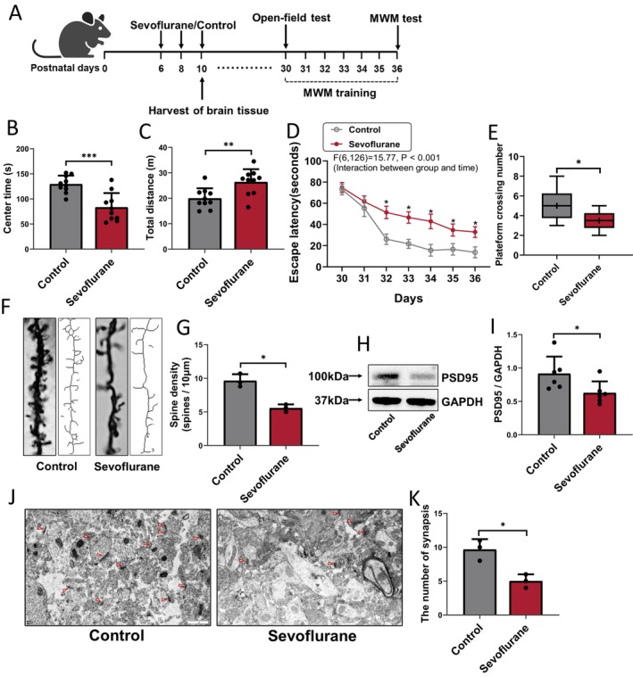
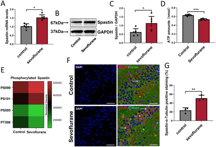
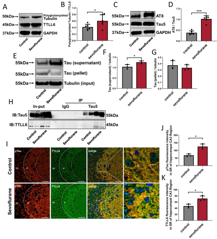
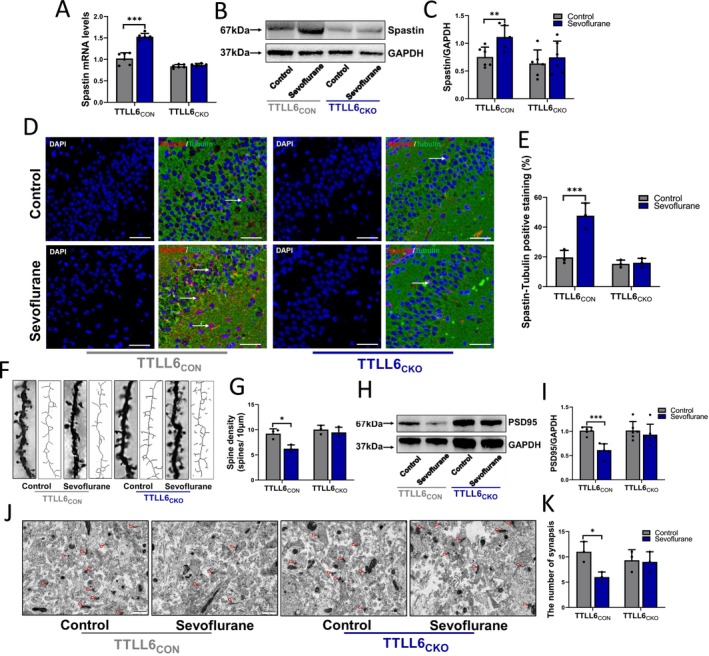
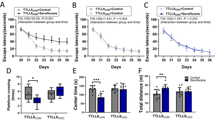
Aims: Repeated sevoflurane exposure during the neonatal stage may induce Tau phosphorylation, dendritic spine loss, and neurocognitive impairment in the developing brain. Tubulin tyrosine ligase like-6 (TTLL6), which aggregates in dendrites due to Tau missorting, regulates microtubule stability via α-tubulin polyglutamylation. Meanwhile, Spastin modulates dendritic spine formation by severing microtubules. We hypothesize that repeated sevoflurane treatment impairs dendritic spine remodeling in neonatal mice by enhancing TTLL6-mediated tubulin polyglutamylation and increasing Spastin expression, leading to cognitive dysfunction in their pre-adolescent stage.

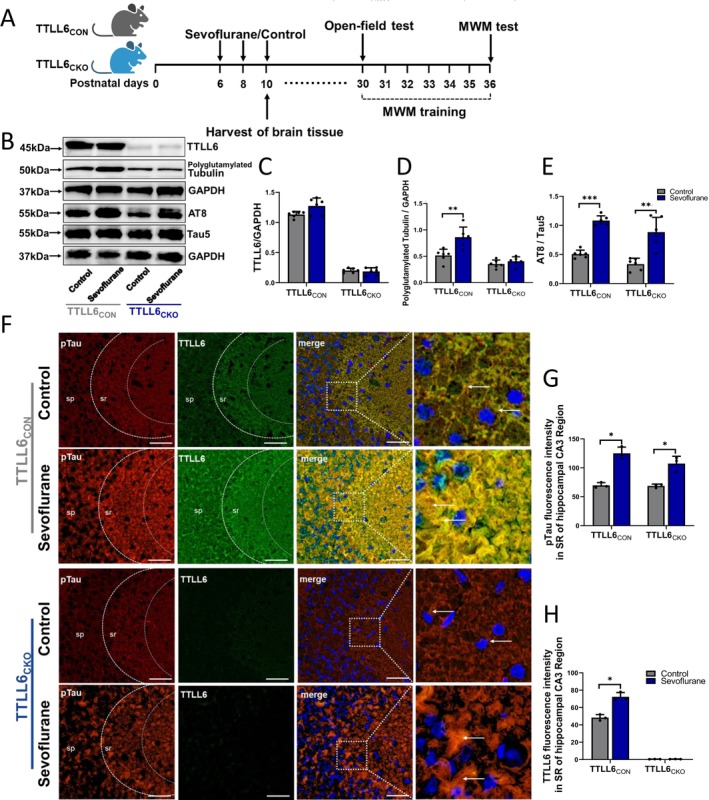
Methods: Six-day-old wild type (WT), TTLL6 brain conditional knockout (TTLL6CKO), TTLL6-flox (TTLL6CON) and Tau-knockout mice were treated with 3% sevoflurane for 2 h daily on postnatal days (P) 6, 8, and 10. Levels of Tau, phosphorylated Tau (pTau), TTLL6, polyglutamylated tubulin, ATP, Spastin, PSD95, Tau-TTLL6 interaction, Tau-TTLL6 missorting, dendritic spine remodeling, and behavioral alterations were compared across these groups.

Results: Repeated sevoflurane exposure during brain development in neonatal mice could reduce dendritic spine density, synapse number, PSD95, and ATP levels, while increasing pTau, polyglutamylated tubulin, Tau-TTLL6 missorting from axons to the somatodendritic compartment, and Spastin levels, leading to cognitive impairment later in their pre-adolescent stage (P30). However, these changes were ameliorated in the TTLL6CKO mice.

Conclusions: Repeated neonatal sevoflurane exposure results in synaptic impairment through TTLL6-mediated tubulin polyglutamylation and increased Spastin expression, causing pre-adolescent cognitive dysfunction in mice. This process is initiated by Tau phosphorylation and missorting from axons to somatodendritic compartments.

Keywords: cognitive impairment; neonatal mice; sevoflurane; synaptic plasticity; tubulin polyglutamylation.


Conflict of interest statement

Transparency statement: The design, data collection, analysis, and interpretation study adhere to rigorous scientific standards. All relevant data and methodologies have been clearly described in this manuscript, and any reproducible data are available upon request. There has been no selective reporting, and all hypotheses were based on a prespecified analysis plan.

The authors declare no conflicts of interest.

Figures

FIGURE 1
FIGURE 1
Effects of repeated sevoflurane exposure in neonatal mice on cognitive and synaptic plasticity functions in developing brain. (A) Experimental design: Neonatal mice (P6) were administered 3% sevoflurane +60% O2 or 60% O2 alone for 2 h every day for 3 days (P6, P8, and P10). Hippocampus tissues were collected on P10 at the conclusion of the sevoflurane or control condition. Behavioral tests were conducted from P30 to P36 (open‐field test: P30; MWM test: P36; MWM training: P30–P36). Summary of (B) centre time and (C) total distance in the open‐field test after control or repetitive sevoflurane treatment; n = 10 mice/group. Navigation test and probe trial were respectively used to compare the (D) escape latency (P30–P36) and (E) platform crossing number (P36) between the two groups (control and sevoflurane); n = 10 mice/group. (F) Representative original photomicrographs and skeletonized dendrites images, and (G) qualification of Golgi‐stained dendrites in the CA3 region, n = 3 mice/group. (H) PSD95 protein expression and (I) qualification in the hippocampus after control or repetitive sevoflurane treatment; n = 6 mice/group. (J) Electron microscope showing the number of synapses (red triangles mark the postsynaptic density (PSD), serving as a marker for synapse counting), scale bar: 1 μm. (K) Qualification of synapsis numbers, n = 3 mice/group. *p < 0.05, **p < 0.01, ***p < 0.001.
FIGURE 2
FIGURE 2
Effects of repeated sevoflurane exposure on Spastin expression in the hippocampus of neonatal mice. (A) Spastin mRNA and (B) Spastin protein expression and (C) qualification in the hippocampus after control or repetitive sevoflurane treatment; n = 6 mice/group. (D) ATP expression levels in the hippocampus of neonatal mice in Control and Sevoflurane groups; n = 3 mice/group. (E) Phosphorylated sites of Spastin expression after control or repetitive sevoflurane treatment; n = 6 mice/group. (F) Immunofluorescence of the protein labeled by Spastin (red) and α‐tubulin (green) in the CA3 area of the hippocampus after control or repetitive sevoflurane treatment, Spastin‐α‐tubulin positive staining (pink) was marked with white arrows, scale bar: 50 μm, blue is DAPI. (G) Qualification of Spastin‐α‐tubulin positive staining (%) in different groups; n = 3 biological replicates. *p < 0.05, **p < 0.01, ***p < 0.001.
FIGURE 3
FIGURE 3
Effects of repeated sevoflurane exposure on Tau and TTLL6 missorting in hippocampus of neonatal mice. (A) Expression of polyglutamylated Tubulin and TTLL6 in the hippocampus after control or repetitive sevoflurane treatment. (B) Qualification of polyglutamylated tubulin/TTLL6 in different groups; n = 6 mice/group. (C) Expression of AT8 and Tau5 in the hippocampus after control or repetitive sevoflurane treatment. (D) Qualification of AT8/Tau5 in different groups; n = 6 mice/group. (E) Results of microtubule binding assay in the hippocampus of neonatal mice in Control and Sevoflurane groups, (F) the supernatanted protein representing microtubule‐unbound Tau, (G) the pelleted protein representing microtubule‐bond Tau; n = 3 mice/group. (H) Immunoprecipitation analysis of Tau5 and TTLL6 interaction in hippocampus cells. IgG: Negative control; Input: Total protein and positive control; IB: Immunoblotting; IP: Immunoprecipitation; n = 3 biological replicates. (I) Immunofluorescence of the protein labeled by pTau (red) and TTLL6 (green) in the CA3 area of hippocampus after control or repetitive sevoflurane treatment, repeated sevoflurane treatment increased pTau‐TTLL6 co‐expressed levels (yellow) in cell body and dendrites (marked with write arrows); blue is DAPI, scale bar: 50 μm. Qualification of (J) pTau and (K) TTLL6 fluorescence intensity in SR area of CA3 region in different groups; n = 3 biological replicates. SR, Stratum radiatum area: Composed mostly of apical dendrites of pyramidal neurons; SP: Stratum pyramidale: Composed mostly of tightly arranged pyramidal cells. *p < 0.05, **p < 0.01, ***p < 0.001.
FIGURE 4
FIGURE 4
Effects of repeated sevoflurane exposure on related protein expression of Tau‐TTLL6 missorting in TTLL6CON or TTLL6CKO neonatal mice. (A) Experimental design: TTLL6CON (TTLL6f/f) and TTLL6CKO (Camk2‐Cre+; TTLL6f/f) mice were administered 3% sevoflurane + 60% O2 or 60% O2 alone for 2 h every day for 3 days (P6, P8, and P10). Hippocampus tissues were collected at the conclusion of the repetitive sevoflurane inhalation or control condition. Behavioral tests were conducted from P30 to P36 (open‐field test: P30; MWM test: P36; MWM training: P30–P36). (B) Expression of TTLL6, polyglutamylated Tubulin, AT8, and Tau5 in the hippocampus after control or repetitive sevoflurane treatment. Qualification of (C) TTLL6, (D) polyglutamylated Tubulin, and (E) AT8/Tau5 in different groups; n = 6 mice/group. (F) Immunofluorescence of the protein labeled by pTau (red) and TTLL6 (green) in the CA3 area of hippocampus after control or repetitive sevoflurane treatment. pTau‐TTLL6 co‐expressed levels (yellow) was marked with white arrows; blue is DAPI, scale bar: 50 μm. Qualification of (G) pTau and (H) TTLL6 fluorescence intensity in the SR area of the CA3 region in different groups; n = 3 biological replicates. SR, Stratum radiatum area: Composed mostly of apical dendrites of pyramidal neurons; SP, Stratum pyramidale: Composed mostly of tightly arranged pyramidal cells. *p < 0.05, **p < 0.01, ***p < 0.001.
FIGURE 5
FIGURE 5
Effects of repeated sevoflurane exposure on Spastin expression and synaptic plasticity functions in TTLL6CON or TTLL6CKO, neonatal mice. (A) Spastin mRNA and (B) Spastin protein expression and (C) qualification in the hippocampus after control or repetitive sevoflurane treatment; n = 6 mice/group. (D) Immunofluorescence of the protein labeled by Spastin (red) and α‐tubulin (green) in the CA3 area of hippocampus after control or repetitive sevoflurane treatment, Spastin‐α‐tubulin positive staining (pink) was marked with write arrows, scale bar: 50 μm, blue is DAPI. (E) Qualification of Spastin‐α‐tubulin positive staining (%) in different groups; n = 3 biological replicates. (F) Representative original photomicrographs and skeletonized dendrites images, and (G) qualification of Golgi‐stained dendrites in the CA3 region, n = 3 mice/group. (H) PSD95 protein expression and (I) qualification in the hippocampus after control or repetitive sevoflurane treatment; n = 6 mice/group. (J) Electron microscope showing the number of synapses (red triangles mark the postsynaptic density (PSD), serving as a marker for synapse counting), scale bar: 1 μm. (K) Qualification of synapsis numbers, n = 3 mice/group. *p < 0.05, **p < 0.01, ***p < 0.001.
FIGURE 6
FIGURE 6
Effects of repeated sevoflurane exposure during the neonatal stage on cognitive functions in TTLL6CON or TTLL6CKO pre‐adolescent mice. Behavioral tests were conducted from P30 to P36 (MWM test: P36; MWM training: P30‐P36; open‐field test: P30). Qualification of escape latency (P30‐P36) (A) between Control and Sevoflurane groups in TTLL6CON mice, (B) between Control in TTLL6CON mice and Control in TTLL6CKO mice, and (C) between Control and Sevoflurane groups in TTLL6CKO mice. Qualification of (D) platform crossing number (P36) in MWM, (E) centre time, and (F) total distance in the open‐field test after control or repetitive sevoflurane treatment; n = 10 mice/group. *p < 0.05, **p < 0.01, ***p < 0.001.

Similar articles

References

    1. Wong‐Kee‐You A. M. B., Loveridge‐Easther C., Mueller C., Simon N., and Good W. V., “The Impact of Early Exposure to General Anesthesia on Visual and Neurocognitive Development,” Survey of Ophthalmology 68, no. 3 (2023): 539–555. - PubMed
    1. Sun M., Xie Z., Zhang J., and Leng Y., “Mechanistic Insight Into Sevoflurane‐Associated Developmental Neurotoxicity,” Cell Biology and Toxicology 38, no. 6 (2022): 927–943. - PMC - PubMed
    1. Yu Y., Yang M., Zhuang X., et al., “Neurotoxic 18‐kDa Apolipoprotein E Fragment Production Contributes to Anesthetic Sevoflurane‐Induced Tau Phosphorylation and Neuroinflammation In Vitro,” Human & Experimental Toxicology 41 (2022): 9603271221102519. - PubMed
    1. Warner D. O., Zaccariello M. J., Katusic S. K., et al., “Neuropsychological and Behavioral Outcomes After Exposure of Young Children to Procedures Requiring General Anesthesia: The Mayo Anesthesia Safety in Kids (MASK) Study,” Anesthesiology 129, no. 1 (2018): 89–105. - PMC - PubMed
    1. Wilder R. T., Flick R. P., Sprung J., et al., “Early Exposure to Anesthesia and Learning Disabilities in a Population‐Based Birth Cohort,” Anesthesiology 110, no. 4 (2009): 796–804. - PMC - PubMed

MeSH terms

LinkOut - more resources