

Molecular cloning and host range analysis of three cytomegaloviruses from Mastomys natalensis
- PMID: 40202317
- PMCID: PMC12090778
- DOI: 10.1128/jvi.02147-24
Molecular cloning and host range analysis of three cytomegaloviruses from Mastomys natalensis
Abstract
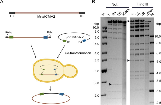
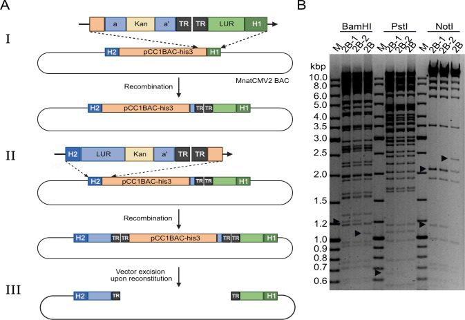
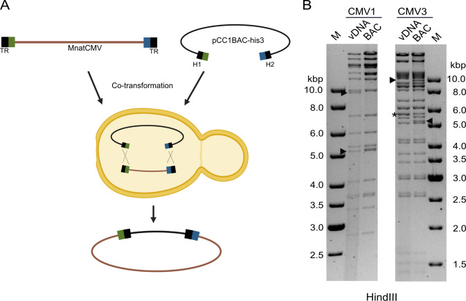
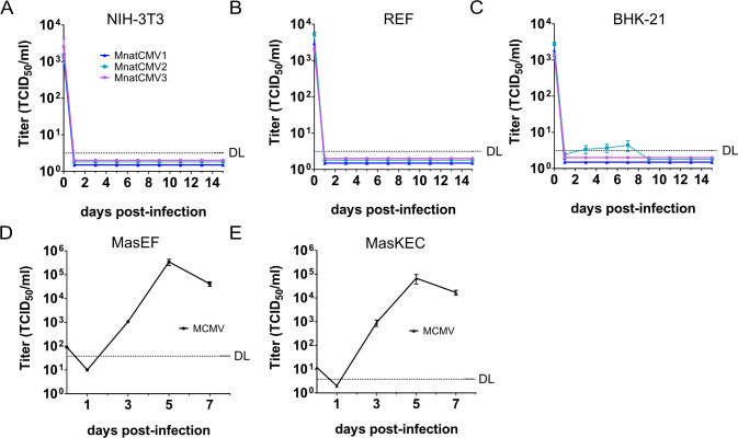
Herpesvirus-based vectors are attractive for use as conventional or transmissible vaccines against emerging zoonoses in inaccessible animal populations. In both cases, cytomegaloviruses (CMVs) as members of the subfamily Betaherpesvirinae are particularly suitable for vaccine development as they are highly specific for their natural host species, infect a large proportion of their host population, and cause mild infections in healthy individuals. The Natal multimammate mouse (Mastomys natalensis) is the natural reservoir of Lassa virus, which causes deadly hemorrhagic fever in humans. M. natalensis was recently reported to harbor at least three different cytomegaloviruses (MnatCMV1, MnatCMV2, and MnatCMV3). Herein, we report the molecular cloning of three complete MnatCMV genomes in a yeast and bacterial artificial chromosome (YAC-BAC) hybrid vector. Purified viral genomes were cloned in yeast by single-step transformation-associated recombination (STAR cloning) and subsequently transferred to Escherichia coli for further genetic manipulation. The integrity of the complete cloned viral genomes was verified by sequencing, and the replication fitness of viruses reconstituted from these clones was analyzed by replication kinetics in M. natalensis fibroblasts and kidney epithelial cells. We also found that neither parental nor cloned MnatCMVs replicated in mouse and rat fibroblasts, nor did they show sustained replication in baby hamster kidney cells, consistent with the expected narrow host range for these viruses. We further demonstrated that an exogenous sequence can be inserted by BAC-based mutagenesis between open reading frames M25 and m25.1 of MnatCMV2 without affecting replication fitness in vitro, identifying this site as potentially suitable for the insertion of vaccine target antigen genes.IMPORTANCECytomegaloviruses (CMVs) recently discovered in the Natal multimammate mouse (Mastomys natalensis) are widespread within the M. natalensis population. Since these rodents also serve as natural hosts of the human pathogen Lassa virus (LASV), we investigated the potential suitability of M. natalensis CMVs (MnatCMVs) as vaccine vectors. We describe the cloning of three different MnatCMV genomes as bacterial artificial chromosomes (BACs). The replicative capacity and species specificity of these BAC-derived MnatCMVs were analyzed in multiple cell types. We also identified a transgene insertion site within one of the MnatCMV genomes suitable for the incorporation of vaccine target antigens. Together, this study provides a foundation for the development of MnatCMVs as transmissible MnatCMV-based LASV vaccines to reduce LASV prevalence in hard-to-reach M. natalensis populations and, thereby, zoonotic transmission to humans.
Keywords: DNA virus; Lassa virus; Mastomys natalensis; bacterial artificial chromosome; cytomegalovirus; herpesvirus; muromegalovirus; recombination; yeast artificial chromosome.
Conflict of interest statement
The authors declare a conflict of interest. Michael A. Jarvis is the founder and a shareholder of The Vaccine Group Ltd.
Figures
Similar articles
-
Isolation and genome sequencing of cytomegaloviruses from Natal multimammate mice (Mastomys natalensis).J Gen Virol. 2023 Aug;104(8):001873. doi: 10.1099/jgv.0.001873. J Gen Virol. 2023. PMID: 37643006 Free PMC article.
-
Inoculation route-dependent Lassa virus dissemination and shedding dynamics in the natural reservoir - Mastomys natalensis.Emerg Microbes Infect. 2021 Dec;10(1):2313-2325. doi: 10.1080/22221751.2021.2008773. Emerg Microbes Infect. 2021. PMID: 34792436 Free PMC article.
-
Multiple DNA viruses identified in multimammate mouse (Mastomys natalensis) populations from across regions of sub-Saharan Africa.Arch Virol. 2020 Oct;165(10):2291-2299. doi: 10.1007/s00705-020-04738-9. Epub 2020 Aug 4. Arch Virol. 2020. PMID: 32754877 Free PMC article.
-
Cloning of herpesviral genomes as bacterial artificial chromosomes.Rev Med Virol. 2003 Mar-Apr;13(2):111-21. doi: 10.1002/rmv.380. Rev Med Virol. 2003. PMID: 12627394 Review.
-
Mapping the zoonotic niche of Lassa fever in Africa.Trans R Soc Trop Med Hyg. 2015 Aug;109(8):483-92. doi: 10.1093/trstmh/trv047. Epub 2015 Jun 17. Trans R Soc Trop Med Hyg. 2015. PMID: 26085474 Free PMC article. Review.
References
-
- Krug LT, Pellett PE. 2021. The family herpesviridae: a brief Introduction, p 212–234. In Howley PM, Knipe DM, Cohen JL, Damania BA (ed), Fields Virology, 7th ed. Wolters Kluwer, Philadelphia.
-
- Brune W. 2013. Molecular Basis of Cytomegalovirus Host Species Specificity, p 322–329. In Reddehase MJ (ed), Cytomegaloviruses: From Molecular Pathogenesis to Intervention. Caister Academic Press, Norfolk, UK.
-
- Calvignac-Spencer S, Kouadio L, Couacy-Hymann E, Sogoba N, Rosenke K, Davison AJ, Leendertz F, Jarvis MA, Feldmann H, Ehlers B. 2020. Multiple DNA viruses identified in multimammate mouse (Mastomys natalensis) populations from across regions of sub-Saharan Africa. Arch Virol 165:2291–2299. doi:10.1007/s00705-020-04738-9 - DOI - PMC - PubMed
MeSH terms
Grants and funding
LinkOut - more resources
Full Text Sources
