. 2025 Apr 9;21(4):e1011407.
doi: 10.1371/journal.pgen.1011407. eCollection 2025 Apr.

Genetic risk for neurodegenerative conditions is linked to disease-specific microglial pathways

Affiliations

Genetic risk for neurodegenerative conditions is linked to disease-specific microglial pathways

Aydan Askarova et al. PLoS Genet. .

Abstract

Genome-wide association studies have identified thousands of common variants associated with an increased risk of neurodegenerative disorders. However, the noncoding localization of these variants has made the assignment of target genes for brain cell types challenging. Genomic approaches that infer chromosomal 3D architecture can link noncoding risk variants and distal gene regulatory elements such as enhancers to gene promoters. By using enhancer-to-promoter interactome maps for human microglia, neurons, and oligodendrocytes, we identified cell-type-specific enrichment of genetic heritability for brain disorders through stratified linkage disequilibrium score regression. Our analysis suggests that genetic heritability for multiple neurodegenerative disorders is enriched at microglial chromatin contact sites, while schizophrenia heritability is predominantly enriched at chromatin contact sites in neurons followed by oligodendrocytes. Through Hi-C coupled multimarker analysis of genomic annotation (H-MAGMA), we identified disease risk genes for Alzheimer's disease, Parkinson's disease, multiple sclerosis, amyotrophic lateral sclerosis and schizophrenia. We found that disease-risk genes were overrepresented in microglia compared to other brain cell types across neurodegenerative conditions and within neurons for schizophrenia. Notably, the microglial risk genes and pathways identified were largely specific to each disease. Our findings reinforce microglia as an important, genetically informed cell type for therapeutic interventions in neurodegenerative conditions and highlight potentially targetable disease-relevant pathways.


Conflict of interest statement

The authors have declared that no competing interests exist.

Figures

Fig 1
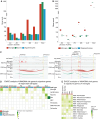
Fig 1. Microglia enhancer-to-promoter interactions were enriched for disease-risk variants across multiple neurodegenerative conditions.
A) Doughnut plots of classifications of PLAC-seq interactions identified in human microglia, neurons and oligodendrocytes [40] with the total number of interactions shown in the center. ‘Promoters’, PLAC-seq bins that overlap a H3K4me3 and H3K27ac peak within 2,000 bp of a transcriptional start site (TSS). ‘Enhancers’, PLAC-seq bins that overlap H3K27ac peaks distal to the TSS. ‘H3K4me3’, PLAC-seq bins that overlap H3K4me3 peaks distal to TSS. ‘ATAC’, PLAC-seq bins that overlap chromatin accessible regions devoid of H3K4me3 and H3K27ac. B) Percent distribution of the number of enhancers interacting with individual promoters (top plot) and the number of promoters interacting with individual enhancers (bottom plot). C) Distribution plot of the proportion of distances between midpoints of promoters and midpoints of enhancers that interact based on chromatin interaction PLAC-seq data. D) Heatmap of partitioned heritability sLDSC regression analysis of PLAC-seq-derived functional annotations: (i) total PLAC-seq bins, (ii) promoter & enhancer PLAC-seq bins, (iii) promoter PLAC-seq bins and (iv) enhancer PLAC-seq bins for microglia, neurons and oligodendrocytes in AD [29,30], PD [27] (excluding 23andMe), MS [33], ALS [31], and schizophrenia [32]. Shown are LDSC enrichment p-values with Benjamini–Hochberg FDR correction for the number of diseases and cell types (-log10(q)). Disease enrichment was considered insignificant if the coefficient z-score was negative and assigned a 0.0 -log10(p) score. OLs, oligodendrocytes; SCZ, schizophrenia.
Fig 2
Fig 2. Microglial disease risk genes were identified for distal GWAS variants using chromatin loops.
A) The number of disease risk genes identified in microglia, neurons and oligodendrocytes using H-MAGMA and GWAS for AD, PD (excluding 23andMe), MS, ALS, and schizophrenia. Gene-to-SNP associations were assigned for SNPs that were located within the promoter or exon of a gene, and within enhancers that were linked to genes through PLAC-seq interactions. B) The number of disease risk genes identified using the sampled down loops for microglia, neurons and oligodendrocytes with H-MAGMA for AD, PD (excluding 23andMe), MS, ALS, and schizophrenia. To account for differences in chromatin interactions between cell types, the number of enhancer-to-promoter interactions was randomly sampled down to 60,000 loops 10 times. Dunn’s test (non-parametric) between cell types within each group: AD (Jansen 2019): microglia-neurons (**), microglia-oligodendrocytes (****), neurons-oligodendrocytes (ns); AD (Kunkle 2019): microglia-neurons (*), microglia-oligodendrocytes (****), neurons-oligodendrocytes (*); PD: microglia-neurons (ns), microglia-oligodendrocytes (*****), neurons-oligodendrocytes (**); MS: microglia-neurons (****), microglia-oligodendrocytes (**), neurons-oligodendrocytes (ns); ALS: microglia-neurons (****), microglia-oligodendrocytes (**), neurons-oligodendrocytes (ns); schizophrenia: microglia-neurons (*), microglia-oligodendrocytes (*), neurons-oligodendrocytes (****). C) UCSC browser visualization of the AD INPP5D locus and the PD MED12L locus. Top track, GWAS SNPs (line indicates GWAS p-value –log10(5e-8) =7.301); middle tracks H3K27ac and H3K4me3 ChIP-seq; bottom track, PLAC-seq interactomes [40]. Yellow, promoters of risk genes identified only by H-MAGMA; pink, promoters of risk genes identified by H-MAGMA and MAGMA. D) EWCE analysis identified cell type enrichment of H-MAGMA disease risk genes from Fig 2A using single-cell RNA-seq from human brain cortical tissue from Tsartsalis et al. (2024) [58]. E) EWCE analysis identified microglia subtype enrichment of microglia H-MAGMA disease risk genes using single-cell RNA-seq of human microglia subtypes from Sun et al. (2023) [59]. EWCE heatmap colour scale represents the number of standard deviations from the mean level of expression found in the target gene lists relative to the bootstrapped mean. SCZ, schizophrenia; OLs, oligodendrocytes.; EWCE p-values *p <0.05, **p<5e-8, ***p=0.
Fig 3
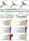
Fig 3. Microglia disease-risk genes impacted disease-specific pathways.
A) UpSet visualization of unique and intersecting H-MAGMA disease-risk gene numbers between AD, PD (excluding 23andMe), MS, ALS and schizophrenia for each cell type. B) Heatmaps of H-MAGMA identified risk genes based on enhancer-to-promoter interactions from PLAC-seq data for AD, PD (excluding 23andMe), MS, ALS (H-MAGMA p<5e-8) and schizophrenia (H-MAGMA p<5e-12) for microglia, neurons and oligodendrocytes. Shown are H-MAGMA FDR corrected p-values (-log10(q)). C) Gene ontology pathway analysis of microglial risk genes identified by H-MAGMA for AD, PD (excluding 23andMe), MS, ALS, and schizophrenia; shown are top 20 pathways. SCZ, schizophrenia.

Similar articles

References

    1. Kurland LT, Mulder DW. Epidemiologic investigations of amyotrophic lateral sclerosis. 2. Familial aggregations indicative of dominant inheritance. I. Neurology. 1955;5(3):182–96. doi: 10.1212/wnl.5.3.182 - DOI - PubMed
    1. Al-Chalabi A, Fang F, Hanby MF, Leigh PN, Shaw CE, Ye W, et al.. An estimate of amyotrophic lateral sclerosis heritability using twin data. J Neurol Neurosurg Psychiatry. 2010;81(12):1324–6. doi: 10.1136/jnnp.2010.207464 - DOI - PMC - PubMed
    1. Graham AJ, Macdonald AM, Hawkes CH. British motor neuron disease twin study. J Neurol Neurosurg Psychiatry. 1997;62(6):562–9. doi: 10.1136/jnnp.62.6.562 - DOI - PMC - PubMed
    1. Gatz M, Reynolds CA, Fratiglioni L, Johansson B, Mortimer JA, Berg S, et al.. Role of genes and environments for explaining Alzheimer disease. Arch Gen Psychiatry. 2006;63(2):168–74. doi: 10.1001/archpsyc.63.2.168 - DOI - PubMed
    1. Wingo TS, Lah JJ, Levey AI, Cutler DJ. Autosomal recessive causes likely in early-onset Alzheimer disease. Arch Neurol. 2012;69(1):59–64. doi: 10.1001/archneurol.2011.221 - DOI - PMC - PubMed

MeSH terms