. 2025 Apr 11;11(15):eadt9695.
doi: 10.1126/sciadv.adt9695. Epub 2025 Apr 9.

Invention of an oral medication for cardiac Fabry disease caused by RNA mis-splicing

Affiliations

Invention of an oral medication for cardiac Fabry disease caused by RNA mis-splicing

Tomonari Awaya et al. Sci Adv. .

Abstract

Pathogenic RNA splicing variants have emerged as promising therapeutic targets due to their role in disease while preserving coding sequences. In this study, we developed RECTAS-2.0, a small molecule designed to correct RNA mis-splicing caused by the GLA c.639+919G>A mutation, which leads to the inclusion of a 57-nucleotide poison exon, resulting in later-onset Fabry disease, particularly prevalent in East Asia. RECTAS-2.0 restored normal GLA mRNA splicing and α-galactosidase activity in patient-derived B-lymphoblastoid cell lines and induced pluripotent stem cell-derived cardiomyocytes. Furthermore, oral administration of RECTAS-2.0 effectively corrected splicing in a transgenic mouse model, demonstrating its substantial splice-switching activity and safety for clinical application. RECTAS-2.0 demonstrated potential applicability to other genetic disorders that involve similar exon competition. These findings underscore the therapeutic potential of RECTAS-2.0 for Fabry disease and highlight its broader implications for RNA splicing-targeted therapies in genetic disorders.


Figures

Fig. 1.
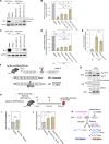
Fig. 1.. RECTAS suppresses the pathogenic GLA poison exon inclusion caused by the c.639+919G>A mutation.
(A) Diagram illustrating the c.639+919G>A mutation-dependent poison exon, leading to NMD and α-GAL loss in FD. (B to D) RECTAS suppresses poison exon inclusion in FD patient–derived B-LCLs. RT-PCR analysis shows reduced poison exon inclusion in two FD B-LCLs (B), while no effect is observed in healthy donor cells (C). Treatment with RECTAS (10, 30 μM, 24 hours) significantly restores α-GAL enzyme activity in patient B-LCLs in a dose-dependent manner (D). (E to I) RECTAS restores normal GLA splicing and reduces Gb3 accumulation in FD iPSC-CMs. Representative fluorescent images of iPSC-CMs from wild-type (wt) and mutant (mut) patients with FD (E) and colocalization of Gb3 with LAMP1 lysosomal marker in iPSC-CMs/mut (F). RT-PCR analysis confirms significant poison exon skipping in iPSC-CMs/mut treated with RECTAS (30 μM, 72 hours) (G), leading to an increase in α-GAL protein levels, as shown by Western blot (H). Gb3 accumulation is significantly reduced following RECTAS treatment (I). (J to L) Transcriptomic analysis of RECTAS-induced alternative splicing events in iPSC-CMs. RNA-seq analysis of FD iPSC-CMs treated with RECTAS (30 μM, 72 hours) identifies 682 alternative splicing events (FDR < 0.10, |dPSI| ≥ 0.20, read count ≥8) (J), categorized into different splicing types (K). The GLA poison exon skipping shows a ΔPSI of 26.8% and is highlighted in red (L). Scale bars, 20 μm [(E) and (F). Mean ± SD; n.s., P ≥ 0.05, **P < 0.01, and ***P < 0.001 by Student’s t test [n = 3 (D) and 5 (I)].
Fig. 2.
Fig. 2.. RECTAS prevents poison exon inclusion by promoting SRSF6-dependent exon 5 recognition in GLA.
(A) Diagram of the SPREADD splicing reporter for GLA c.639+919G>A mutation. (B and C) RECTAS, but not kinetin, suppresses poison exon inclusion. Representative fluorescent images (B) and quantification plot (C). (D) RNA oligonucleotide sequences for the pulldown assay. SRSF6 binding sites predicted by ESE Finder are highlighted for E5_#2. (E) Western blot of pulldown assay shows SRSF6 binding to E5_#2. (F) RECTAS effects on GLA splicing examined with or without siRNA knockdown of SRSF6/SRSF8. (G and H) Effects of SRSF6 binding site mutations on RECTAS-induced exon skipping. (I and J) In vitro splicing assay for GLA exon 4 and poison exon. Pre-mRNA constructs (I) and splicing results (J). Mutation status (G or A at c.639+919) and Mg2+ concentration (0.5 or 2 mM) are indicated. optU1, U>G mutation at poison exon SD to optimize U1snRNA pairing at the −1 position. (K and L) Effects of exon 5 SA optimization on RECTAS-induced poison exon skipping. Mean ± SD; n.s., P ≥ 0.05, **P < 0.01, and ***P < 0.001 by Student’s t test {n = 4 [(C), (F), (H), and (L)]}.
Fig. 3.
Fig. 3.. Identification of RECTAS-2.0 through phenotypic screening for GLA (c.639+919G>A) splicing correction.
(A) Process of the phenotypic screening for GLA (c.639+919G>A). HeLa cells expressing GLA (c.639+919G>A) SPREADD reporter were treated with 418 originally synthesized RECTAS analogs at 10 μM, as well as RECTAS at 10 μM and vehicle only (0.1% DMSO), for 18 hours. GLA splicing rates were quantified on the basis of GFP/RFP fluorescent intensities, and pharmacokinetics and metabolic stability were further considered to identify candidate compounds. (B) A plot of the relative GLA poison exon (Ψ) skipping rate, according to GFP/RFP fluorescent intensities for 418 new compounds, as well as RECTAS and DMSO. Circles, mean values; bars, SE; n = 4 for each sample. The full statistical data are presented in data S2. (C) Chemical structures of RECTAS and RECTAS-2.0 (cpd5819). (D) Treatment of RECTAS-2.0 (1, 3, and 10 μM), RECTAS (10 μM), and DMSO (0.1%), using HeLa cells expressing GLA (c.639+919G>A) splicing Reporter. n.s., P ≥ 0.05; *P < 0.05 and ***P < 0.001 by Student’s t test. n = 4 for each group. (E) Structures of eight compounds assessed for heart delivery rates. (F) Relative activity for GLA (c.639+919G>A mutation) and heart Kp-adjusted activity (indicating expected efficacies in heart tissue when administered p.o.) are plotted for the eight compounds indicated in (D).
Fig. 4.
Fig. 4.. RECTAS-2.0 exhibits therapeutic effects in FD models with the c.639+919 G>A splicing mutation.
(A to E) Effects of RECTAS treatment in FD patient–derived iPSC-CMs. iPSC-CMs/wt and iPSC-CMs/mut were treated with RECTAS (10 μM), RECTAS-2.0 (1 or 10 μM), or solvent only (0.1% DMSO), and analyzed by RT-PCR (A), qRT-PCR (B), Western blotting (C), GLA enzyme activity (D), and lysosomal Gb3 accumulation (E). (F) A diagram indicating transgenic mouse, named Tg(GLA:c.639+919G>A), in which the GLA coding sequence with full-length intron 4 harboring the c.639+919 G>A mutation was integrated into the genome. (G) Peripheral blood cells from Tg(GLA:c.639+919G>A) were cultured ex vivo and treated with CHX for 6 hours to assess the effect of NMD. GLA, Gla, and Actb were detected with the following primer sets: oAM85+oAM95, oAM508+oAM509, and oAM365+oAM366, respectively (primer sequences are indicated in data S3). (H to K) Study of RECTAS-2.0 administration for GLA poison exon suppression. The experimental scheme (H), as well as data quantifying the cardiac expression of normally spliced hGLA after the administration of RECTAS (I) or RECTAS-2.0 (J), is shown. (K) Summary of the current study. RECTAS or RECTAS-2.0 administration prevents GLA transcripts from undergoing NMD by inhibiting inclusion of the poison exon created by the c.639+919G>A mutation of the GLA gene. Data are presented as mean ± SD; n.s., P ≥ 0.05, **P < 0.01, ***P < 0.001, and ****P < 0.0001 by Student’s t test; n = 5 in (B), (D), (E), (I), and (J), n = 10 in (I), and n = 7 in (J).

References

    1. Kim J., Hu C., El Achkar C. M., Black L. E., Douville J., Larson A., Pendergast M. K., Goldkind S. F., Lee E. A., Kuniholm A., Soucy A., Vaze J., Belur N. R., Fredriksen K., Stojkovska I., Tsytsykova A., Armant M., Di Donato R. L., Choi J., Cornelissen L., Pereira L. M., Augustine E. F., Genetti C. A., Dies K., Barton B., Williams L., Goodlett B. D., Riley B. L., Pasternak A., Berry E. R., Pflock K. A., Chu S., Reed C., Tyndall K., Agrawal P. B., Beggs A. H., Grant P. E., Urion D. K., Snyder R. O., Waisbren S. E., Poduri A., Park P. J., Patterson A., Biffi A., Mazzulli J. R., Bodamer O., Berde C. B., Yu T. W., Patient-customized oligonucleotide therapy for a rare genetic disease. N. Engl. J. Med. 381, 1644–1652 (2019). - PMC - PubMed
    1. Kim J., Woo S., de Gusmao C. M., Zhao B., Chin D. H., Di Donato R. L., Nguyen M. A., Nakayama T., Hu C. A., Soucy A., Kuniholm A., Thornton J. K., Riccardi O., Friedman D. A., El Achkar C. M., Dash Z., Cornelissen L., Donado C., Faour K. N. W., Bush L. W., Suslovitch V., Lentucci C., Park P. J., Lee E. A., Patterson A., Philippakis A. A., Margus B., Berde C. B., Yu T. W., A framework for individualized splice-switching oligonucleotide therapy. Nature 619, 828–836 (2023). - PMC - PubMed
    1. Boisson B., Honda Y., Ajiro M., Bustamante J., Bendavid M., Gennery A. R., Kawasaki Y., Ichishima J., Osawa M., Nihira H., Shiba T., Tanaka T., Chrabieh M., Bigio B., Hur H., Itan Y., Liang Y., Okada S., Izawa K., Nishikomori R., Ohara O., Heike T., Abel L., Puel A., Saito M. K., Casanova J.-L., Hagiwara M., Yasumi T., Rescue of recurrent deep intronic mutation underlying cell type-dependent quantitative NEMO deficiency. J. Clin. Investig. 129, 583–597 (2019). - PMC - PubMed
    1. Shibata S., Ajiro M., Hagiwara M., Mechanism-based personalized medicine for cystic fibrosis by suppressing pseudo exon inclusion. Cell Chem. Biol. 27, 1472–1482.e6 (2020). - PubMed
    1. Ohara H., Hosokawa M., Awaya T., Hagiwara A., Kurosawa R., Sako Y., Ogawa M., Ogasawara M., Noguchi S., Goto Y., Takahashi R., Nishino I., Hagiwara M., Branchpoints as potential targets of exon-skipping therapies for genetic disorders. Mol. Ther. Nucleic Acids 33, 404–412 (2023). - PMC - PubMed