. 2025 May;24(5):100965.
doi: 10.1016/j.mcpro.2025.100965. Epub 2025 Apr 7.

Quantitative Proteomics and Phosphoproteomics Analysis of Patient-Derived Ovarian Cancer Stem Cells

Affiliations

Quantitative Proteomics and Phosphoproteomics Analysis of Patient-Derived Ovarian Cancer Stem Cells

Giulia Franciosa et al. Mol Cell Proteomics. 2025 May.

Abstract

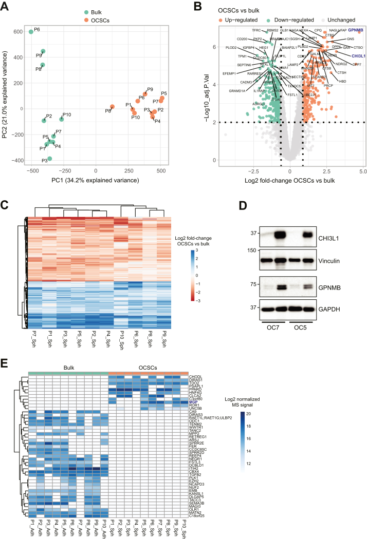
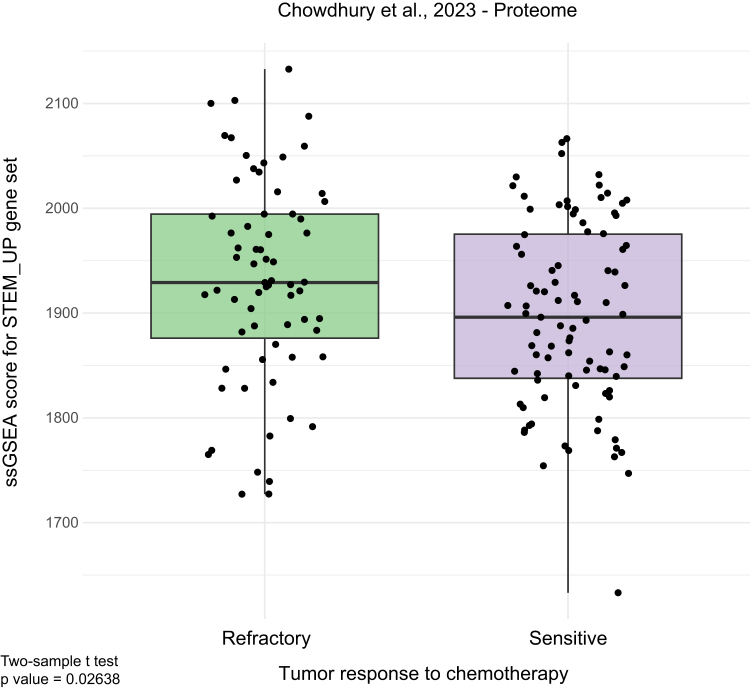
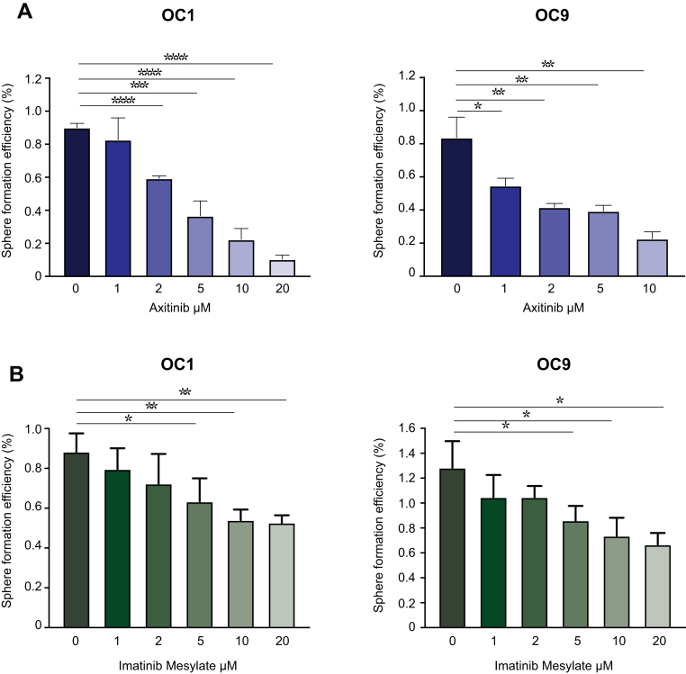
High-grade serous ovarian carcinoma (HGSOC) is the deadliest gynecologic cancer. Key to the progression and ultimate lethality of this subtype is the intra-tumoral heterogeneity, which is defined as the coexistence of different cell types and populations within a single tumor. Among those, ovarian cancer stem cells (OCSCs) are a distinct subpopulation of tumor cells endowed with stem-like properties, which can survive current standard therapies, resulting in tumor recurrence. Here, we generated ex vivo primary OCSC-enriched three-dimensional (3D) spheres from 10 distinct treatment naive patient-derived adherent (2D) cultures. We used state-of-the-art quantitative mass spectrometry to characterize the molecular events associated with OCSCs by analyzing their proteome and phosphoproteome. Our data revealed a stemness-related protein signature, shared within a heterogeneous patient cohort, which correlates with chemo-refractoriness in a clinical proteomics dataset. Moreover, we identified targetable deregulated kinases and aberrant PDGF receptor activation in OCSCs. Pharmacological inhibition of PDGFR in adherent OC cells reduced the stemness potential, measured by sphere formation assay. Overall, we provide a valuable resource to identify new OCSC markers and putative targets for OCSC-directed therapies.

Keywords: HGSOC; cancer stem cells; ovarian cancer; phosphoproteomics; proteomics.


Conflict of interest statement

Conflict of interest The authors declare that they have no conflicts of interest with the contents of this article.

Figures

None
Graphical abstract
Fig. 1
Fig. 1
Proteomic profiling of a cancer stem cell model in ovarian cancer. A, representative images of primary tumor cells from an HGSOC patient, cultured for 7 days either as 2D, adherent cells (left) or as 3D, spheres (right). Scale bar, 200 μm. B, experimental workflow employed in this study. C, number of IDs obtained in this study. D, mRNA expression of stem-associated genes in OCSCs normalized on their adherent counterpart (dashed line) in two patient-derived cultures. Comparisons between experimental groups were performed by unpaired, two-sided, two-sample Student's t test. ∗ = p < 0.05; ∗∗ = p < 0.01; ∗∗∗ = p < 0.005. E, gene set enrichment analysis (GSEA) using the curated gene sets CGP (chemical and genetic perturbations). Proteins were ranked by Log2 fold-change OCSCs/bulk. DDA = data-dependent acquisition; DIA: data-independent acquisition; GndCl = guanidinium chloride; SPD = samples per day; TiIMAC = titanium IMAC; TMT = tandem mass tags.
Fig. 2
Fig. 2
Proteomic analysis of ovarian cancer stem cells. A, principal component analysis (PCA) of the proteomic data. B, Volcano plot analysis of the proteomic data. Proteins colored in orange or green are considered up- or down-regulated, respectively. Proteins were ranked by Limma t statistics, and the for the top 30 on each side the gene name is displayed. C, heatmap of log2 fold-changes OCSCs vs bulk for significantly regulated proteins. D, immunoblot analysis for GPNMB and CHI3L1 in whole cell lysates from two primary cell lines. Vinculin and GAPDH served as loading controls. E, heatmap of log2 MS intensities for proteins unique to one condition (bulk or OCSCs). Adj.P.Val = Benjamini-Hochberg adjusted p value; PC = principal component.
Fig. 3
Fig. 3
Characterization of proteins altered by stemness. A, single-sample gene set enrichment analysis (ssGSEA) of the proteome dataset from Yang et al. The gene set STEM_UP includes proteins significantly upregulated in OCSCs compared to bulk plus proteins unique to OCSCs. The squared dot in red represents the mean. The p value was calculated through one-way ANOVA. 2i = combination of MEK and GSK3 inhibition; LIF = leukemia inhibitory factor. B and C, functional STRING protein networks of proteins belonging to the gene ontology terms and pathways highlighted in blue in supplemental Figs. S7 and S8. Nodes with missing values correspond to proteins unique to OCSCs (B) or bulk (C).
Fig. 4
Fig. 4
Augmented stemness signature in HGSOC tumors refractory to chemotherapy. Single-sample gene set enrichment analysis (ssGSEA) of the proteome dataset from Chowdhury et al. The gene set STEM_UP includes all proteins up-regulated in OCSCs compared to bulk plus proteins unique to OCSCs. The p value was calculated through unpaired, two-sided, two-sample Student's t test.
Fig. 5
Fig. 5
Phosphoproteomic analysis of ovarian cancer stem cells. A, Volcano plot analysis of the phosphoproteomic data. Phosphorylation sites colored in orange or green are considered up- or down-regulated, respectively. Significantly regulated phosphosites from the PDGF receptors are labeled. B, heatmap of log2 fold-changes in OCSCs vs bulk for each individual patient pair for the PDGFR sites highlighted in A. C, immunoblot analysis for phosphorylated and total PDGFRB in whole cell lysates from two primary ovarian cancer cell lines. D, RoKAI inference of kinase activity from the phosphoproteomic data. Kinases with a number of substrates ≥ 3 and p value ≤ 0.05 are shown.
Fig. 6
Fig. 6
PDGFR role in stemness. Sphere formation assay of two primary ovarian cancer cell lines, after treatment with the PDGF receptor inhibitors Axitinib (A) and Imatinib Mesylate (B).

References

    1. Torre L.A., Trabert B., DeSantis C.E., Miller K.D., Samimi G., Runowicz C.D., et al. Ovarian cancer statistics, 2018. CA Cancer J. Clin. 2018;68:284–296. - PMC - PubMed
    1. Bamodu O.A., Chung C.-C., Pisanic T.R., 2nd, Wu A.T.H. The intricate interplay between cancer stem cells and cell-of-origin of cancer: implications for therapeutic strategies. Front. Oncol. 2024;14 - PMC - PubMed
    1. Lupia M., Cavallaro U. Ovarian cancer stem cells: still an elusive entity? Mol. Cancer. 2017;16:64. - PMC - PubMed
    1. Martinez-Val A., Guzmán U.H., Olsen J.V. Obtaining complete human proteomes. Annu. Rev. Genomics Hum. Genet. 2022 - PubMed
    1. Zhang H., Liu T., Zhang Z., Payne S.H., Zhang B., McDermott J.E., et al. Integrated proteogenomic characterization of human high-grade serous ovarian cancer. Cell. 2016;166:755–765. - PMC - PubMed

LinkOut - more resources