. 2025 Apr 10;16(1):269.
doi: 10.1038/s41419-025-07597-x.

Monocyte-macrophage membrane expression of IL-1R2 is a severity biomarker in sepsis

Affiliations

Monocyte-macrophage membrane expression of IL-1R2 is a severity biomarker in sepsis

Domenico Supino et al. Cell Death Dis. .

Abstract

Interleukin-1 (IL-1)/IL-1 receptor family consists of activators and inhibitors which play a key role in inflammation, emergency myelopoiesis, and myeloid cell activation. The latter includes the IL-1R2 decoy receptor. To investigate the expression and significance of IL-1R2 in sepsis, we conducted high-dimensional flow cytometry of circulating cells from patients stratified according to the Sequential Sepsis-Related Organ Failure Assessment (SOFA) score. Here we report that the IL-1 decoy receptor is selectively upregulated on the plasma membrane of leukocytes and, in particular, monocytes from septic patients, and downregulated in septic shock. Flow cytometry combined with transcriptomic analysis of publicly available datasets indicated that IL-1R2 is associated with the differentiation of monocytes to a population of circulating monocytic cells with macrophage features (Mono/Mφ). In vitro stimulation of monocytes from healthy donors with Colony Stimulating Factors (CSFs), in particular GM-CSF and Lipopolysaccharides (LPS), induced IL-1R2+ Mono/Mφ, which recapitulated the characteristics of sepsis-associated monocytic cells, including low expression of HLA-DR, high levels of macrophage markers such as MS4A4A and CD63, immune checkpoints, immunosuppressive molecules and selected scavenger receptors. Membrane-associated IL-1R2 and MS4A4A correlated with immunological markers, cytokine storm, and clinical parameters (e.g., SOFA score, creatinine, survival), reflecting the infection severity in hospitalized patients.Thus, in sepsis IL-1R2 is expressed in a subset of circulating monocytes co-expressing mature macrophage and immune dysfunction features with clinical significance.


Conflict of interest statement

Competing interests: The authors declare no competing interests. Ethical approval and Consent for publication: This study complied with the provisions of the Declaration of Helsinki and was approved by the Institutional Review Board of Humanitas Research Hospital (Approval n° 820/18). Patients were enrolled only after the signature of a written informed consent. In this case, the patient was unable to provide consent, this was obtained from their relatives. Confidentiality of patient data was preserved, and no patient identifiers were used in the dataset.

Figures
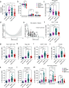
Fig. 1
Fig. 1. IL-1R2 expression and characterization of leukocytes in septic conditions.
ad Frequency of circulating monocytes (a), CD14+CD16-, CD14+CD16+ and CD14-CD16+ monocyte subsets (b), and HLA-DR protein expression (c) in PBMCs from the entire cohort. d Association between disease severity (SOFA score) and HLA-DR expression on monocytes in the entire cohort. The gray area represents the 95% confidence interval. e Correlation of monocyte HLA-DR expression with SOFA score in non-sepsis plus sepsis subgroups. Flow cytometry analysis of IL-1R2 expression in CD3+ T cells (f), Tconv CD4+ (g), Treg (h), CD8+ T cells (i), B cells (j), NK cells (k), total monocytes (l), and CD14+CD16, CD14+CD16+ and CD14CD16+ monocyte subsets (m). am Bars represent mean ± SEM. Kruskal–Wallis with Dunn’s multiple comparison test (ab, fm) or one-way ANOVA with Tukey multiple comparison test (c). *p < 0.05, **p < 0.01, ***p < 0.001, ****p < 0.0001. Healthy donors: n = 19; Non-sepsis patients: n = 23; Sepsis patients: n = 69; Septic Shock patients: n = 25.
Fig. 2
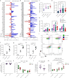
Fig. 2. Association of IL-1R2 to Mono/Mφ functional signatures in monocytic cells in sepsis.
a, b Diagrams showing the expression in monocytes or macrophages of urosepsis monocytic cells (MS1) signatures. DEGs of MS1 Monocytes versus All Monocytes (a) or versus MS2 Monocytes (b) are shown. c CD63 protein expression in monocytes. MS4A4A protein expression in monocytes (d) and in CD14+CD16, CD14+CD16+, and CD14CD16+ monocyte subsets in the entire cohort (e). Flow cytometry analysis of IL-1R2 (f), CD63 (g), and MS4A4A (h) expression in unstimulated monocytes, GM-CSF- and M-CSF-induced Mono/Mφ. i Representative polychromatic plot showing HLA-DR, CD14, and CD16 expression in GM-CSF- and M-CSF-induced monocytic cells. j Frequency of HLA-DR+ CD14+ subset in M-CSF-induced Mono/Mφ after stimulation with LPS. k Frequency of HLA-DR+, CD14+CD16 and CD14+CD16+ subsets in GM-CSF-generated Mono/Mφ after stimulation with LPS. Expression of IL-1R2 in polarized M-CSF- (l) and GM-CSF-induced (m) Mono/Mφ normalized on the respective unstimulated monocytes. cm Bars represent mean ± SEM. One-way ANOVA with Tukey multiple comparison test (c); Kruskal–Wallis with Dunn’s multiple comparison (d, e); Two-tailed Student’s t-test (FH, JM); *p < 0.05, **p < 0.01, ***p < 0.001, ****p < 0.0001. c n = 3–5 per group. d, e Healthy donors: n = 19; Non-sepsis patients: n = 23; Sepsis patients: n = 69; Septic Shock patients: n = 25. fh One representative experiment out of two with similar results is shown. n = 3 healthy donors, with technical duplicates of Mono/Mφ. m n = 6, pooled data from two experiments are shown.
Fig. 3
Fig. 3. Markers of human IL-1R2high Mono/Mφ cells and clinical significance.
a Gating strategy to identify IL-1R2high and IL-1R2low Mono/Mφ. Expression of ARG-1 (b), IL-10 (c), CD204 (d), PD-L1 (e), and SIRPα (f) in monocytes, not polarized IL-1R2high and IL-1R2low Mono/Mφ and Mono/Mφ stimulated with LPS. GM-CSF Mono/Mφ in the left panels; M-CSF in the right panels. bf Data are shown as mean ± SEM. bf One-way ANOVA with Tukey multiple comparison test, *p < 0.05, **p < 0.01, ***p < 0.001, ****p < 0.0001. n = 4 with technical duplicates of Mono/Mφ for 3 donors. Pooled data from two experiments are shown.
Fig. 4
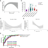
Fig. 4. Association of monocyte membrane-IL-1R2 with mortality.
a Association between the SOFA score and IL-1R2 expression on monocytes in the entire cohort. The gray area represents the 95% confidence interval. b Expression of IL-1R2 in monocytes from non-sepsis, sepsis, and septic shock patients with positive and adverse outcomes. Association of IL-1R2 in monocytes with survival in non-sepsis + sepsis (c) and septic shock (d) cohorts by using proportional hazards additive models with three degrees of freedom. The y-axis showed the partial contribution of IL-1R2 as a survival predictor. A value of 0 on the y-axis indicated no contribution to the model’s outcome. Positive values indicated that the predictor was associated with a higher outcome (mortality) within the cohort. e ROC curve analysis of IL-1R2. Blue line: ROC curve of healthy donors and non-sepsis patients; green line: ROC curve of healthy donors and septic patients; red line: ROC curve of non-sepsis and septic patients. b Two-tailed Mann–Whitney U-test. *p < 0.05. ae Healthy donors: n = 19; Non-sepsis patients: n = 23; Sepsis patients: n = 69; Septic shock patients: n = 25.

References

    1. Singer M, Deutschman CS, Seymour CW, Shankar-Hari M, Annane D, Bauer M, et al. The third international consensus definitions for sepsis and septic shock (Sepsis-3). Jama. 2016;315:801–10. - PMC - PubMed
    1. Rubio I, Osuchowski MF, Shankar-Hari M, Skirecki T, Winkler MS, Lachmann G, et al. Current gaps in sepsis immunology: new opportunities for translational research. Lancet Infect Dis. 2019;19:e422–e36. - PubMed
    1. van der Poll T, van de Veerdonk FL, Scicluna BP, Netea MG. The immunopathology of sepsis and potential therapeutic targets. Nat Rev Immunol. 2017;17:407–20. - PubMed
    1. Cecconi M, Evans L, Levy M, Rhodes A. Sepsis and septic shock. Lancet. 2018;392:75–87. - PubMed
    1. van der Poll T, Shankar-Hari M, Wiersinga WJ. The immunology of sepsis. Immunity. 2021;54:2450–64. - PubMed