. 2025 Apr 9;16(1):3360.
doi: 10.1038/s41467-025-58726-1.

Long-term physical exercise facilitates putative glymphatic and meningeal lymphatic vessel flow in humans

Affiliations

Long-term physical exercise facilitates putative glymphatic and meningeal lymphatic vessel flow in humans

Roh-Eul Yoo et al. Nat Commun. .

Abstract

Regular voluntary exercise has been shown to increase waste transport through the glymphatic system in mice. Here, we investigate the impact of physical exercise on both upstream and downstream brain waste clearance in healthy volunteers via noninvasive MR imaging. Putative glymphatic influx, evaluated using intravenous contrast-enhanced dynamic T1 mapping, increases significantly at the putamen after 12 weeks of long-term exercise using a cycle ergometer. The putative meningeal lymphatic vessel size and flow, measured by intravenous contrast-enhanced black-blood imaging and IR-ALADDIN technique, increase significantly after long-term exercise. Plasma proteomics reveals significant changes in inflammation-related and immune-related proteins (down-regulated: S100A8, S100A9, PSMA3, and DEFA1A3; up-regulated: J chain) after long-term exercise, which correlate with putative glymphatic influx or mLV flow. Our results suggest that increased glymphatic and mLV flow may be the potential mechanism underlying the neuroprotective effects of exercise on cognition, highlighting the importance of long-term, regular exercise.


Conflict of interest statement

Competing interests: The authors declare no competing interests.

Figures
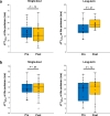
Fig. 1
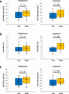
Fig. 1. Changes in ΔT1 values (ΔT1influx and ΔT1efflux) at the putamen after exercise in each group (Single-bout [n = 21], long-term [n = 16]).
Changes in ΔT1influx (a) and ΔT1efflux (b) values at the putamen after exercise are shown for each group. The line across the box denotes the median value, and the boundaries of the box represent the 25th and 75th percentiles. The whiskers indicate the minimum and maximum values. The P values were calculated using the two-sided Wilcoxon signed-rank test. Source data are provided as a source data file.
Fig. 2
Fig. 2. Changes in the mLV ROI size after exercise in representative participants in the single-bout and long-term exercise groups.
2D coronal IR-ALADDIN baseline images (a, c) and reconstructed 3D BB images (b, d) depict an increase in the mLV ROI size after exercise in the long-term exercise group (c, d) but not in the single-bout exercise group (a, b). mLV meningeal lymphatic vessels, ROI region-of-interest.
Fig. 3
Fig. 3. Changes in the mLV ROI size from BB imaging after exercise in each group (single-bout [n = 19], long-term [n = 12]).
The line across the box denotes the median value, and the boundaries of the box represent the 25th and 75th percentiles. The whiskers indicate the minimum and maximum values. The P values were calculated using the two-sided Wilcoxon signed-rank test. Source data are provided as a Source Data file. BB black-blood, mLV meningeal lymphatic vessels, ROI region-of-interest.
Fig. 4
Fig. 4. Changes in mLV imaging metrics (mLV ROI size, mLV PSC, and mLV flow) from IR-ALADDIN after exercise in each group (Single-bout [n = 21], long-term [n = 15]).
Changes in the mLV ROI size (a), mLV PSC (b), and mLV flow (c) after exercise are shown for each group. The line across the box denotes the median value, and the boundaries of the box represent the 25th and 75th percentiles. The whiskers indicate the minimum and maximum values. The P values were calculated using the two-sided Wilcoxon signed-rank test. Source data are provided as a Source Data file. mLV meningeal lymphatic vessels, PSC percent signal change, ROI region-of-interest.
Fig. 5
Fig. 5. Heatmap displaying the correlation coefficients between differentially expressed proteins (DEPs) and MR imaging metrics of glymphatic and mLV flow. ROImLVs were calculated from BB imaging, whereas PSCmLVs and FlowmLVs were derived from IR-ALADDIN imaging.
The P values were calculated using the two-sided Spearman’s correlation test. Asterisks indicate statistical significance of P < 0.05. The raw data of mass spectrometry proteomics have been deposited to the ProteomeXchange Consortium via the PRIDE partner repository with the dataset identifier PXD058255. mLV meningeal lymphatic vessels, PSC percent signal change, ROI region-of-interest.
Fig. 6
Fig. 6. Illustration of the study design, including single-bout and long-term exercise schedules and pre- and post-exercise examinations.
IPAQ international physical activity questionnaire.
Fig. 7
Fig. 7. Schematic image of IR-ALADDIN imaging protocol and ROIs of the dorsal parasagittal mLVs on IR-ALADDIN.
a The positions of nine coronal IR-ALADDIN images are shown as blue stripes. b, c IR-ALADDIN baseline images demonstrate the ROIs of the mLVs (green) and SSS (red). The ROIs of mLVs adjacent to the SSS were segmented based on the distance from the SSS (yellow circle) and PSC on IR-ALADIDN. mLV meningeal lymphatic vessels, PSC percent signal change, ROI region-of-interest.

References

    1. Iliff, J. J. et al. A paravascular pathway facilitates CSF flow through the brain parenchyma and the clearance of interstitial solutes, including amyloid β. Sci. Transl. Med.4, 147ra111 (2012). - PMC - PubMed
    1. Louveau, A. et al. Structural and functional features of central nervous system lymphatic vessels. Nature523, 337–341 (2015). - PMC - PubMed
    1. Aspelund, A. et al. A dural lymphatic vascular system that drains brain interstitial fluid and macromolecules. J. Exp. Med.212, 991–999 (2015). - PMC - PubMed
    1. Jessen, N. A., Munk, A. S. F., Lundgaard, I. & Nedergaard, M. The glymphatic system: a beginner’s guide. Neurochem. Res.40, 2583–2599 (2015). - PMC - PubMed
    1. Hershenhouse, K. S., Shauly, O., Gould, D. J. & Patel, K. M. Meningeal lymphatics: a review and future directions from a clinical perspective. Neurosci. Insights14, 1179069519889027 (2019). - PMC - PubMed