. 2025 Apr;57(4):872-887.
doi: 10.1038/s12276-025-01432-1. Epub 2025 Apr 10.

Role of phospholipase Cη1 in lateral habenula astrocytes in depressive-like behavior in mice

Affiliations

Role of phospholipase Cη1 in lateral habenula astrocytes in depressive-like behavior in mice

Sukwoon Song et al. Exp Mol Med. 2025 Apr.

Abstract

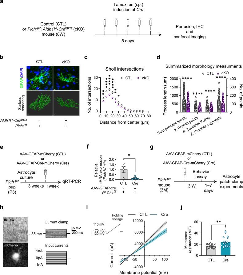
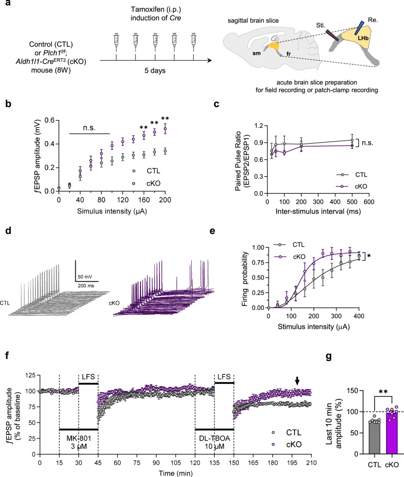
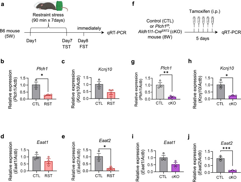
Phospholipase C (PLC) enzymes play crucial roles in intracellular calcium-signaling transduction. Several brain PLC subtypes have been extensively studied, implicating them in psychiatric disorders such as depression, epilepsy and schizophrenia. However, the role of the recently identified PLCη remains largely unknown. We found that PLCη1 is prominently expressed in lateral habenula (LHb) astrocytes. Here, to investigate its physiological role, we generated astrocyte-specific PLCη1 conditional knockout (cKO) mice (Plch1f/f; Aldh1l1-CreERT2). In these cKO mice, we observed a reduction in cellular morphological complexity metrics, such as total process length, as well as a decrease in the passive membrane conductance of LHb astrocytes. Additionally, neuronal function was impacted by the cKO, as the synaptic efficacy and firing rates of LHb neurons increased, while extrasynaptic long-term depression was impaired. Both tonic α-amino-3-hydroxy-5-methyl-4-isoxazolepdlropionic acid receptor/N-methyl-D-aspartate receptor (AMPAR/NMDAR) currents and extracellular glutamate levels were reduced. Interestingly, chemogenetic activation of astrocytes restored the reduced tonic AMPAR/NMDAR currents in cKO mice. Furthermore, LHb astrocyte-specific deletion of PLCη1 via AAV-GFAP-Cre injection induced depressive-like behaviors in mice, which were reversed by chemogenetic activation of LHb astrocytes. Finally, we found that restraint stress exposure decreased Plch1 mRNA expression in the LHb. These findings suggest that PLCη1 could be a potential therapeutic target for depression and highlight the critical role of astrocytes in the etiology of neuropsychiatric disorders.


Conflict of interest statement

Competing interests: The authors report no competing interests.

Figures

Fig. 1
Fig. 1. Expression of PLCη1 in mouse LHb astrocytes.
a FISH analysis of Plch1 mRNA on coronal mouse brain sections reveals localized expression in the thalamic and habenula regions, compared with cortical and hippocampal areas (CA1-3 hippocampal subfields, DG dentate gyrus). b A schematic diagram illustrating the preparation of tamoxifen-induced Plch1 cKO or CTL mice for western blot experiment. c Western blot analysis of LHb tissue lysates shows a significant reduction in PLCη1 protein in Plch1 cKO mice (CTL, 1 ± 0.072, n = 3; cKO, 0.694 ± 0.044, n = 3; P = 0.022; where n indicates the number of pooled sample vials, each containing samples from three nonoverlapping animals). d A schematic diagram illustrating the preparation of tamoxifen-induced Plch1 cKO or CTL mice for western blot experiment. e FISH analysis in the LHb for the combination of Plch1 with S100b (astrocytic marker). The dashed lines distinguish the LHb region from the medial habenula (MHb), thalamus and ventricles. Enlarged images (white boxes) show co-localized astrocyte areas indicated by smaller line boxes. f Quantification of Plch1-positive astrocytes in the LHb. The bar graph shows the percentage of Plch1-positive cells within the S100b-positive astrocytic population in CTL and Plch1 cKO mice. Plch1 expression was significantly reduced in the astrocytes of Plch1 cKO mice (34.44 ± 3.38%; n = 6) compared with CTL mice (69.28 ± 2.06%; n = 5; P < 0.0001), indicating effective astrocyte-specific deletion of Plch1. Data are presented as mean ± s.e.m. n is the number of analyzed cells per group. g A schematic diagram illustrating the preparation of AAV-induced Plch1 cKO or CTL mice for FISH experiments. h FISH analysis in the LHb for the combination of Plch1 with S100b (astrocytic marker) in the AAV-induced cKO model. The dashed lines indicate the LHb region, and enlarged images (white boxes) highlight co-localized astrocyte areas. i Quantification of Plch1-positive astrocytes in the LHb for the AAV-induced cKO model. The bar graph shows a significant reduction in the percentage of Plch1-positive cells within the S100b-positive astrocytic population in Cre mice (21.67 ± 5.43%; n = 5) compared with CTL (69.74 ± 5.71%; n = 5; P < 0.0001). j A 3D reconstruction of the LHb using IMARIS 9.0 software. DAPI (nuclei), Plch1 mRNA and S100b (astrocytic marker) signals were detected and analyzed. k Quantification of the fraction of Plch1-positive cells among S100b-positive cells were significantly lower in the Cre (0.206 ± 0.022, n = 6) groups than CTL (0.284 ± 0.022, n = 6), P = 0.035. l Quantification of the fraction of Plch1-positive cells among S100b-negative cells. There was no significant differences between groups (CTL, 0.229 ± 0.020; Cre, 0.199 ± 0.016; P = 0.284) *P < 0.05, ***P < 0.001 and ****P < 0.0001. Data are presented as mean ± s.e.m. n refers to the number of animals, unless otherwise stated. Six pairs of left and right hemispheric LHb images were used for analysis for each animal.
Fig. 2
Fig. 2. Deletion of PLCη1 leads to structural and functional changes in LHb astrocytes.
a A schematic diagram of tamoxifen-induced cKO or CTL mice. b Representative confocal images and surface renderings of 3D reconstruction. Scale bar, 5 µm. c A Sholl intersection plot showing intersections within a 5–70 µm radius from the center of GFAP filament in the LHb (CTL: n = 87, N = 7; cKO: n = 88, N = 7; two-way ANOVA interaction effect, P = 0.006; distance from the center effect, P < 0.0001; group effect, P < 0.0001; post hoc multiple comparison, P = 0.003 for 5 µm, P < 0.0001 for 10–20 µm, P = 0.068 for 25 µm and P = 0.010 for 90 µm. n indicates the number of cells and N refers to the total animals). d Detailed measurement results demonstrating that astrocyte process complexity is significantly reduced in Plch1 cKO mice (total process length: CTL, 760.966 ± 39.756 µm; cKO, 532.648 ± 27.388 µm, P < 0.0001; number of branch points: CTL, 67.943 ± 3.543; cKO, 47.318 ± 2.570, P < 0.0001; number of terminal points: CTL, 69.862 ± 3.672; cKO, 48.670 ± 2.633, P < 0.0001; number of process segments: CTL, 135.552 ± 7.224; cKO, 94.091 ± 5.172, P < 0.0001). e A schematic illustration of the cultured astrocytes used for estimation of viral efficacy of deleting Plch1 in Plch1f/f cells. f qRT–PCR shows a significant reduction of PLCη1 mRNA (CTL, 0.993 ± 0.311, n = 4; Cre, 0.120 ± 0.081, n = 4, P = 0.035; n refers a cultured cells from each independent animal). g A schematic illustration of CTL and Cre mice used in the astrocyte patch-clamp experiment. h Identification of LHb astrocytes were carried by fluorescence visualization of mCherry signal before whole-cell patch and confirming the absence of action potential against positive current injection in current-clamp during whole-cell patch. i The voltage ramp (the voltage command is depicted in the inset) protocol was applied to cell and the I–V relationship was established. j The LHb astrocyte membrane resistances calculated from the I–V curve were significantly bigger in Cre mice (CTL, 16.472 ± 0.953, n = 15, N = 4; Cre, 23.807 ± 2.230, n = 17, N = 4, P = 0.0072). **P < 0.001. Data are presented as mean ± s.e.m.
Fig. 3
Fig. 3. Deletion of astrocytic PLCη1 increases LHb synaptic efficacy and response firing probability in Plch1 cKO mice.
a The experimental scheme illustrates the preparation of the CTL and Plch1 cKO mice. b, c Assessment of basal transmission at the SM–LHb synapse in CTL and Plch1 cKO mice: an input–output relationship measurement between the current intensity (0–200 μA) and evoked ƒEPSP amplitude (mV) shows enhanced synaptic efficacy in cKO mice (CTL, n = 4, N = 4; cKO, n = 7, N = 7; n indicates number of brain tissue and N refers to the total animals; two-way ANOVA, group factor, P < 0.0001, post hoc multiple comparison, P = 0.0100 for 80 μA, 0.0068 for 90 μA and 0.0013 for 100 μA) (b) and the paired pulse ratio of ƒEPSP amplitudes across varied interstimulus intervals (25, 50, 100, 200 and 500 ms) shows no significant difference between CTL and Plch1 cKO mice (CTL, n = 4, N = 4; cKO, n = 5, N = 5; n and N as in b) (c). d Superimposed traces of firing EPSPs evoked by varying (20–180 µA) SM stimulation are shown. e The mean firing probability for each stimulus strength is shown. The sigmoidal fits to each dataset exhibited statistically significant differences. IC50 of CTL (216.891 ± 77.956 µA, 95% confidence interval, n = 7, N = 5), was higher than cKO (142.424 ± 10.112 µA, 95% confidence interval, n = 6, N = 5; n refers the number of cells measured and N refers the total number of animals). f After masking synaptic NMDAR by MK-801, DL-TBOA treatment combined with LFS induced an extrasynaptic LTD in the CTL mice but did not induced in Plch1 cKO mice. Time courses of the ƒEPSP amplitude are displayed. g The means ƒEPSP amplitude of the last 10 min (arrow) were compared (CTL, 78.739 ± 2.102%, n = 7, N = 7; cKO, 97.508 ± 4.319%, n = 7, N = 7, P = 0.002; n refers the number of slices recorded and N refers the total number of animals). **P < 0.05 and **P < 0.01. Data are presented as mean ± s.e.m.
Fig. 4
Fig. 4. Deletion of astrocytic PLCη1 reduces tonic AMPAR/NMDAR current and impairs extrasynaptic LTD in the LHb.
a The experimental scheme depicting the preparation of CTL and Plch1 cKO mice and brain slices for electrophysiology. Tonic currents were estimated on a coronal brain slice and extrasynaptic LTD was evaluated on a sagittal brain slice. b Representative traces show attenuated tonic AMPAR/NMDAR in the Plch1 cKO mice. Each 10 s portion (gray parts of traces) was compared before and after the drug treatment (dashed gray lines and brackets between the lines). Scale bars, 50 s and 5 pA. c The summarized mean tonic AMPAR/NMDAR current plot shows a significant difference between CLT and Plch1 cKO mice (CTL, 4.141 ± 0.497 pA, n = 14, N = 8; cKO, 1.622 ± 0.453 pA; n = 9, N = 6; P = 0.002; n refers the number of cells measured and N refers the total number of animals). d Representative traces show that the magnitude of tonic GABAAR current is not different in CTL and Plch1 cKO mice. e The summarized plot of tonic GABAAR current shows no significant difference between CLT and Plch1 cKO mice (CTL, 2.867 ± 0.676 pA, n = 10, N = 6; cKO, 3.126 ± 1.024 pA, n = 8, N = 6; P = 0.830; n and N as in c). f A schematic representation of the microdialysis. g Quantification of extracellular glutamate levels in the LHb. Microdialysis followed by glutamate assay revealed a significant reduction in extracellular glutamate in Plch1 cKO mice compared with CTL mice (CTL, 8.578 ± 0.375 ng/μl, n = 3; cKO, 4.724 ± 0.831 ng/μl, n = 5, P = 0.0015; n refers the number of samples and a sample is derived from an animal). h A schematic representation of the CNO-inducible astrocytes in cKO mouse LHb. i mCherry expression of viral infection was confirmed after behavioral tests. j Representative traces of tonic AMPAR/NMDAR currents of both groups. Tonic currents were restored in Plch1 cKO mice following CNO application in cKO-Gq but not in cKO-CTL. Scale bars, 50 s and 5 pA. k Quantification of tonic AMPAR/NMDAR currents. With 10 min of CNO pretreatment, cKO-Gq mice showed significantly more tonic AMPAR/NMDAR current than cKO-CTL mice (cKO-CTL, 2.944 ± 0.907 pA, n = 9, N = 5; cKO-Gq, 10.14 ± 2.33 pA, n = 9, N = 5, P = 0.011; n refers the number of cells recorded and N refers the number of animal). *P < 0.05 and **P < 0.01. Data are presented as mean ± s.e.m.
Fig. 5
Fig. 5. Deletion of astrocytic PLCη1 in the LHb affects depressive-like behavior.
a The experimental scheme depicts the preparation of CTL and Cre virus-injected mice for behavioral assessments. b The injection sites were confirmed to be in the LHb after behavioral assays to exclude any off-target subjects from analysis. c A schematic representation of the mobile and immobile states of mice during the FST. d Bar graphs displaying the mean of immobility duration for CTL and Cre mice (CTL, 82.28 ± 10.06 s, N = 7; Cre 122.66 ± 12.35 s, N = 6, P = 0.02). e A schematic representation of the mobile and immobile states of mice during the TST. f Bar graphs displaying the mean of immobility duration for CTL and Cre mice (CTL, 82.28 ± 10.06 s, N = 7; Cre 122.66 ± 12.35 s, N = 6, P = 0.027). g A schematic representation of the chemogenetic experiment. Plch1f/fmice were injected with AAV-GFAP-Cre combined with either DIO-mCherry (cKO-CTL) or DIO-hM3D(Gq) (cKO-Gq) to knock down Plch1 and induce astrocyte-specific expression of the DIO constructs in the LHb. h Fluorescence imaging following the behavioral experiments conducted to confirm viral infection in the LHb. i During the TST, the immobility time is significantly reduced in the CNO session of cKO-Gq compared with cKO-CTL and Veh session of cKO-Gq (Cre-CTL, Veh, 152.57 ± 21.12 s; Cre-CTL, CNO, 154.36 ± 18.49; Cre-Gq, Veh, 150.43 ± 23.88 s; Cre-Gq, CNO, 75.63 ± 5.66 s), No significant differences were observed in the cKO-CTL group between vehicle and CNO treatment. j The immobility time during FST is significantly reduced in the CNO session of cKO-Gq compared with the Veh session cKO-Gq, while there was no difference across sessions in cKO-Cre mice (Cre-CTL, Veh, 64.71 ± 8.61 s; Cre-CTL, CNO, 65.63 ± 8.09 s; Cre-Gq, Veh, 100.29 ± 12.1 9 s; Cre-Gq, CNO, 46.79 ± 10.11 s). N = 7 and 6 for Cre-CTL and Cre-Gq, respectively. *P < 0.05, **P < 0.01, ***P < 0.001 and ****P < 0.0001. N refers to the total number of animals. Data are presented as mean ± s.e.m.
Fig. 6
Fig. 6. Acute stress reduces PLCη1, Kir4.1, GLAST and GLT-1 mRNA expression in the LHb of wild-type mice and astrocytic Plch1 cKO mimics the mRNA expression patterns.
a Schematic representation of the evaluation of mRNA expression changes in LHb following restraint stress. be Relative mRNA expression levels of Plch1 (PLCη1), Kcnj10 (Kir4.1), Eaat1 (GLAST) and Eaat2 (GLT-1) in LHb from CTL and RST mice, as assessed by qRT–PCR: Plch1 (CTL, 1.030 ± 0.183 n = 3; RST, 0.290 ± 0.025, P = 0.016) expression was significantly reduced in RST mice compared with CTL (b); there were no significant changes in Kcnj10 (CTL, 1.033 ± 0.182, n = 3; RST, 0.450 ± 0.156, n = 3, P = 0.717) (c) or Eaat1 (CTL, 1.020 ± 0.149, n = 3; RST, 0.695 ± 0.178, n = 3, P = 0.232) (d) and Eaat2 (CTL, 1.040 ± 0.203, n = 3; RST, 0.185 ± 0.068, n = 3, P = 0.016) also exhibited significant decreases in the RST mice relative to CTL (e). f Schematic representation of the mRNA espression changes in LHb of CTL and Plch1 cKO mice. gj Analogous mRNA expression levels from Plch1 cKO mice compared with CTL: similar to the RST model, Plch1 cKO mice demonstrated substantial downregulation of Plch1 mRNA (CTL, 1.023 ± 0.155, n = 3; cKO, 0.170 ± 0.040, n = 3; P = 0.025) (g) and Kcnj10 (CTL, 1.027 ± 0.175, n = 3; cKO, 0.240 ± 0.029, n = 3) (h); Eaat1 (CTL, 1.020 ± 0.150, n = 3; 0.533 ± 0.128, n = 3, P = 0.071) (i) did not show significant changes and Eaat2 (CTL, 1.010 ± 0.099, n = 3; cKO, 0.143 ± 0.015, n = 3) expression was significantly reduced (j). *P < 0.05, **P < 0.01 and ****P < 0.0001. n indicates the number of vials and three nonoverlapping animals were pooled in each vial. Data are presented as mean ± s.e.m.

Similar articles

Cited by

References

    1. Cocco, L., Follo, M. Y., Manzoli, L. & Suh, P. G. Phosphoinositide-specific phospholipase C in health and disease. J. Lipid Res.56, 1853–1860 (2015). - PMC - PubMed
    1. Rhee, S. G. & Choi, K. D. Regulation of inositol phospholipid-specific phospholipase C isozymes. J. Biol. Chem.267, 12393–12396 (1992). - PubMed
    1. Rebecchi, M. J. & Pentyala, S. N. Structure, function, and control of phosphoinositide-specific phospholipase C. Physiol. Rev.80, 1291–1335 (2000). - PubMed
    1. Lisman, J., Yasuda, R. & Raghavachari, S. Mechanisms of CaMKII action in long-term potentiation. Nat. Rev. Neurosci.13, 169–182 (2012). - PMC - PubMed
    1. Lankford, K. L. & Letourneau, P. C. Evidence that calcium may control neurite outgrowth by regulating the stability of actin filaments. J. Cell Biol.109, 1229–1243 (1989). - PMC - PubMed

Substances

LinkOut - more resources