Clinical and MRI features contributing to the clinico-radiological dissociation in a large cohort of people with multiple sclerosis
- PMID: 40204954
- PMCID: PMC11982092
- DOI: 10.1007/s00415-025-12977-6
Clinical and MRI features contributing to the clinico-radiological dissociation in a large cohort of people with multiple sclerosis
Abstract
Background: People with Multiple Sclerosis (PwMS) often show a mismatch between disability and T2-hyperintense white matter (WM) lesion volume (LV), that in general is referred to as the clinico-radiological paradox.
Objectives: This study aimed to understand how an extensive clinical, neuropsychological, and MRI analysis could better elucidate the clinico-radiological dissociation in a large cohort of PwMS.
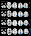
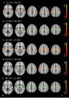
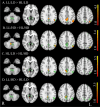
Methods: Clinical scores, such as Expanded Disability Status Scale (EDSS), 9 Hole Peg Test (9HPT), 25-foot Walking Test (25-FWT), Paced Auditory Serial Addition Test at 3 s (PASAT3), Symbol digit Modalities Test (SDMT), demographics, and 3 T-MRI of 717 PwMS and 284 healthy subjects (HS) were downloaded from the INNI database. Considering medians of LV and EDSS scores, PwMS were divided into four groups: low LV and disability (LL/LD); high LV and low disability (HL/LD); low LV and high disability (LL/HD); high LV and disability (HL/HD). MRI measures included: volumes of gray matter (GM), WM, cerebellum, basal ganglia and thalamus, spinal cord (SC) area, and functional connectivity of resting-state networks.
Results: The clinico-radiological dissociation involved 36% of our sample. HL/LD showed worse SDMT scores and lower global and deep GM volumes than HS and LL/LD. LL/HD showed lower GM, thalamus, and cerebellum volumes, and SC area than HS, and lower SC area than LL/LD.
Conclusions: A more extensive clinical assessment, including cognitive tests, and MRI evaluation including deep GM and SC, could better describe the real status of the disease and help clinicians in early and tailored treatment in PwMS.
Keywords: Clinico-radiological dissociation; Magnetic resonance imaging (MRI); Multiple Sclerosis.
© 2025. The Author(s).
Figures


References
-
- Kurtzke JF (1983) Rating neurologic impairment in multiple sclerosis: an expanded disability status scale (EDSS). Neurology 33:1444–1452. 10.1212/wnl.33.11.1444 - PubMed
-
- Amato MP, Portaccio E (2007) Clinical outcome measures in multiple sclerosis. J Neurol Sci 259:118–122. 10.1016/j.jns.2006.06.031 - PubMed
-
- Cohen M, Bresch S, Thommel Rocchi O et al (2021) Should we still only rely on EDSS to evaluate disability in multiple sclerosis patients? A study of inter and intra rater reliability. Mult Scler Relat Disord 54:103144. 10.1016/j.msard.2021.103144 - PubMed
-
- Hart BA, Bauer J, Muller HJ et al (1998) Histopathological characterization of magnetic resonance imaging-detectable brain white matter lesions in a primate model of multiple sclerosis: a correlative study in the experimental autoimmune encephalomyelitis model in common marmosets (Callithrix jacchus). Am J Pathol 153:649–663. 10.1016/s0002-9440(10)65606-4 - PMC - PubMed
MeSH terms
Grants and funding
LinkOut - more resources
Full Text Sources
Medical
Research Materials

