

Prostate-specific membrane antigen (PSMA) expression in primary and metastatic renal cell cancer (UroCCR-65 study)
- PMID: 40205264
- PMCID: PMC11981970
- DOI: 10.1186/s13550-025-01232-8
Prostate-specific membrane antigen (PSMA) expression in primary and metastatic renal cell cancer (UroCCR-65 study)
Abstract
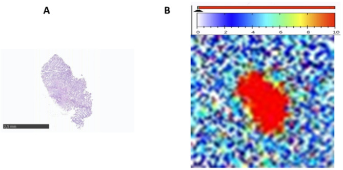
Background: Prostate-specific membrane antigen (PSMA) has been shown to be overexpressed in the neo-vasculature of renal cancers. However, studies investigating the pattern of PSMA expression in primary RCC and RCC metastases according to metastatic sites are rare. 44 frozen samples of RCC, 19 primaries (9 clear cell (cc) RCC, 7 papillary (pap) RCC, and 3 chromophobe (ch) RCC) and 25 (24 samples have ccRCC histology and one is unclassified) unpaired metastases (8 from adrenals, 8 from bones, 2 from lungs, 2 from liver and 5 others (1 lymph node, 1 pancreas, 1 brain, 1 gallbladder and 1 muscle)), were available from the UroCCR project (NCT03293563). PSMA expression was assessed by autoradiography using [177Lu]Lu-PSMA-617 as binding agent and the specific binding (total binding-non-specific binding) was calculated and expressed as a percentage of total binding. A patient suffering from metastatic ccRCC was also administered [68Ga]Ga-PSMA-11 to evaluate PSMA expression.
Results: The mean specific binding was 28.9 ± 40.4% for primary renal cancer and 65.0 ± 38.9% for metastasis. Regarding histology, high PSMA expression was depicted in 33.3% of ccRCC, 33.3% of chRCC and 57.1% of papRCC. PSMA was more frequently expressed in primary samples of papRCC histology with renal capsule invasion (p = 0.0286). A higher PSMA-specific binding and a higher number of samples with high PSMA-expression were depicted in metastatic samples. Bone metastasis showed lower binding than other metastatic sites combined (p = 0.0005). The patient suffering from metastatic ccRCC showed high [68Ga]Ga-PSMA-11 uptake on known distant metastases and additional site uncovered.
Conclusion: PSMA showed high expression in metastases of ccRCC.
Clinical trial registration: NCT, NCT03293563, prospectively registered September 20, 2017, http://www.
Clinicaltrials: gov .
Keywords: Clear cell RCC; Kidney cancer; Metastatic RCC; PSMA; Primary RCC.
© 2025. The Author(s).
Conflict of interest statement
Declarations. Ethics approval and consent to participate: The UroCCR project (NCT03293563), which is IRB-approved and has the CNIL authorization number DR-2013-206. The UroCCR database received approval from Comité de Protection des Personnes Sud-Ouest et Outre-mer III (DC 2012/108) and favorable opinion of the Comité Consultatif sur le Traitement de l’Information en matière de Recherche dans le domaine de la Santé (CCTIRS). Consent for publication: Written informed consent was obtained for publication. Competing interests: The authors have no relevant financial or non-financial interests to disclose.
Figures


References
-
- Baccala A, Sercia L, Li J, Heston W, Zhou M. Expression of prostate-specific membrane antigen in tumor-associated neovasculature of renal neoplasms. Urology. 2007;70:385–90. - PubMed
-
- Woythal N, Arsenic R, Kempkensteffen C, Miller K, Janssen J-C, Huang K, et al. Immunohistochemical validation of PSMA expression measured by 68Ga-PSMA PET/CT in primary prostate cancer. J Nucl Med. 2018;59:238–43. - PubMed
Associated data
Grants and funding
LinkOut - more resources
Full Text Sources
Medical
Research Materials
Miscellaneous
