Review
. 2025 Apr 9;23(1):411.
doi: 10.1186/s12967-025-06430-5.

Fluorescent probes in autoimmune disease research: current status and future prospects

Affiliations
Review

Fluorescent probes in autoimmune disease research: current status and future prospects

Junli Chen et al. J Transl Med. .

Abstract

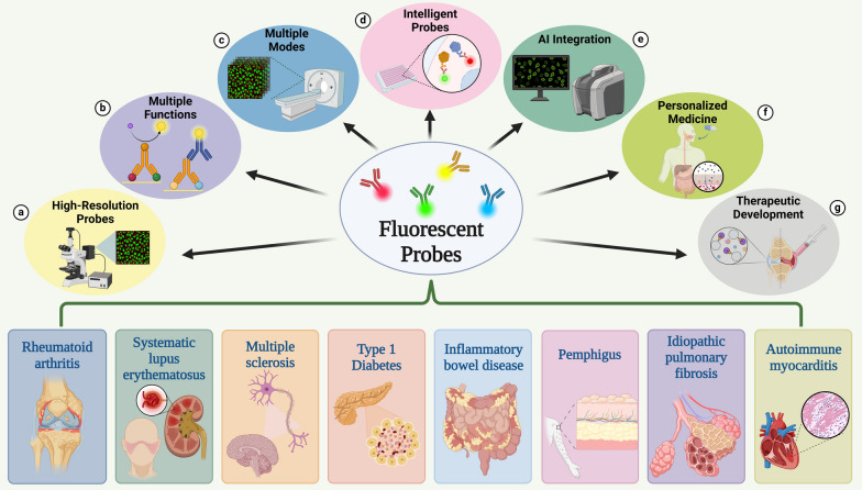
Autoimmune diseases (AD) present substantial challenges for early diagnosis and precise treatment due to their intricate pathogenesis and varied clinical manifestations. While existing diagnostic methods and treatment strategies have advanced, their sensitivity, specificity, and real-time applicability in clinical settings continue to exhibit significant limitations. In recent years, fluorescent probes have emerged as highly sensitive and specific biological imaging tools, demonstrating substantial potential in AD research.This review examines the response mechanisms and historical evolution of various types of fluorescent probes, systematically summarizing the latest research advancements in their application to autoimmune diseases. It highlights key applications in biomarker detection, dynamic monitoring of immune cell functions, and assessment of drug treatment efficacy. Furthermore, this article analyzes the technical challenges currently encountered in probe development and proposes potential directions for future research. With ongoing advancements in materials science, nanotechnology, and bioengineering, fluorescent probes are anticipated to achieve higher sensitivity and enhanced functional integration, thereby facilitating early detection, dynamic monitoring, and innovative treatment strategies for autoimmune diseases. Overall, fluorescent probes possess substantial scientific significance and application value in both research and clinical settings related to autoimmune diseases, signaling a new era of personalized and precision medicine.

Keywords: Autoimmune disease; Early diagnosis; Fluorescent probe; Multimodal imaging; Nanotechnology; Precision medicine.


Conflict of interest statement

Declarations. Competing interests: The authors declare that they have no known competing financial interests or personal relationships that could have appeared to influence the work reported in this paper.

Figures

Fig. 1
Fig. 1
Development history of fluorescent probes (Created in https://BioRender.com)
Fig. 2
Fig. 2
Basic principle of fluorescence probe (Created in https://BioRender.com). a pattern of fluorescence excitation: When fluorescent molecules absorb light energy of a specific wavelength, excited electrons transition from the ground state S0 to the excited state S2. After internal conversion to S1, electrons can return to S0 and produce fluorescence. b FRET mechanism: One fluorescent molecule absorbs light energy and non-radiatively transfers this energy to another fluorescent molecule, resulting in a decrease in the fluorescence intensity of the donor and an increase in the fluorescence intensity of the acceptor [54]. The FRET mechanism can clearly detect interactions between proteins. c PET mechanism: Upon binding of the target molecule to the receptor, the PET process is inhibited, leading to the recovery of the probe's fluorescence [55]. The PET mechanism has significant importance in drug screening and efficacy monitoring [56]. d ICT mechanism: When the donor component interacts with a cation, the electron-donating properties of the probe diminish, causing a blue shift in the absorption spectrum. Conversely, when the acceptor component interacts with a cation, the absorption spectrum exhibits a significant red shift [57]. The ICT mechanism aids in monitoring biochemical processes such as enzyme-catalyzed reactions in vivo [58]
Fig. 3
Fig. 3
Comparison of different types of fluorescent probes (Created in https://BioRender.com) a Organic dye probe: Chemical structures of FITC [67], rhodamine B [82] and fluorescein [83]. The bar graph indicates that FITC has a low sensitivity to the quencher (Ksv = 9.8L/mol), suggesting its high stability in complex environments [67]. b Quantum dots: Schematic representation of ZnSe/CdS/ZnS, InP QDs and CQDs structures. The bar graph compares the quantum yield (QY) of different quantum dots (ZnSe/CdS/ZnS QY = 82% [74], InP QDs QY = 81% [84], CQDs QY = 32.4% [85]), demonstrating their excellent optical stability. c Fluorescent protein: Schematic representation of GFP chemical structure and RFP, YFP, and BFP structures [86]. The bar graph compares the quantum yield (QY) of various fluorescent proteins (GFP QY = 60%, RFP QY = 79%, EYFP QY = 61%, EBFP QY = 31% [86]), proving their good optical stability. d Nanopariticles: Schematic diagram of pDNA/DSP-NPs preparation. It exhibits a very high drug loading capacity, with a drug loading efficiency (DIE) of up to 10.54 ± 2.09 wt% [81]. e Based on the design characteristics of probes in the literature and practical application scenarios, a radar chart compares the performance of organic dyes, quantum dots, fluorescent proteins, and nanoparticles across six key performance indicators (such as sensitivity, specificity, optical stability, biocompatibility, tissue penetration depth, and cost)
Fig. 4
Fig. 4
Comparison of probes for different targets of RA (Created in https://BioRender.com) a Pathogenesis of RA: The pathogenesis of RA involves the presentation of autoimmune antigens by APC, which activate T cells and initiate an immune response. Additionally, activated B cells, macrophages, synovial fibroblasts, and various inflammatory mediators play significant roles in this process [97]. b T cell targets: A schematic diagram of the detection of activated T cells in early RA tissues using IRDye-680RD-OX40 mAb. The ROC curve constructed using the RP/LP fluorescence intensity ratio achieved an AUC of 1.0, indicating that the probe can completely discriminate between the collagen-induced arthritis model (CIA) and the healthy control group, demonstrating high sensitivity and high specificity (redrawn based on data from the literature [105]). c B cell targets:A schematic diagram of the distribution of B cells using Cy5-labeled anti-CD20 antibodies. Immunofluorescence images: Left: healthy control group (few blue fluorescent spots); Right: RA model group (a large number of green fluorescent spots, clustered in the synovial area of the joint). It indicates that the density of B cells in the lesion area is 3.2 times higher than that in the healthy group (p < 0.001). (Redrawn based on data from the literature [107].) d Small molecule targets: A schematic diagram for the quantitative monitoring of ONOO using Ratio-A. The curve graph demonstrates a linear relationship between the logarithm of fluorescence intensity and ONOO concentration (0-10 μM) (y = 0.2221x-0.4495, R2 = 0.9931), intuitively showing the high sensitivity and linear response of Ratio-A to ONOO. (redrawn based on data from the literature [123]). e Cytokine targets: A schematic diagram of TNF-α gene silencing using psi-tGC-NPs. The bar graph shows that both the MTX group (P < 0.01) and the psi-tGC-NPs group (P < 0.05) significantly inhibit the expression of TNF-α mRNA. This intuitively reflects the potential of the nanoparticle delivery system in gene silencing, comparable to the efficacy of MTX (redrawn based on data from the literature [133])
Fig. 5
Fig. 5
The prospect of fluorescent probes (Created in https://BioRender.com) a High-Resolution Probes: The utilization of high-resolution fluorescent probes significantly enhances spatiotemporal resolution and deep tissue penetration, facilitating high-precision imaging. b Multiple Functions: Multifunctional fluorescent probes enable the simultaneous detection of multiple biomarkers, thereby improving detection efficiency. c Multiple Modes: Multimodal fluorescent probes amalgamate various detection methods, compensating for the limitations inherent in different imaging modalities and achieving synergistic effects. d Intelligent Probes: Intelligent fluorescent probes operate at the cellular and molecular levels to dynamically detect specific small molecules and changes within the cellular microenvironment. e AI Integration: The integration of artificial intelligence has accelerated the development of fluorescent probe design, application performance, and data analysis. f Personalized Medicine: Patient-specific probes address diverse requirements for personalized diagnosis and therapeutic monitoring. g Therapeutic Development: Fluorescent probes facilitate research and screening of new drugs and synergize with nanotechnology to advance the development of fluorescent nanoprobes that integrate diagnosis and treatment

Similar articles

References

    1. Sanz I, Lund F. Complexity and heterogeneity - the defining features of autoimmune disease. Curr Opin Immunol. 2019;61:iii–vi. - PubMed
    1. Fugger L, Jensen LT, Rossjohn J. Challenges, progress, and prospects of developing therapies to treat autoimmune diseases. Cell. 2020;181:63–80. - PubMed
    1. Song Y, Li J, Wu Y. Evolving understanding of autoimmune mechanisms and new therapeutic strategies of autoimmune disorders. Signal Transduct Target Ther. 2024;9:263. - PMC - PubMed
    1. Miller FW. The increasing prevalence of autoimmunity and autoimmune diseases: an urgent call to action for improved understanding, diagnosis, treatment, and prevention. Curr Opin Immunol. 2023;80:102266. - PMC - PubMed
    1. Mohamed-Ahmed O, Shang L, Wang L, Chen Z, Kartsonaki C, Bragg F. Incidence and prevalence of autoimmune diseases in China: a systematic review and meta-analysis of epidemiological studies. Glob Epidemiol. 2024;8:100158. - PMC - PubMed

LinkOut - more resources