. 2025 Apr 10;13(1):73.
doi: 10.1186/s40478-025-01990-5.

Transient receptor potential vanilloid 4 blockage attenuates pyroptosis in hippocampus of mice following pilocarpine‑induced status epilepticus

Affiliations

Transient receptor potential vanilloid 4 blockage attenuates pyroptosis in hippocampus of mice following pilocarpine‑induced status epilepticus

Lihan Liu et al. Acta Neuropathol Commun. .

Abstract

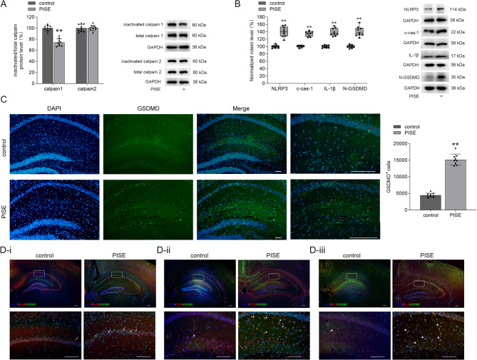
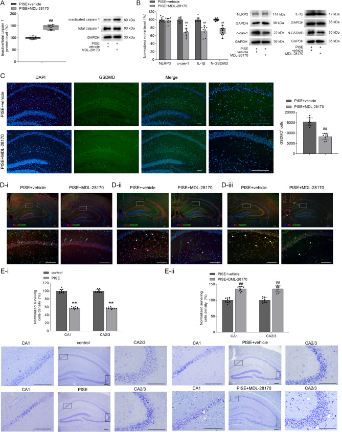
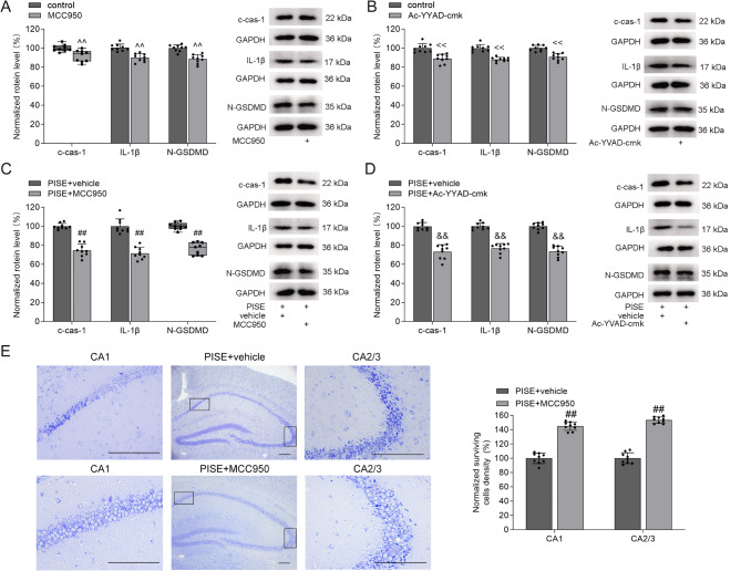
Pyroptosis contributes to the neuronal damage that occurs during epilepsy. Calcium-activated neutral protease (calpain) dissociates cysteinyl aspartate specific proteinase-1 (caspase-1, cas-1) from the cytoskeleton, and the activated cas-1 is responsible for the production of N-terminus of gasdermin D (N-GSDMD), the final executor of pyroptosis. Blocking transient receptor potential vanilloid 4 (TRPV4) can reduce neuronal injury in temporal lobe epilepsy (TLE) model mice. This study investigated the role of TRPV4 in pyroptosis during TLE. In the hippocampus of pilocarpine-induced status epilepticus (PISE) mice, the ratio of inactive calpain 1 protein level to its total protein level (inactive/total calpain 1) significantly decreased, while the ratio of inactive calpain 2 protein level to its total protein level remained unchanged. The protein levels of NLRP3, cleaved cas-1 (c-cas-1), interleukin (IL)-1β, and N-GSDMD increased, with more GSDMD-immunofluorescence-positive (GSDMD+) cells and fewer surviving pyramidal neurons observed in the hippocampus of PISE mice. Calpain inhibition with MDL-28170 reversed these changes, except for the elevated NLRP3 levels. Inhibitors targeting NLRP3 (MCC950) and cas-1 (Ac-YVAD-cmk) blocked the increase in c-cas-1, IL-1β, and N-GSDMD levels in the hippocampus of PISE mice. TRPV4 inhibition via HC-067047 increased the inactive/total calpain 1 ratio, decreased NLRP3, c-cas-1, IL-1β, and N-GSDMD protein levels, reduced GSDMD+ cells number, and improved pyramidal neuron survival in the hippocampus of PISE mice. Conversely, TRPV4 activation with GSK1016790A decreased the inactive/total calpain 1 ratio, elevated NLRP3, c-cas-1, IL-1β, and N-GSDMD levels, and increased GSDMD+ cells number in the hippocampus. In the hippocampus of GSK1016790A-injected mice, the inactive/total calpain 1 ratio was increased by MDL-28170, and c-cas-1, IL-1β, and N-GSDMD protein levels were markedly attenuated by MDL-28170, MCC950, and Ac-YVAD-cmk, respectively. In conclusion, TRPV4 inhibition mitigates pyroptosis in PISE mice by downregulating the calpain 1-NLRP3/cas-1-GSDMD pathway, ultimately reducing neuronal damage.

Keywords: Calpain; Caspase-1; Gasdermin D; Pyroptosis; Temporal lobe epilepsy; Transient receptor potential vanilloid 4.


Conflict of interest statement

Declarations. Ethics approval and consent to participate: This study was approved by the Ethics Committee of Nanjing Medical University (No. IACUC2009007), and all animal experiments were performed in accordance with the Guidelines for Laboratory Animal Research set by Nanjing Medical University. All efforts were made to minimize animal suffering and to reduce the number of animals used. Competing interests: The authors declare no competing interests.

Figures

Fig. 1
Fig. 1
Changes in calpain 1–NLRP3/cas-1–GSDMD pathway in the hippocampus of PISE mice. (A) The inactive/total calpain 1 ratio markedly decreased (independent-samples t test, t = 9.42, P < 0.01) while the inactive/total calpain 2 ratio did not change (independent-samples t test, t = − 0.16, P = 0.87) in the hippocampus of PISE mice. (B) The protein levels of NLRP3 (Mann–Whitney U test, P < 0.01), c-cas-1 (Mann–Whitney U test, P < 0.01), IL-1β (Mann–Whitney U test, P < 0.01), and N-GSDMD (Mann–Whitney U test, P < 0.01) increased markedly in the hippocampus of PISE mice. (C) The number of GSDMD+ cells (red arrow) increased markedly in the hippocampus in PISE mice (independent-samples t test, t = − 18.01, P < 0.01). (D) GSDMD+/NeuN+ (D-i, white arrow), GSDMD+/GFAP+ (D-ii, white star) and GSDMD+/Iba-1+ (D-iii, white triangle) cells in the hippocampus of control and PISE mice. Scale = 50 μm. ×4 objective for the top row in D-i, D-ii and D-iii; ×10 objective for C-DAPI, C-GSDMD, C-Merge; ×20 objective for C-enlarge and the down row in D-i, D-ii and D-iii. **P < 0.01 vs. control
Fig. 2
Fig. 2
Effect of calpain inhibitor MDL-28170 on calpain 1–NLRP3/cas-1–GSDMD pathway in the hippocampus of PISE mice. (A) The calpain inhibitor MDL-28170 markedly increased the inactive/total calpain 1 rato in the hippocampus of PISE mice (Mann–Whitney U test, P < 0.01). (B) MDL-28170 did not affect NLRP3 protein levels in the hippocampus of PISE mice (independent-samples t test, t = 0.74, P = 0.47), but decreased c-cas-1 (independent-samples t test, t = 7.81, P < 0.01), IL-1β (independent-samples t test, t = 6.30, P < 0.01), and N-GSDMD protein levels (Mann–Whitney U test, P < 0.01). (C) MDL-28170 increased GSDMD+ cells (red arrow) number in the hippocampus of PISE mice (independent-samples t test, t = 8.83, P < 0.01). (D) GSDMD+/NeuN+ (D-i, white arrow), GSDMD+/GFAP+ (D-ii, white star) and GSDMD+/Iba-1+ (D-iii, white triangle) cells in the hippocampus of vehicle- and MDL-28179-treated PISE mice. (E) The numbers of surviving pyramidal neurons reduced in the hippocampal CA1 (independent-samples t test, t = 22.76, P < 0.01) and CA2/3 (independent-samples t test, t = 28.02, P < 0.01) area of PISE mice (E-i). Administration of MDL-28170 to PISE mice increased the numbers of surviving pyramidal cells in the hippocampal CA1 (independent-samples t test, t = 6.88, P < 0.01) and CA2/3 area (independent-samples t test, t = 6.15, P < 0.01) (E-ii). Scale = 50 μm. ×4 objective for the top row in D-i, D-ii and D-iii; ×10 objective for C-DAPI, C-GSDMD, C-Merge, and the middle column in E-i and E-ii; ×20 objective for C-enlarge, the down row in D-i, D-ii, and D-iii; ×40 objective for the left and right columns in E-i and E-ii. ##P < 0.01 vs. PISE + vehicle (ip.), **P < 0.01 vs. control
Fig. 3
Fig. 3
Effects of NLRP3 and cas-1 inhibitors on c-cas-1, IL-1β, and N-GSDMD protein levels in the hippocampus of PISE mice. A and C. NLRP3 inhibitor MCC950 decreased c-cas-1 (A, Mann–Whitney U test, P < 0.01; C, independent-samples t test, t = 10.47, P < 0.01), IL-1β (A, independent-samples t test, t = 5.75, P < 0.01; C, independent-samples t test, t = 8.50, P < 0.01), and N-GSDMD protein levels (A, independent-samples t test, t = 5.98, P < 0.01; C, Mann–Whitney U test, P < 0.01) in the hippocampus of control and PISE mice. B and D. Cas-1 inhibitor Ac-YVAD-cmk decreased c-cas-1 (B, independent-samples t test, t = 5.04, P < 0.01; D, independent-samples t test, t = 9.50, P < 0.01), IL-1β (B, independent-samples t test, t = 5.91, P < 0.01; D, independent-samples t test, t = 11.54, P < 0.01), and N-GSDMD protein levels (B, independent-samples t test, t = 8.14, P < 0.01; D, independent-samples t test, t = 12.08, P < 0.01) in the hippocampus of control and PISE mice. E. Administration of MCC950 to PISE mice increased the numbers of surviving pyramidal neurons in the hippocampal CA1 (independent-samples t test, t = 10.01, P < 0.01) and CA2/3 area (independent-samples t test, t = 14.50, P < 0.01). Scale = 50 μm. ×10 objective for the middle column in E; ×40 objective for the left and right columns in E. ^^P < 0.01 vs. control (ip.), <<P < 0.01 vs. control (icv.), ##P < 0.01 vs. PISE + vehicle (ip.), &&P < 0.01 vs. PISE + vehicle (icv.)
Fig. 4
Fig. 4
Effect of TRPV4 antagonist on calpain 1–NLRP3/cas-1–GSDMD pathway in the hippocampus of PISE mice. A and C. Administration of TRPV4 antagonist HC-067047 increased inactive/total calpain 1 ratio in the hippocampus of control (A, Mann–Whitney U test, P < 0.01) and PISE mice (C, independent-samples t test, t = − 17.09, P < 0.01). B and D. Administration of HC-067047 decreased NLRP3 (B, HC-067047: independent-samples t test, t = 6.49, P < 0.01; D, PISE + HC-067047: Mann–Whitney U test, P < 0.01), c-cas-1 (B, HC-067047: independent-samples t test, t = 6.53, P < 0.01; D, PISE + HC-067047: independent-samples t test, t = 9.38, P < 0.01), IL-1β (B, HC-067047: independent-samples t test, t = 4.23, P < 0.01; D, PISE + HC-067047: independent-samples t test, t = 12.31, P < 0.01), and N-GSDMD protein levels (B, HC-067047: independent-samples t test, t = 5.28, P < 0.01; D, PISE + HC-067047: Mann–Whitney U test, P < 0.01). E and F. Administration of HC-067047 decreased GSDMD+ cells (red arrow) numbers in the hippocampus of control (E-i, HC-067047: independent-samples t test, t = 4.34, P < 0.01) and PISE mice (F-i, PISE + HC-067047: Mann–Whitney U test, P < 0.01; white star). GSDMD+/NeuN+ (white arrow), GSDMD+/GFAP+ (white star) and GSDMD+/Iba-1+ (white triangle) cells in the hippocampus in control (E-ii, E-iii and E-iv) and PISE mice (F-ii, F-iii and F-iv) treated with either vehicle or HC-067047. G. HC-067047 treatment increased the numbers of surviving neurons in the hippocampal CA1 (G-i, independent-samples t test, t = 5.14, P < 0.01; G-ii, independent-samples t test, t = 10.12, P < 0.01) and CA2/3 (G-i, independent-samples t test, t = 2.35, P = 0.03; G-ii, independent-samples t test, t = 14.20, P < 0.01) area of control (G-i) and PISE mice (G-ii). Scale = 50 μm. ×4 objective for the top row in E-ii, E-iii, E-vi, F-ii, F-iii, F-vi; ×10 objective for E-i-DAPI, E-i-GSDMD, E-i-Merge, F-i-DAPI, F-i-GSDMD, F-i-Merge, and the middle columns in G-i and G-ii; ×20 objective for E-i-enlarge, F-i-enlarge, the down row in E-ii, E-iii, E-vi, F-ii, F-iii and F-vi; ×40 objective for the left and right columns in G-i, and G-ii. <P < 0.05, <<P < 0.01 vs. control (icv.), &&P < 0.01 vs. PISE + vehicle (icv.)
Fig. 5
Fig. 5
Effect of TRPV4 agonist on calpain 1-NLRP3/cas-1-GSDMD pathway in the hippocampus. A. The TRPV4 agonist GSK1016790A decreased inactive/total calpain 1 ratio (independent-samples t test, t = 12.72, P < 0.01) but did not change inactive/total calpain 2 ratio (independent-samples t test, t = 0.19, P = 0.86) in the hippocampus. B and C. GSK1016790A increased NLRP3 (B, Mann–Whitney U test, P < 0.01), c-cas-1 (B, Mann–Whitney U test, P < 0.01), IL-1β (B, Mann–Whitney U test, P < 0.01), and N-GSDMD (B, Mann–Whitney U test, P < 0.01) protein levels, and GSDMD+ cells (red arrow) number (C, independent-samples t test, t = − 20.96, P < 0.01) in the hippocampus. D. GSDMD+/NeuN+ (D-i, white arrow), GSDMD+/GFAP+ (D-ii, white star) and GSDMD+/Iba-1+ (D-iii, white triangle) cells in the hippocampus of control and GSK1016790A-injected mice. E and F. The calpain inhibitor MDL-28170 increased inactive/total calpain 1 ratio (E, Mann–Whitney U test, P < 0.01) and decreased c-cas-1 (F, independent-samples t test, t = 8.73, P < 0.01), IL-1β (F, independent-samples t test, t = 9.35, P < 0.01), and N-GSDMD protein levels (F, independent-samples t test, t = 13.52, P < 0.01) but did not affect NLRP3 protein levels (F, independent-samples t test, t = − 0.20, P = 0.85) in the hippocampus of GSK1016790A-injected mice. G and H. The NLRP3 inhibitor MCC950 (G) and cas-1 inhibitor Ac-YVAD-cmk (H) decreased c-cas-1 (G, independent-samples t test, t = 8.17, P < 0.01; H, Mann–Whitney U test, P < 0.01), IL-1β (G, independent-samples t test, t = 11.62, P < 0.01; H, Mann–Whitney U test, P < 0.01) and N-GSDMD protein levels (G, Mann–Whitney U test, P < 0.01; H, independent-samples t test, t = 13.38, P < 0.01) in the hippocampus in GSK1016790A-injected mice. Scale = 50 μm. ×4 objective for the top row in D-i, D-ii and D-iii; ×10 objective for C-DAPI, C-GSDMD and C-Merge; ×20 objective for C-enlarge, the down row in D-i, D-ii and D-iii. <<P < 0.01 vs. Control (icv.), $$P < 0.01 vs. GSK1016790A + vehicle, %%P < 0.01 vs. GSK1016790A + vehicle

Similar articles

References

    1. Henshall DC, Chen J, Simon RP (2000) Involvement of caspase-3-like protease in the mechanism of cell death following focally evoked limbic seizures. J Neurochem 74(3):1215–1223. 10.1046/j.1471-4159.2000.741215.x - PubMed
    1. Wang Z, Zhou L, An D, Xu W, Wu C, Sha S et al (2019) TRPV4-induced inflammatory response is involved in neuronal death in pilocarpine model of Temporal lobe epilepsy in mice. Cell Death Dis 10(6):386. 10.1038/s41419-019-1612-3 - PMC - PubMed
    1. Chen L, Wang Y, Chen Z (2020) Adult neurogenesis in epileptogenesis: an update for preclinical finding and potential clinical translation. Curr Neuropharmacol 18(6):464–484. 10.2174/1570159x17666191118142314 - PMC - PubMed
    1. Mohseni-Moghaddam P, Roghani M, Khaleghzadeh-Ahangar H, Sadr SS, Sala C (2021) A literature overview on epilepsy and inflammasome activation. Brain Res Bull 172:229–235. 10.1016/j.brainresbull.2021.05.001 - PubMed
    1. Zheng X, Chen W, Gong F, Chen Y, Chen E (2021) The role and mechanism of pyroptosis and potential therapeutic targets in sepsis: A review. Front Immunol 12:711939. 10.3389/fimmu.2021.711939 - PMC - PubMed

Publication types

MeSH terms

LinkOut - more resources