. 2025 Apr 9;17(1):36.
doi: 10.1186/s13073-025-01461-5.

Genomic and functional analysis of rmp locus variants in Klebsiella pneumoniae

Affiliations

Genomic and functional analysis of rmp locus variants in Klebsiella pneumoniae

Margaret M C Lam et al. Genome Med. .

Abstract

Background: Klebsiella pneumoniae is an opportunistic pathogen and a leading cause of healthcare-associated infections in hospitals, which are frequently antimicrobial resistant (AMR). Exacerbating the public health threat posed by K. pneumoniae, some strains also harbour additional hypervirulence determinants typically acquired via mobile genetic elements such as the well-characterised large virulence plasmid KpVP-1. The rmpADC locus is considered a key virulence feature of K. pneumoniae and is associated with upregulated capsule expression and the hypermucoid phenotype, which can enhance virulence by contributing to serum resistance. Typically such strains have been susceptible to all antimicrobials besides ampicillin; however, the recent emergence of AMR hypermucoid strains is concerning.

Methods: Here, we investigate the genetic diversity, evolution, mobilisation and prevalence of rmpADC, in a dataset of 14,000 genomes from isolates of the Klebsiella pneumoniae species complex, and describe the RmST virulence typing scheme for tracking rmpADC variants for the purposes of genomic surveillance. Additionally, we examine the functionality of representatives for variants of rmpADC introduced into a mutant strain lacking its native rmpADC locus.

Results: The rmpADC locus was detected in 7% of the dataset, mostly from genomes of K. pneumoniae and a very small number of K. variicola and K. quasipneumoniae. Sequence variants of rmpADC grouped into five distinct lineages (rmp1, rmp2, rmp2A, rmp3 and rmp4) that corresponded to unique mobile elements, and were differentially distributed across different populations (i.e. clonal groups) of K. pneumoniae. All variants were demonstrated to produce enhanced capsule production and hypermucoviscosity.

Conclusions: These results provide an overview of the diversity and evolution of a prominent K. pneumoniae virulence factor and support the idea that screening for rmpADC in K. pneumoniae isolates and genomes is valuable to monitor the emergence and spread of hypermucoid K. pneumoniae, including AMR strains.

Keywords: Klebsiella pneumoniae; Genomic surveillance; Hypermucoid; Hypermucoviscosity; Hypervirulence; Virulence; Virulence plasmids.


Conflict of interest statement

Declarations. Ethics approval and consent to participate: Not applicable. Consent for publication: Not applicable. Competing interests: The authors declare no competing interests.

Figures
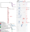
Fig. 1
Fig. 1
Maximum-likelihood phylogenetic tree of rmp sequence types (RmSTs). Lineages are labelled and tips are coloured by the associated mobile genetic element according to the legend. Column labels are as follows for a particular RmST: presence or absence of truncations in the rmpA, rmpD and rmpC, detection within a hypervirulent (blue) or MDR (red) clone (definitions of clones from Wyres et al. [25]) or non-K. pneumoniae species (Kv, Klebsiella variicola; Kqs, Klebsiella quasipneumoniae subsp. similipneumoniae). Clones included in ‘Other MDR’: CG14, CG17, CG29, CG101, CG147, CG152 and CG307. The number of genomes from which each RmST was detected is shown in the bar graph on the right-hand side
Fig. 2
Fig. 2
Comparison of upstream and downstream regions of rmp associated with different mobile variants. Arrows represent coding sequences and those corresponding to genes of interest are labelled and coloured by functionality as per the legend. Other labelled loci include arsCBR (arsenic resistance), virB1-virB11 (virB-type 4 secretion system) and ybtSX (part of the yersiniabactin locus). Shading corresponds to regions of similarity (amino acid sequence identity ≥ 30%) as identified by clinker
Fig. 3
Fig. 3
Hypermucoviscosity and capsule production of ectopically expressed rmp loci from the five different lineages. A Mucoviscosity assay and B uronic acid assay of KPPR1S, ΔwcaJ and Δrmp strains with vector (pMWO-078) or lineage-specific pRmp (see Table 1). Data from two technical replicates and three biological replicates were obtained following a 5.5-h induction of KPPR1S expression plasmid-borne rmp genes as described in Methods. One-way ANOVA with Tukey’s post-test was used to determine significance. *, P < 0.05; ***, P < 0.001; ****, P < 0.0001; comparisons not indicated were not significant
Fig. 4
Fig. 4
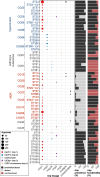
Distribution of rmp across K. pneumoniae sequence types with ≥5 rmp+ genomes. Rows indicated K. pneumoniae sequence types (STs; as labelled), and grouped and labelled by clonal group (CG) where applicable; those corresponding to hypervirulent, pathotype subspecies or multidrug-resistant (MDR) clones are labelled accordingly. Other clones are considered ‘generalist’ clones. The bubble plot shows the number of rmp+ genomes assigned to a particular rmp lineage, coloured by the type of mobile element. The first bar plot shows prevalence of rmp (i.e. black: present, grey: absent) and the second shows functional status of the locus (i.e. black: intact and functional, red: non-functional due to deletions or truncations)
Fig. 5
Fig. 5
Distribution of rmp within K locus groups and KpSC clonal groups (CG) or species. Each circle represents an rmp+ genome with a confident K locus call (917 genomes) and is coloured by the functionality of the rmp locus as per the figure legend. Twenty-five K loci with 4 or fewer genomes were collapsed into ‘Other’: KL6, KL10, KL12, KL14, KL15, KL17, KL19, KL21, KL23, KL24, KL25, KL27, KL30, KL34, KL35, KL39, KL45, KL55, KL63, KL102, KL108, KL127, KL137, KL147 and KL149. Clonal group labels are coloured blue and red to indicate hypervirulent and MDR lineages, respectively. Kq: Klebsiella quasipneumoniae, Kv: Klebsiella variicola

References

    1. Podschun R, Ullmann U. Klebsiella spp. as nosocomial pathogens: epidemiology, taxonomy, typing methods, and pathogenicity factors. Clin Microbiol Rev. 1998;11:589–603. - PMC - PubMed
    1. Shon AS, Bajwa RPS, Russo TA. Hypervirulent (hypermucoviscous) Klebsiella pneumoniae: a new and dangerous breed. Virulence. 2013;4:107–18. - PMC - PubMed
    1. Russo TA, Marr CM. Hypervirulent Klebsiella pneumoniae. Clin Microbiol Rev. 2019;32:e00001–19. - PMC - PubMed
    1. Lee IR, et al. Differential host susceptibility and bacterial virulence factors driving Klebsiella liver abscess in an ethnically diverse population. Sci Rep. 2016;6:29316. - PMC - PubMed
    1. Choby JE, Howard-Anderson J, Weiss DS. Hypervirulent Klebsiella pneumoniae – clinical and molecular perspectives. J Intern Med. 2020;287:283–300. - PMC - PubMed

LinkOut - more resources