. 2025 Mar 26:16:1573135.
doi: 10.3389/fphar.2025.1573135. eCollection 2025.

Senecio scandens Buch.-Ham. polysaccharides exert anti-atopic dermatitis effects by modulating gut microbiota and the MAPK/NF-κB pathway

Affiliations

Senecio scandens Buch.-Ham. polysaccharides exert anti-atopic dermatitis effects by modulating gut microbiota and the MAPK/NF-κB pathway

Zhi-Qin Hu et al. Front Pharmacol. .

Abstract

This study aims to extract polysaccharides from Senecio scandens Buch.-Ham. (SSP) using alcohol and water extraction and investigate whether they can be delivered orally to treat atopic dermatitis (AD). In vivo investigations demonstrated that SSP notably improved inflammation in mice, reducing ear swelling, scratching frequency, mast cell infiltration, and epidermal thickness. Furthermore, it lowered the levels of associated inflammatory markers, increased the production of skin barrier-associated proteins, and restored gut microbial diversity, which altered the composition of bacterial communities. In vitro experiments demonstrated that SSP could diminish the levels of inflammatory factors in the human immortal keratinocyte line (HaCaT) and suppress the MAPK/NF-κB signaling pathway. Our results suggest SSP exerts anti-AD effects and regulates the gut-skin axis in mice. The anti-inflammatory mechanism involves the MAPK/NF-κB signaling pathway. It is being tested for development into an effective drug for AD.

Keywords: Senecio scandens Buch.-Ham.; anti-inflammatory; atopic dermatitis; gut–skin axis; polysaccharides.


Conflict of interest statement

The authors declare that the research was conducted in the absence of any commercial or financial relationships that could be construed as a potential conflict of interest.

Figures

FIGURE 1
FIGURE 1
Outline of the methodology used in the animal experimentation.
FIGURE 2
FIGURE 2
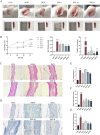
Impact of SSP on the symptoms and pathology in mice. (A) Ear and spleen changes. (B) Dermatitis severity scores (n = 5), the thickness differences between the left and right ears (n = 3), and the number of scratching behaviors (n = 3–5). (C, D) Ear histopathologic features. Epidermal thickening was assessed by HE staining (magnification, ×100), and mast cell infiltration was assessed by toluidine blue staining (magnification, ×200). (E) Ear weight changes (n = 3). (F) Epidermal thickness of HE-stained tissues (n = 6). (G) Expression of mast cells observed through toluidine blue-stained tissues (n = 3). MOD: MC903 (2 nmol/20 μL). SSP-L: SSP (20 mg/kg). SSP-M: SSP (50 mg/kg). SSP-H: SSP (100 mg/kg). Data were presented as the mean ± SEM. ### P < 0.001 vs. CON group; * p < 0.05, ** p < 0.01, and *** p < 0.001 vs. MOD group.
FIGURE 3
FIGURE 3
Impact of SSP on serum inflammatory factors and skin barrier protein expressions in mice. (A–D) IgE, MDC, TARC, and RANTES levels in the serum (n = 6). (E–G) FLG and LOR expression levels in the skin (n = 3). MOD: MC903 (2 nmol/20 μL). SSP-L: SSP (20 mg/kg). SSP-M: SSP (50 mg/kg). SSP-H: SSP (100 mg/kg). Data were presented as the mean ± SEM. ## P < 0.01, ### p < 0.001 vs. CON group; * p < 0.05, ** p < 0.01, and *** p < 0.001 vs. MOD group.
FIGURE 4
FIGURE 4
Impact of SSP on relative mRNA expressions in the ear. (A–F) Relative amounts of TSLP, IL-4, IL-13, IL-1β, IL-6, and IFN-γ mRNA expression (n = 3). MOD: MC903 (2 nmol/20 μL). SSP-L: SSP (20 mg/kg). SSP-M: SSP (50 mg/kg). SSP-H: SSP (100 mg/kg). Data were presented as the mean ± SEM. # P < 0.05, ### p < 0.001 vs. CON group; * p < 0.05, ** p < 0.01, and *** p < 0.001 vs. MOD group.
FIGURE 5
FIGURE 5
Gut microbial composition and alpha diversity in mice. (A, B) Pan/core curve of the samples (n = 6). (C) Venn diagram that showed OTU composition (n = 6). (D–G) Ace, Chao, coverage, and Sobs indices of OTU levels (n = 6). MOD: MC903 (2 nmol/20 μL). SSP: SSP (100 mg/kg). Mothur (version v.1.30.2) was used for the alpha diversity index analysis. The boot (version 1.3.18) and stats (version 3.3.1) packages in R language (version 3.3.1) were used for inter-index difference testing. * P < 0.05 and ** p < 0.01 vs. CON group.
FIGURE 6
FIGURE 6
Gut microbial beta diversity in mice. (A) Hierarchical clustering results (n = 6). (B–E) PCoA, NMDS, PLS-DA, and ANOSIM analysis results (n = 6). The corresponding box represents the within-group difference distance, and the box of the between group represents the between-group difference distance. MOD: MC903 (2 nmol/20 μL). SSP: SSP (100 mg/kg). QIIME was used for calculating the beta diversity distance matrix, and then R language (version 3.3.1) was used for conducting statistical analysis and plotting.
FIGURE 7
FIGURE 7
Gut microbial differences in community composition and species differences analysis in mice. (A, B) Relative microbial abundance in mice at the phylum level (n = 6). (C, D) Relative microbial abundance in mice at the genus level (n = 6). (E) LEfSe multi-level species discriminant analysis (n = 6). MOD: MC903 (2 nmol/20 μL). SSP: SSP (100 mg/kg). LEfSe software was used for LEfSe multi-level species discriminant analysis. ## p < 0.01 vs. CON group; * p < 0.05; ** p < 0.01 vs. MOD group.
FIGURE 8
FIGURE 8
Impact of SSP on relative mRNA expressions in HaCaT cells. (A-E) Relative amounts of IL-1β, IL-6, MDC, TARC, and RANTES mRNA expression (n = 3). MOD: TNF-α/IFN-γ (10 ng/mL). SSP-L: SSP (6.25 μg/mL). SSP-M: SSP (25 μg/mL). SSP-H: SSP (100 μg/mL). Data were presented as the mean ± SEM. ### p < 0.001 vs. CON group; * p < 0.05, ** p < 0.01, and *** p < 0.001 vs. MOD group.
FIGURE 9
FIGURE 9
SSP’s effect on the MAPK/NF-κB pathway in HaCaT cells. (A) Expressions of the proteins p-ERK, p-JNK, p-p38, ERK, JNK, and p38 in HaCaT cells (n = 3). (B) Expression of the proteins p-p65 and p65 in HaCaT cells (n = 3). (C–F) Relative protein expression levels of p-ERK/ERK, p-JNK/JNK, p-p38/p38, and p-p65/p65 (n = 3). MOD: TNF-α/IFN-γ (10 ng/mL). SSP-L: SSP (6.25 μg/mL). SSP-M: SSP (25 μg/mL). SSP-H: SSP (100 μg/mL). Data were presented as the mean ± SEM. # P < 0.05, ### p < 0.001 vs. CON group; * p < 0.05, ** p < 0.01, *** p < 0.001 vs. MOD group.

Similar articles

Cited by

References

    1. Anders H.-J., Andersen K., Stecher B. (2013). The intestinal microbiota, a leaky gut, and abnormal immunity in kidney disease. Kidney Int. 83, 1010–1016. 10.1038/ki.2012.440 - DOI - PubMed
    1. Balakirski G., Novak N. (2022). Atopic dermatitis and pregnancy. J. Allergy Clin. Immunol. 149, 1185–1194. 10.1016/j.jaci.2022.01.010 - DOI - PubMed
    1. Ben Abdallah H., Vestergaard C. (2022). Atopic dermatitis, hypertension and cardiovascular disease. Br. J. Dermatol. 186, 203–204. 10.1111/bjd.20802 - DOI - PubMed
    1. Chen B.-R., Hsu K.-T., Li T.-L., Chan Y.-L., Wu C.-J. (2021). Topical application of fucoidan derived from Cladosiphon okamuranus alleviates atopic dermatitis symptoms through immunomodulation. Int. Immunopharmacol. 101, 108362. 10.1016/j.intimp.2021.108362 - DOI - PubMed
    1. Chen M.-C., Chen C.-S., Wu Y.-W., Yang Y.-Y. (2020). Poly-γ-Glutamate microneedles as transdermal immunomodulators for ameliorating atopic dermatitis-like skin lesions in Nc/Nga mice. Acta Biomater. 114, 183–192. 10.1016/j.actbio.2020.07.029 - DOI - PubMed

LinkOut - more resources