Review
. 2025 Mar 17:27:1124-1139.
doi: 10.1016/j.csbj.2025.03.023. eCollection 2025.

Structure, function and evolution of the bacterial DinG-like proteins

Affiliations
Review

Structure, function and evolution of the bacterial DinG-like proteins

Kaiying Cheng. Comput Struct Biotechnol J. .

Abstract

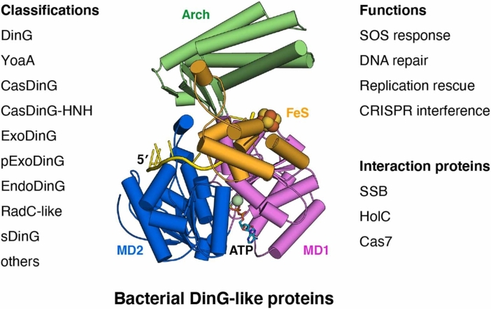
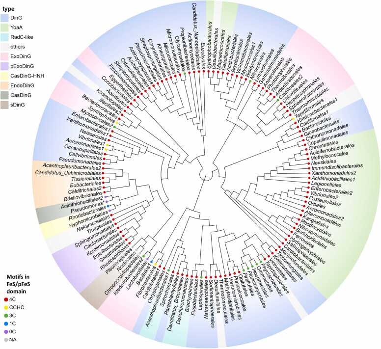
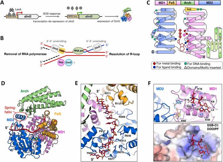
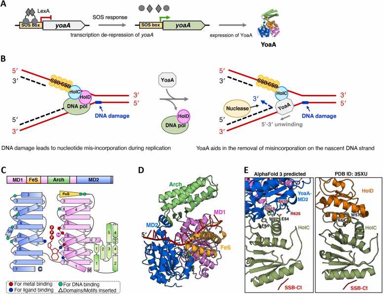
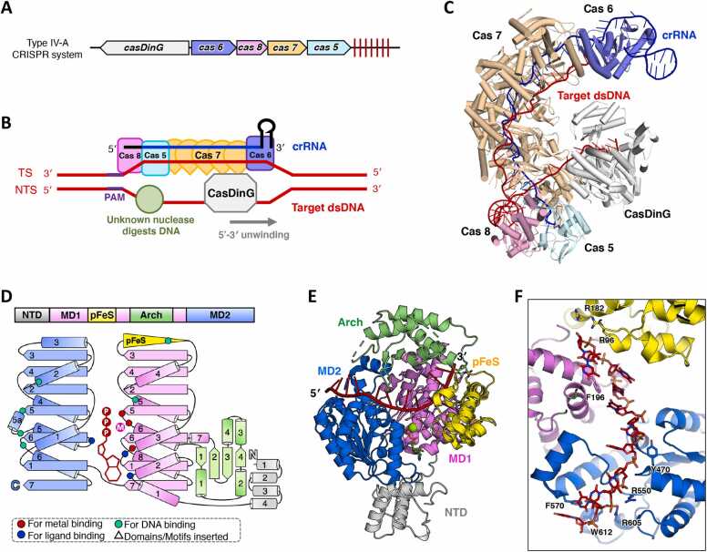
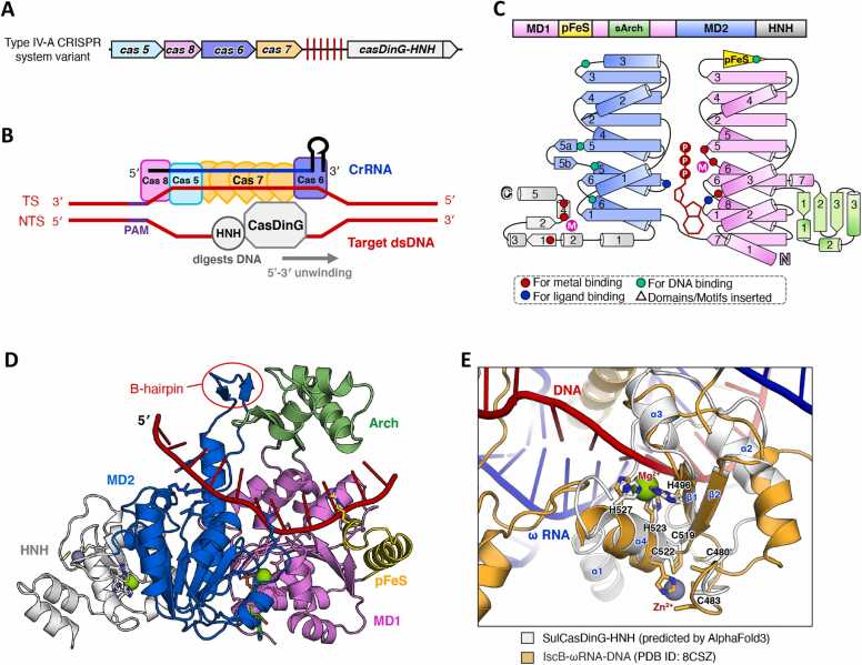
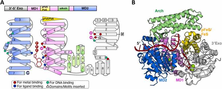
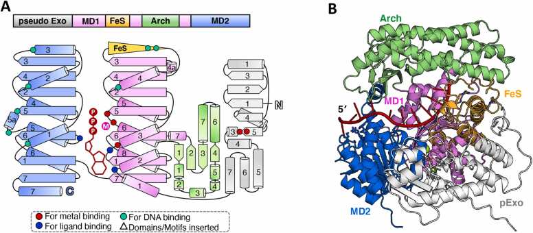
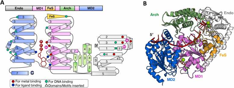
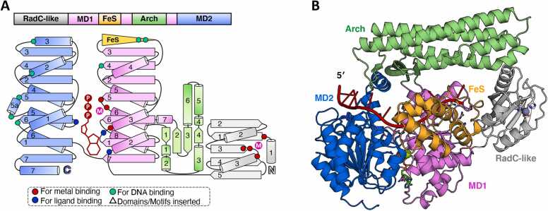
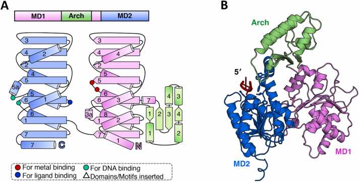
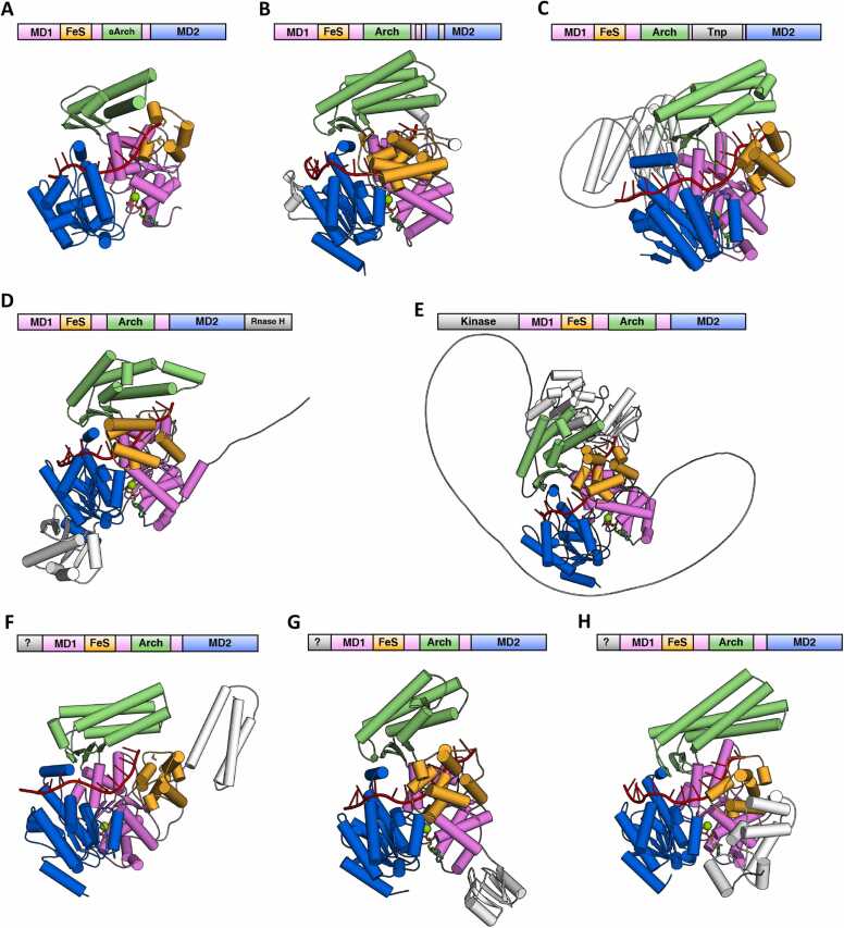
The damage-inducible G (DinG)-like proteins represent a widespread superfamily 2 (SF2) of DNA helicases, exhibiting remarkable diversity in domain architecture, substrate specificity, regulatory mechanisms, biological functions, interaction partners, and taxonomic distribution. Many characterized DinG-like proteins play critical roles in bacterial stress responses and immunity, including the SOS response, DNA repair, and phage interference. This review aims to provide a summary of bacterial DinG-like proteins, categorizing them into subgroups such as DinG, YoaA, CasDinG, CasDinG-HNH, ExoDinG, pExoDinG, EndoDinG, RadC-like DinG, sDinG, and others. This classification provides an analysis of sequence-structure-function relationships within this superfamily. Further sequence clustering revealed inter-cluster relationships and subgroup heterogeneity, suggesting potential functional divergence. Integrating sequence analysis, domain architecture, structural data, and genomic context enabled functional predictions for these DinG-like protein subgroups, shedding light on their evolutionary and biological significance.

Keywords: CRISPR interference; DNA repair; DinG; SOS response; Structure.


Conflict of interest statement

The authors declare that they have no known competing financial interests or personal relationships that could have appeared to influence the work reported in this paper.

Figures

None
Graphical abstract
Fig. 1
Fig. 1
Phylogenetic tree of the DinG-like proteins from different bacteria orders. The DinG-like protein sequences used for constructing the phylogenetic tree were derived from representative bacteria across various bacterial orders. The leaves of the phylogenetic tree are labeled with the names of the bacterial orders, and the corresponding species names and protein sequences can be found in Table S1. Multiple sequence alignment and phylogenetic tree construction was performed using Clustal Omega. The phylogenetic tree was visualized using the ChiPlot online program. Various color blocks indicate the different subgroups of DinG-like proteins. The dots of varying colors denote the presence and number of conserved cysteines in FeS or pFeS, as well as their potential to coordinate with FeS clusters.
Fig. 2
Fig. 2
Summary of the biological function and structure of DinG. (A) The cartoon model illustrates the induction of DinG by the SOS response. During the SOS response, there is a de-repression of dinG transcription and an increase in DinG protein expression, which is controlled by the LexA repressor. (B) The cartoon model depicts how DinG participates in rescuing transcription-blocked replication forks. In highly transcribed regions, DinG assists in resolving R-loops and removing RNA polymerase, likely with the assistance of Rep and UvrD proteins, to ensure efficient replication. (C) The domain arrangement and the topology diagram of EcDinG. EcDinG was selected for structural analysis within DinG subgroup proteins. The MD1, MD2, Arch, and FeS domains were colored violet, marine, lime green, and orange, respectively. Key residues involved in metal ion coordination, ATP binding, and substrate interaction were highlighted as red dots, blue dots, and cyan dots, respectively. (D) The overall structure of the EcDinG–ssDNA–ADPBeF3 complex. The structure (PDB ID: 6FWS) was shown in cartoon form, with each domain colored as described in (C). ssDNA and ADPBeF3·Mg2+ were colored red and green, respectively. (E) A zoomed-in view of the DNA binding mode of EcDinG. The DNA and the key residues involved in DNA interaction were shown as sticks and labelled. (F) AlphaFold 3 predicted EcDinG–SSB-Ct interaction. Upper, zoomed-in view of the predicted EcDinG–SSB-Ct interaction interface. The predicted binding pocket (α1 and α2 of the EcDinG MD1 domain) was illustrated as cartoon. SSB-Ct peptide (DDDIPF) and the potential residues essential for SSB-Ct interaction were depicted as sticks. The input information used for model prediction is detailed in the supplementary material. Bottom, an analysis of the electrostatic properties of the SSB-Ct binding pocket on EcDinG. The electrostatic potential of the SSB interaction interface on the MD1 of EcDinG was determined using APBS, which was then projected onto the solvent-accessible surface of the structure at contouring levels of ± 5 kT (depicted in blue/red). Amino acids of SSB-Ct were represented as sticks, colored red, and labelled.
Fig. 3
Fig. 3
Summary of the biological function and predicted structure of YoaA. (A) The cartoon model illustrates the induction of YoaA. During the SOS response, there is a de-repression of yoaA transcription and an increase in DinG protein expression, which is regulated by the LexA repressor. (B) The cartoon model depicts how YoaA assists in genomic stability. During DNA replication, DNA damage leads to nucleotide mis-incorporation. HolC–SSB recruits YoaA helicase, which unwinds the 3′ nascent strand, allowing nuclease access for the removal of the mis-incorporated nucleotides. (C) The domain arrangement and the topology diagram of EcYoaA. EcYoaA was selected for structural analysis within the YoaA subgroup proteins. The MD1, MD2, Arch, and FeS domains were colored violet, marine, lime green, and orange, respectively. Key residues involved in metal ion coordination, ATP binding, and substrate interaction were highlighted as red dots, blue dots, and cyan dots, respectively. (D) The overall structure of EcYoaA–ssDNA–ATP complex. The AlphaFold3 predicted structure was shown in cartoon form, with each domain colored as described in (C). ssDNA and ATP·Mg2+ were colored red and green, respectively. The input information used for model prediction is detailed in the supplementary material. (E) Structural comparison between EcYoaA–HolC–SSB-Ct complex and EcHolD–HolC–SSB-Ct complex. A zoomed-in view of the protein-protein interfaces in the AlphaFold3 predicted EcYoaA–HolC–SSB-Ct complex structure and the experimentally determined EcHolD–HolC–SSB-Ct complex structure (PDB ID: 3XSU) is shown. The (predicted) key residues involved in protein-protein interactions were shown as sticks and labelled. The input information used for model prediction is detailed in the supplementary material.
Fig. 4
Fig. 4
Summary of the biological function and structure of CasDinG. (A) The casDinG related gene operon with a CRISPR array in Type IV-A CRISPR system. (B) A simplified cartoon model illustrating the involvement of CasDinG in interference within a Type IV-A CRISPR system. (C) The overall CryoEM structure of PaCasDinG bound to the Csf–crRNA–dsDNA complex (PDB ID:7XG3). The Cas5, Cas6, Cas7, Cas8 and CasDinG subunit were colored cyan, slate, wheat, pink, and white, respectively. Target dsDNA and crRNA were colored red and blue, respectively. (D) The domain arrangement and the topology diagram of PaCasDinG. PaCasDinG was selected for structural analysis within the CasDinG subgroup proteins. The MD1, MD2, Arch, pFeS, and NTD domains were colored violet, marine, lime green, yellow, and white, respectively. Key residues involved in metal ion coordination, ATP binding, and substrate interaction were highlighted as red dots, blue dots, and cyan dots, respectively. (E) The overall structure of PaCasDinG–ssDNA–ATP complex. The NTD truncated complex structure (PDB ID: 7XF1) and the AlphaFold3 predicted NTD were combined and shown in cartoon form, with each domain colored as described in (D). ssDNA and ATP·Mg2+ were colored red and green, respectively. (F) A zoomed-in view of the DNA binding mode of PaCasDinG. The DNA and the key residues involved in DNA interaction were shown as sticks and labelled.
Fig. 5
Fig. 5
Summary of the biological function and predicted structure of CasDinG-HNH. (A) The casDinG-HNH related gene operon with a CRISPR array in a Type IV-A CRISPR system variant. (B) A simplified cartoon model illustrating the involvement of CasDinG-HNH in interference within a Type IV-A CRISPR system variant. (C) The domain arrangement and the topology diagram of CasDinG-HNH. CasDinG-HNH from Sulfitobacter sp. JL08 was selected for structural analysis within the CasDinG-HNH subgroup proteins. The MD1, MD2, Arch, pFeS, and HNH domains were colored violet, marine, lime green, yellow, and white, respectively. Key residues involved in metal ion coordination, ATP binding, and substrate interaction were highlighted as red dots, blue dots, and cyan dots, respectively. (D) The overall structure of AlphaFold3 predicted SuCasDinG-HNH–ssDNA–ATP complex. The structure was shown in cartoon form, with each domain colored as described in (C). ssDNA and ATP·Mg2+, Zn2+ were colored red, green, and purple blue respectively. The input information used for model prediction is detailed in the supplementary material, while the DNA/RNA hybrid duplex that could not be modeled with high-quality has been omitted from this figure for clarity. (E) Structural comparison of the HNH domain between SuCasDinG-HNH and IscB–ωRNA–DNA. A zoomed-in view of the catalytic sites within the HNH, as predicted by AlphaFold3 for SuCasDinG-HNH, alongside the crystallographic structure of IscB in complex with ωRNA and DNA (PDB ID: 8CSZ). Key residues implicated in metal catalysis and Zn-finger coordination in SuCasDinG-HNH are depicted as sticks and labeled. ωRNA and DNA were shown as cartoon, colored blue and red, respectively.
Fig. 6
Fig. 6
Summary of the predicted structure information of ExoDinG. (A) The domain arrangement and the topology diagram of ExoDinG. ExoDinG from B. subtilis was selected for structural analysis within the ExoDinG subgroup proteins. The MD1, MD2, Arch, pFeS/FeS, and 3′–5′ Exonuclease domains were colored violet, marine, lime green, yellow, and white, respectively. Key residues involved in metal ion coordination, ATP binding, and substrate interaction were highlighted as red dots, blue dots, and cyan dots, respectively. (B) The overall structure of the BsExoDinG–ssDNA–ATP complex, as predicted by AlphaFold3, is depicted in cartoon representation, with each domain colored as described in (A). ssDNA and ATP·Mg2+ were colored red and green, respectively. The input information used for model prediction is detailed in the supplementary material.
Fig. 7
Fig. 7
Summary of the predicted structure information of pExoDinG. (A) The domain arrangement and the topology diagram of pExoDinG. pExoDinG from C. vibrioides was selected for structural analysis within the pExoDinG subgroup proteins. The MD1, MD2, Arch, FeS, and pseudo exonuclease domains were colored violet, marine, lime green, orange, and white, respectively. Key residues involved in metal ion coordination, ATP binding, and substrate interaction were highlighted as red dots, blue dots, and cyan dots, respectively. (B) The overall structure of the CvpExoDinG–ssDNA–ATP complex, as predicted by AlphaFold3, was shown in cartoon form, with each domain colored as described in (A). ssDNA and ATP·Mg2+ were colored red and green, respectively. The input information used for model prediction is detailed in the supplementary material, while the elements (including one ssDNA strand and Mg2+ near the supposed nuclease site) that could not be modeled with high-quality have been omitted from this figure for clarity.
Fig. 8
Fig. 8
Summary of the predicted structure information of EndoDinG. (A) The domain arrangement and the topology diagram of EndoDinG. EndoDinG from B. bacteriovorus HD100 was selected for structural analysis within the EndoDinG subgroup proteins. The MD1, MD2, Arch, FeS, and 3′–5′ Exonuclease/Endonuclease domains were colored violet, marine, lime green, orange, and white, respectively. Key residues involved in metal ion coordination, ATP binding, and substrate interaction were highlighted as red dots, blue dots, and cyan dots, respectively. (B) The overall structure of the BbEndoDinG–ssDNA–ATP complex, as predicted by AlphaFold3, was shown in cartoon form, with each domain colored as described in (A). ssDNA and ATP·Mg2+ were colored red and green, respectively. The input information used for model prediction is detailed in the supplementary material.
Fig. 9
Fig. 9
Summary of the predicted structure information of RadC-like DinG. (A) The domain arrangement and the topology diagram of RadC-like DinG. RadC-like DinG from D. soudanensis was selected for structural analysis within the RadC-like DinG subgroup proteins. The MD1, MD2, arch, FeS, and RadC-like domains were colored violet, marine, lime green, orange, and white, respectively. Key residues involved in metal ion coordination, ATP binding, and substrate interaction were highlighted as red dots, blue dots, and cyan dots, respectively. (B) The overall structure of the DsRadC-like DinG–ssDNA–ATP complex, as predicted by AlphaFold3, was shown in cartoon form, with each domain colored as described in (A). ssDNA, ATP·Mg2+, Zn2+ were colored red, green, and purple blue respectively. The input information used for model prediction is detailed in the supplementary material.
Fig. 10
Fig. 10
Summary of the predicted structure information of sDinG. (A) The domain arrangement and the topology diagram of sDinG. sDinG from Nostoc sp. PCC 7120 was selected for structural analysis within the sDinG subgroup proteins. The MD1, MD2, and Arch domains were colored violet, marine and lime green, respectively. Key residues involved in metal ion coordination, and substrate interaction were highlighted as red dots, blue dots, and cyan dots, respectively. (B) The overall structure of NossDinG–ssDNA complex, as predicted by AlphaFold3, was shown in cartoon form, with each domain colored as described in (A). ssDNA was colored red. The input information used for model prediction is detailed in the supplementary material, while the elements (include a portion of the ssDNA strand and ATP·Mg2+) that could not be modeled with high-quality have been omitted from this figure for clarity.
Fig. 11
Fig. 11
Summary of the predicted structure information of the other undefined subgroups of DinG-like proteins. The domain arrangements and the predicted DNA and ATP·Mg2+ bound complex structures of DinG-like proteins from bacteria orders Truepera radiovictrix (order Trueperales) (A), Nakamurella multipartite (order Nakamurellales) (B), Humisphaera borealis (order Tepidisphaerales) (C), Caldilinea aerophila (order Caldilineales) (D), Ktedonobacterales bacterium SCAWS-G2 (order Ktedonobacterales) (E), Desulfotalea psychrophila (order Desulfobacterales) (F), Hippea maritima (order Desulfurellales) (G), and Kosmotoga olearia (order Kosmotogales) (H) were depicted. The MD1, MD2, FeS, Arch, and the additional domains were colored violet, marine, lime green, and white, respectively. AlphaFold3 predicted structures were shown as cartoon. The ssDNA and ATP·Mg2+ were colored red and green, respectively.

Similar articles

Cited by

References

    1. White M.F. Structure, function and evolution of the XPD family of iron-sulfur-containing 5′-> 3′ DNA helicases. Biochem Soc Trans. 2009;37(Pt 3):547–551. - PubMed
    1. Liu H., Rudolf J., Johnson K.A., McMahon S.A., Oke M., Carter L., et al. Structure of the DNA repair helicase XPD. Cell. 2008;133(5):801–812. - PMC - PubMed
    1. Wolski S.C., Kuper J., Hanzelmann P., Truglio J.J., Croteau D.L., Van Houten B., et al. Crystal structure of the FeS cluster-containing nucleotide excision repair helicase XPD. PLoS Biol. 2008;6(6) - PMC - PubMed
    1. Kuper J., Wolski S.C., Michels G., Kisker C. Functional and structural studies of the nucleotide excision repair helicase XPD suggest a polarity for DNA translocation. EMBO J. 2012;31(2):494–502. - PMC - PubMed
    1. Fan L., Fuss J.O., Cheng Q.J., Arvai A.S., Hammel M., Roberts V.A., et al. XPD helicase structures and activities: insights into the cancer and aging phenotypes from XPD mutations. Cell. 2008;133(5):789–800. - PMC - PubMed

LinkOut - more resources