Advanced Endodontic Techniques for Treating Root Perforation in a Hypertaurodont Molar: A Case Report
- PMID: 40206778
- PMCID: PMC11981002
- DOI: 10.22037/iej.v20i1.46431
Advanced Endodontic Techniques for Treating Root Perforation in a Hypertaurodont Molar: A Case Report
Abstract
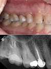
Taurodontism is a dental anomaly characterized by an enlarged pulp chamber and apically displaced pulpal floor. This disorder poses significant challenges in endodontic treatment, especially when perforations occur. The present case study details the endodontic retreatment of a hypertaurodont maxillary second molar in a 36-year-old female patient with a mesial canal perforation. The procedure employed a dental operating microscope for enhanced visualization and precision. Canals were prepared using a crown-down technique, with the perforation site managed using MTA and bioceramic material applied via the second mesiobuccal canal. The remaining canals were obturated using gutta-percha and bioceramic sealer. At the 1-year follow-up, the tooth was functional and asymptomatic, with radiographic evidence of a normal periodontal ligament space. This case demonstrates the efficacy of contemporary endodontic techniques, including bioceramic materials and advanced magnification, in managing the unique challenges posed by taurodontism.
Keywords: Anatomic Variation; Perforation; Root Canal; Taurodontism.
Conflict of interest statement
None.
Figures


References
-
- Toure B, Kane A, Sarr M, Wone M, Fall F. Prevalence of taurodontism at the level of the molar in the black Senegalese population 15 to 19 years of age. Odontostomatol Trop. 2000;23(89):36–9. - PubMed
Publication types
LinkOut - more resources
Full Text Sources
