. 2025 Apr 9:29:0173.
doi: 10.34133/bmr.0173. eCollection 2025.

Nanotension Relief Agent Enhances Tissue Penetration by Reducing Solid Stress in Pancreatic Ductal Adenocarcinoma via Rho/ROCK Pathway Inhibition

Affiliations

Nanotension Relief Agent Enhances Tissue Penetration by Reducing Solid Stress in Pancreatic Ductal Adenocarcinoma via Rho/ROCK Pathway Inhibition

Feiran Yu et al. Biomater Res. .

Abstract

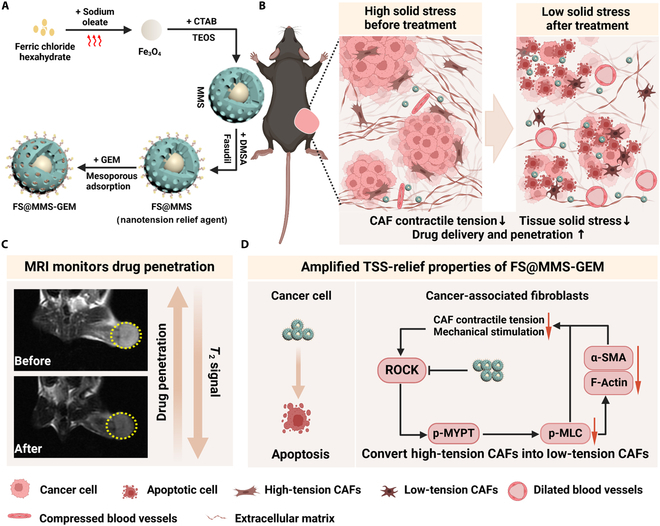
The formidable contractile tension exerted by cancer-associated fibroblasts (CAFs) in pancreatic ductal adenocarcinoma (PDAC) tissue is crucial for maintaining high tissue solid stress (TSS), which impedes the delivery and penetration of chemotherapeutic drugs. To address this obstacle, we constructed a pH-responsive nanotension relief agent (FS@MMS), in which fasudil (FS) was ingeniously conjugated to mesoporous silica encapsulated with magnetic iron oxide (MMS). The nanotension relief agent was demonstrated to inhibit the synthesis of phosphorylated myosin light chain by blocking the Rho/Rho-associated serine/threonine kinase (ROCK) pathway, triggering the swift transformation of high-tension CAFs into low-tension CAFs in PDAC tissue, which relieves TSS and enhances drug penetration in Panc02/NIH-3T3 multicellular tumor spheroids. When the nanotension relief agent was further loaded with the chemotherapeutic drug gemcitabine (GEM), as FS@MMS-GEM, the enhanced permeation of GEM progressively killed tumor cells and amplified their TSS-relief properties, thereby maximizing the anticancer efficacy of chemotherapeutic agents in Panc02/NIH-3T3 coplanted model mice. The magnetic resonance imaging results revealed that the synergistic effect substantially improved drug delivery and penetration efficiency. The developed approach holds great potential for improving chemotherapy efficacy in PDAC and provides a novel therapeutic approach for the treatment of related stroma-rich tumors.


Conflict of interest statement

Competing interests: The authors declare that they have no competing interests.

Figures

Fig. 1.
Fig. 1.
Tissue solid stress (TSS) relief scheme for pancreatic ductal adenocarcinoma (PDAC). (A) Schematic diagram illustrating the generation of FS@MMS-GEM. (B) FS@MMS-GEM treatment reduced the TSS in PDAC tissue and enhanced drug penetration, which increased the anticancer effect of chemotherapeutic agents. (C) Magnetic resonance imaging (MRI) was used to assess the drug penetration efficiency. (D) Fasudil inhibited phosphorylated myosin light chain (p-MLC) synthesis by blocking the Rho/Rho-associated serine/threonine kinase (ROCK) pathway, resulting in the rapid conversion of high-tension cancer-associated fibroblasts (CAFs) to low-tension CAFs, which reduced TSS and increased drug penetration efficiency. The enhanced permeation of gemcitabine (GEM) progressively killed tumor cells and amplified the TSS-relief properties of FS@MMS-GEM. Created with BioRender.com. CTAB, cetyltrimethylammonium bromide; TEOS, tetraethoxysilane; MMS, mesoporous silica encapsulated with magnetic iron oxide; α-SMA, alpha smooth muscle actin; p-MLC, phosphorylated myosin light chain; DMSA, dimercaptosuccinic acid; p-MYPT, phospho-myosin phosphatase.
Fig. 2.
Fig. 2.
Characterization of FS@MMS-GEM. (A and B) Transmission electron microscopy (TEM) image and high-resolution TEM (HRTEM) image of the Fe3O4 nanoparticles. (C) TEM image of the MMS nanocarrier. (D) Size of the MMS nanocarrier. (E) Zeta potentials of MMS, FS@MMS, and FS@MMS-GEM in 10 mM Hepes buffer (pH 7.4). (F) X-ray diffraction (XRD) patterns of the Fe3O4 and MMS nanocarriers. (G) Fourier transform infrared spectroscopy (FTIR) spectra of the Fe3O4 and MMS nanocarriers. (H and I) Release rates of GEM and fasudil from MMS at various pH values and time intervals. (J) Saturation magnetization of the MMS nanocarrier. (K) T2-weighted images and T2 mapping of different concentrations of the nanotension relief agent (FS@MMS) (1.03, 0.51, 0.13, 0.06, 0.03, and 0.002 mM). (L) The fitting curve between 1/T2 and the concentration of Fe was obtained according to T2 mapping. (M) Magnetic resonance (MR) images of Panc02 cells and CAFs incubated with the nanotension relief agent (100 μg/ml) for 0, 0.5, 1, 2, and 4 h. (N) As the incubation time between the cells and nanocarrier increased, the signal intensity decreased gradually. PBS, phosphate-buffered saline.
Fig. 3.
Fig. 3.
Nanotension relief agents (FS@MMS) rapidly transform high-tension CAFs into low-tension CAFs. (A) Prussian blue staining of CAFs incubated with the nanotension relief agents (100 μg/ml) for 0, 1, 2, and 4 h (scale bars: 100 μm). (B, E, and F) Protein expression and semiquantitative analysis of p-MLC and phospho-myosin phosphatase (p-MYPT) in CAFs from different treatment groups. (C) Morphological manifestations of F-actin in CAFs treated with different drugs (scale bars: 50 μm). (D and G) The expression of α-SMA in the different treatment groups (scale bars: 100 μm). Data are presented as mean ± SD (n = 3). ns, not significant; **P < 0.01; ***P < 0.001. GAPDH, glyceraldehyde-3-phosphate dehydrogenase.
Fig. 4.
Fig. 4.
Cytotoxicity of FS@MMS-GEM in vitro. (A) Prussian blue staining of CAFs incubated with FS@MMS-GEM (100 μg/ml) for 0, 1, 2, and 4 h (scale bars: 50 μm). (B) Viability of Panc02 cells incubated with different drug combinations at different concentrations for 24 h. (C) Fluorescence images of Panc02 cells stained with calcein-AM and propidium iodide (PI) after treatment with different formulations (red: dead/late apoptotic cells; green: viable cells; scale bars: 100 μm). (D) Flow cytometric analysis of Panc02 cells after staining with annexin V–fluorescein isothiocyanate (FITC) and PI. (E) Nuclear changes in Panc02 cells induced by different combinations of drugs were observed via TEM; arrows indicate the nanomedicine engulfed by Panc02 cells, and the scale bars represent 1.2 μm. Data are presented as mean ± SD (n = 3). ns, not significant; *P < 0.05; **P < 0.01; ***P < 0.001.
Fig. 5.
Fig. 5.
Amplified TSS improvement of FS@MMS-GEM in Panc02/NIH-3T3 multicellular tumor spheroids (MTSs). (A) FITC–dextran penetration and (B) corresponding 3-dimensional (3D) conversion into Panc02/NIH-3T3 MTSs in the different groups. (E) Fluorescence intensity distribution along the diameter of the Panc02/NIH-3T3 MTSs. (C and F) Images of α-SMA expression and differences in α-SMA expression among the different groups. (D and G) Images of collagen I expression and differences in collagen I expression among the different groups (scale bars: 100 μm). The data are presented as mean ± SD (n = 3). *P < 0.05; ***P < 0.001.
Fig. 6.
Fig. 6.
Evaluation of the penetration efficiency and therapeutic effect of FS@MMS-GEM in PDAC tissue. (A) Diagram of the in vivo treatment process. (B) Real-time MR image of the tumor tissue 2 h before and after the first and last tail vein injections of nanotension relief agents (FS@MMS) and FS@MMS-GEM. (C) Statistical analysis of the changes in signal intensity before and after treatment with the nanotension relief agents and FS@MMS-GEM treatment groups at the first and last time points. (D) At 24 h after the last tail vein injection of nanotension relief agents and FS@MMS-GEM, the tumor tissue was stained with Prussian blue (scale bars: 100 μm). (E) The Fe content in major organs and tumor tissues of mice 24 h after the first injection of normal saline, the nanotension relief agent, or FS@MMS-GEM. (F) Schematic diagram of the method used to measure the TSS in tumors. (G) After treatment for 15 d, the TSS of the tumor tissue in different groups was measured. (H) Images of tumors from each group after treatment. (I) Changes in tumor volume (I: PBS; II: free GEM; III: free fasudil + free GEM; IV: FS@MMS; V: GEM-MMS; VI: FS@MMS-GEM). The data are presented as mean ± SD (n = 5). *P < 0.05; **P < 0.01; ***P < 0.001. NS, normal saline.
Fig. 7.
Fig. 7.
In vivo antitumor effect. (A) Hematoxylin and eosin (H&E) staining of PDAC tissue after treatment (scale bars are 200 μm). (B, D, and F) α-SMA, collagen I and CD31 immunofluorescence staining of PDAC tissue after treatment. (C) Masson staining was performed on PDAC tissue after treatment (scale bars: 100 μm). (E and G) Ki67 and hypoxia-inducible factor-1α (HIF-1α) immunohistochemical staining of PDAC tissue after treatment (scale bars: 100 μm). The data are presented as mean ± SD (n = 5).
Fig. 8.
Fig. 8.
Biosafety evaluation. (A) The results of routine blood, liver, and kidney function biochemical analyses after treatment were normal. (B) H&E staining of the main organs from each group after treatment (scale bars: 200 μm). The data are presented as mean ± SD (n = 3). WBC, white blood cells; RBC, red blood cells; MCHC; mean corpuscular hemoglobin concentration; PLT, platelet; BUN, blood urea nitrogen; ALT, alanine transaminase; AST, aspartate transaminase.

Similar articles

References

    1. Siegel RL, Miller KD, Fuchs HE, Jemal A. Cancer statistics, 2021. CA Cancer J Clin. 2021;71(1):7–33. - PubMed
    1. Sung H, Ferlay J, Siegel RL, Laversanne M, Soerjomataram I, Jemal A, Bray F. Global cancer statistics 2020: GLOBOCAN estimates of incidence and mortality worldwide for 36 cancers in 185 countries. CA Cancer J Clin. 2021;71(3):209–249. - PubMed
    1. Manji GA, Olive KP, Saenger YM, Oberstein P. Current and emerging therapies in metastatic pancreatic cancer. Clin Cancer Res. 2017;23(7):1670–1678. - PubMed
    1. Nia HT, Munn LL, Jain RK. Physical traits of cancer. Science. 2020;370(6516): Article eaaz0868. - PMC - PubMed
    1. Zhou H, Wang M, Zhang Y, Su Q, Xie Z, Chen X, Yan R, Li P, Li T, Qin X, et al. . Functions and clinical significance of mechanical tumor microenvironment: Cancer cell sensing, mechanobiology and metastasis. Cancer Commun. 2022;42(5):374–400. - PMC - PubMed

LinkOut - more resources