. 2024 Sep 24;14(3):103263.
doi: 10.1016/j.eats.2024.103263. eCollection 2025 Mar.

Arthroscopic Tri-anchor Double-Pulley Suture-Bridge Reduction of Greater Tuberosity Fracture

Affiliations

Arthroscopic Tri-anchor Double-Pulley Suture-Bridge Reduction of Greater Tuberosity Fracture

Peiguan Huang et al. Arthrosc Tech. .

Abstract

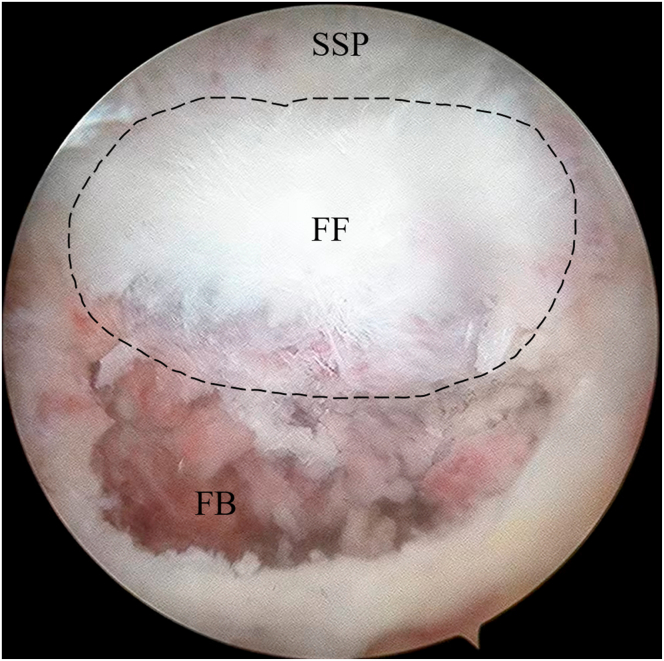
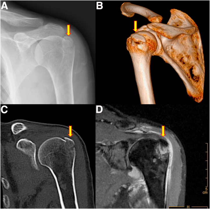
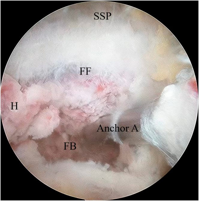
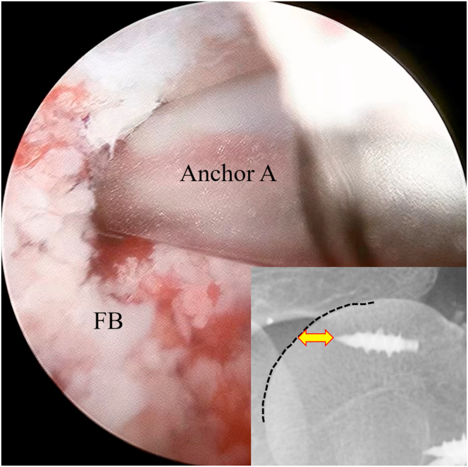
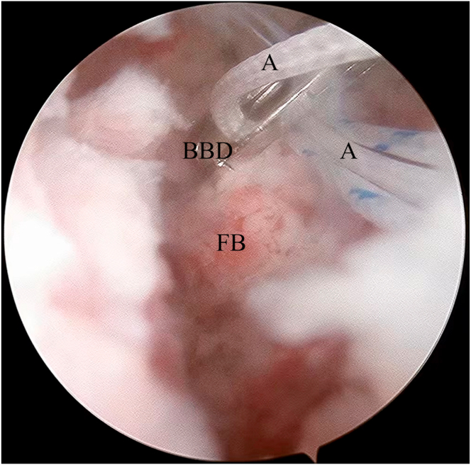
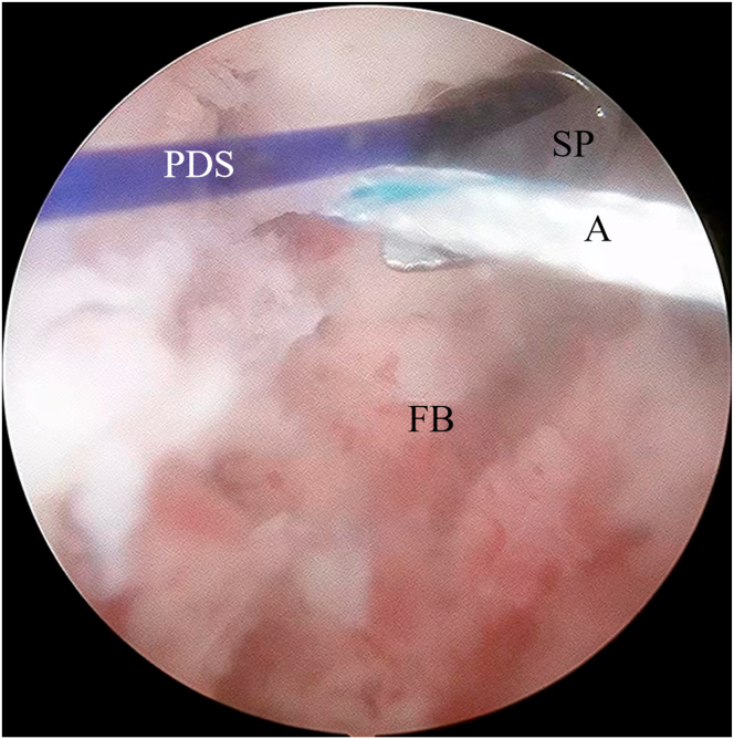
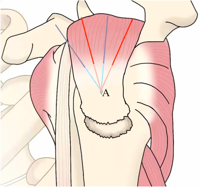
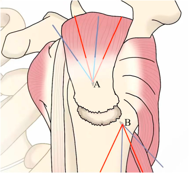
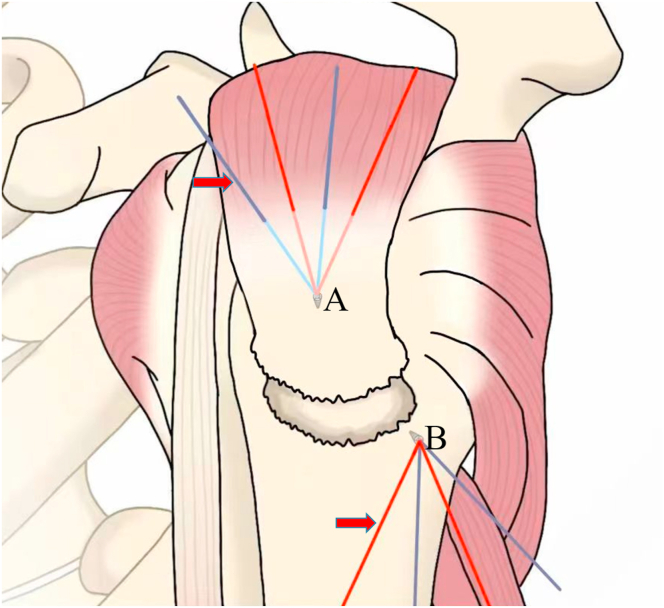
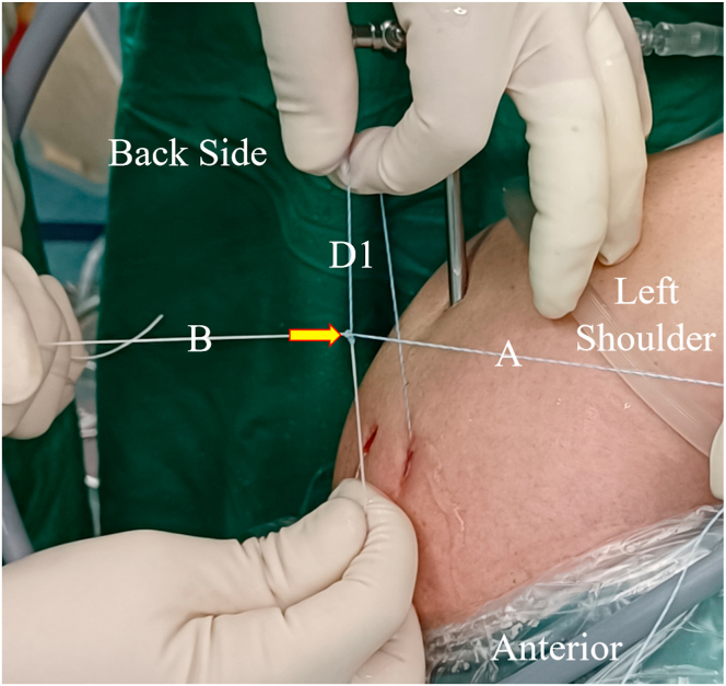
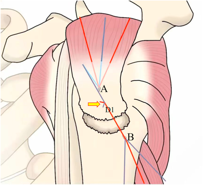
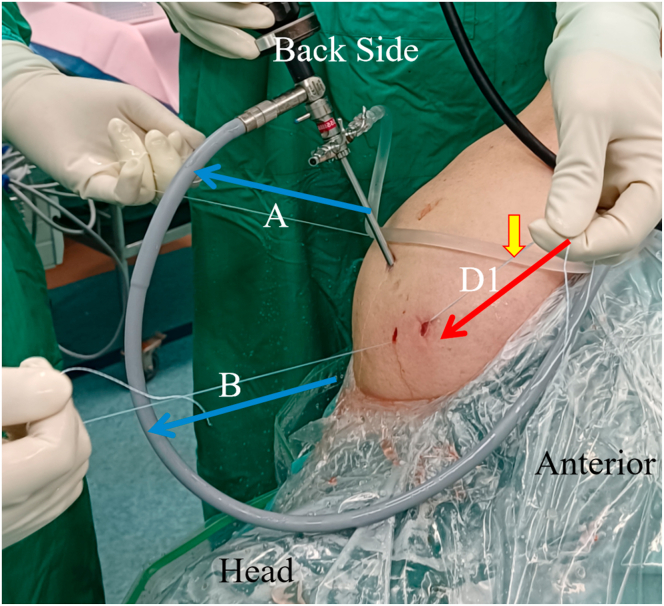
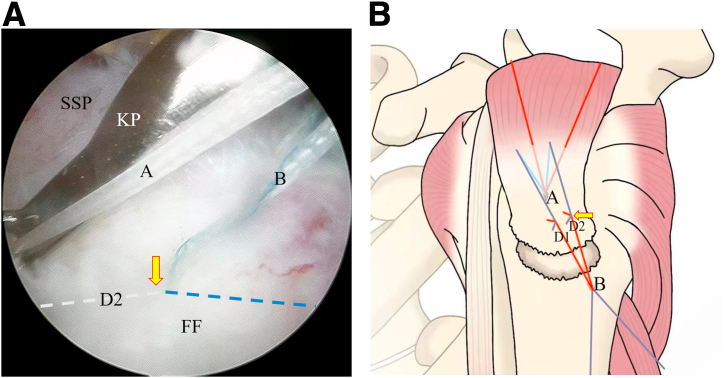
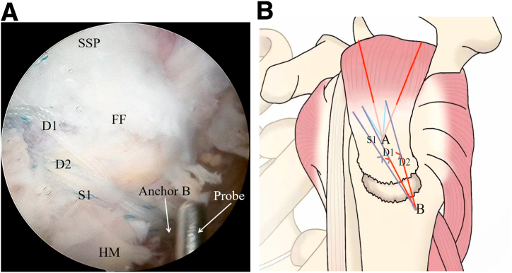
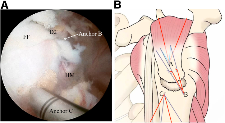
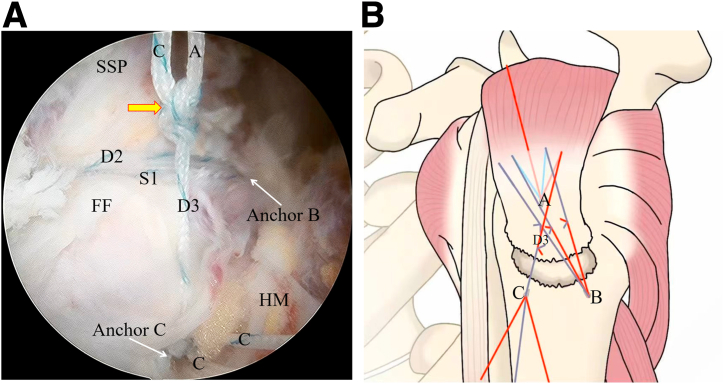
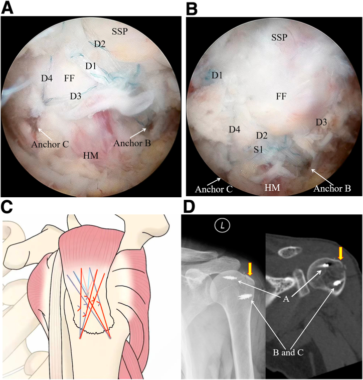
Currently, cannulated screws, plates, and suture bridges are widely applied in the treatment of greater tuberosity fracture; however, further fragmentation of the fracture, risk of loss of fracture reduction, implant impingement, and anchor pullout are the drawbacks. Therefore, we present a pragmatic surgical technique called the arthroscopic tri-anchor double-pulley suture-bridge technique that uses a double-loaded metallic anchor as a lateral-row anchor. In the treatment of greater tuberosity fracture, this hybrid repair including 4 sets of double-pulley suture bridges and 2 sets of single rows can obtain powerful stiffness of the suture construct, the metallic anchor used as a lateral-row anchor can significantly reduce the risk of anchor pullout, the single-row process can lessen the overall surgical time, and implant impingement will not occur postoperatively.


Conflict of interest statement

All authors (P.H., X.W., Y.F., Z.X., Z.L., B.P., C.H.) declare that they have no known competing financial interests or personal relationships that could have appeared to influence the work reported in this paper.

Figures

Fig 1
Fig 1
Arthroscopic image of left shoulder (with patient in lateral decubitus position), viewed through subacromial lateral portal, showing localization of fracture margin, fracture bed (FB), and native anatomic contour (dashed line). It is determined that the supraspinatus tendon (SSP) is attached to the fracture fragment (FF).
Fig 2
Fig 2
Displaced greater tuberosity fracture (arrows) in left shoulder on anteroposterior radiograph (A), computed tomography scan with 3-dimensional reconstruction (B), anteroposterior computed tomography scan (C), and magnetic resonance imaging (oblique coronal view) (D).
Fig 3
Fig 3
Arthroscopic image of left shoulder (with patient in lateral decubitus position), viewed through subacromial lateral portal, showing insertion of double-loaded metallic anchor (anchor A), as medial-row anchor, into medial aspect of fracture bed (FB). (FF, fracture fragment; H, hematoma; SSP, supraspinatus tendon.)
Fig 4
Fig 4
Arthroscopic image of left shoulder (with patient in lateral decubitus position), viewed through subacromial lateral portal, showing insertion of anchor A into medial aspect of fracture bed (FB). Attention should be paid to the depth of anchor insertion. The arrow indicates the distance from the tip of anchor A to the articular surface of the humeral head (dashed line).
Fig 5
Fig 5
Arthroscopic image of left shoulder (with patient in lateral decubitus position), viewed through subacromial lateral portal, showing bird-beak device (BBD), which has been passed through supraspinatus tendon, grasping white strand (A) from anchor A. (FB, fracture bed.)
Fig 6
Fig 6
Arthroscopic image of left shoulder (with patient in lateral decubitus position), viewed through subacromial lateral portal, showing suture passer (SP), which has been passed through supraspinatus tendon, pushing out polydioxanone suture (PDS). (A, suture strand from anchor A; FB, fracture bed.)
Fig 7
Fig 7
Illustration of left shoulder showing passage of medial-row 4 strands from anchor A (A), which are regarded as middle 4 of 6 strands, through tendon with alternating blue and white.
Fig 8
Fig 8
Illustration of left shoulder showing insertion of another double-loaded metallic anchor (anchor B [B]), as posterior lateral-row anchor, into posterior portion of intact humeral metaphysis, which is placed at margin of fracture site. (A, anchor A.)
Fig 9
Fig 9
Illustration of left shoulder showing one blue strand from anchor A (A) that passes through tendon and one white strand from anchor B (B) that will be grasped out of body. The arrows indicate the strands from anchors A and B.
Fig 10
Fig 10
In an extracorporeal manner, 1 blue strand (A) from anchor A and 1 white strand (B) from anchor B are tightly knotted (arrow) and created into the first set of double-pulley suture bridges (D1).
Fig 11
Fig 11
Illustration of left shoulder showing creation of 1 blue strand (A) from anchor A that passes through tendon and 1 white strand (B) from anchor B into first set of double-pulley suture bridges (D1).
Fig 12
Fig 12
As a result of the double-pulley effect, the first set of double-pulley suture bridges (D1) is delivered into the subacromial space in a stepwise manner by pulling the opposite blue (A) and white (B) strands. The yellow arrow indicates the knot; blue arrows, the directions in which the opposite blue and white strands are pulled out; and red arrow, the direction in which the first set of double-pulley suture bridges is delivered into the body.
Fig 13
Fig 13
(A) Arthroscopic image of left shoulder (with patient in lateral decubitus position), viewed through subacromial lateral portal, showing tight knotting of 1 blue strand (A) from anchor A that passes through tendon and 1 white strand (B) from anchor B with knot pusher (KP) in subacromial space and creation into second set of double-pulley suture bridges (DPSBs) (D2, blue and white dashed lines). The yellow arrow indicates the knot. (FF, fracture fragment; SSP, supraspinatus tendon.) (B) Illustration of left shoulder showing creation of 1 blue strand from anchor A (A) that passes through tendon and 1 white strand from anchor B (B) into second set of DPSBs (D2). The arrow indicates the knot. (D1, first set of DPSBs.)
Fig 14
Fig 14
(A) Arthroscopic image of left shoulder (with patient in lateral decubitus position), viewed through subacromial lateral portal, showing first set (D1) and second set (D2) of double-pulley suture bridges and first set of single rows (S1). (Anchor B, location of anchor B insertion; FF, fracture fragment; HM, humeral metaphysis; SSP, supraspinatus tendon.) (B) Illustration of left shoulder showing first set (D1) and second set (D2) of double-pulley suture bridges and first set of single rows (S1). (A, anchor A; B, anchor B.)
Fig 15
Fig 15
(A) Arthroscopic image of left shoulder (with patient in lateral decubitus position), viewed through subacromial lateral portal, showing insertion of third double-loaded metallic anchor (anchor C), as anterior lateral-row anchor, into anterior portion of intact humeral metaphysis (HM), which is placed at margin of fracture site. (Anchor B, location of anchor B insertion; D2, second set of double-pulley suture bridges; FF, fracture fragment; SSP, supraspinatus tendon.) (B) Illustration of left shoulder showing insertion of double-loaded metallic anchor (C), as anterior lateral-row anchor, into anterior portion of intact humeral metaphysis, which is placed at margin of fracture site. (A, anchor A; B, anchor B.)
Fig 16
Fig 16
(A) Arthroscopic image of left shoulder (with patient in lateral decubitus position), viewed through subacromial lateral portal, showing that as a result of the double-pulley effect, the third set of double-pulley suture bridges (DPSBs) (D3) is delivered into the subacromial space in a stepwise manner by pulling the opposite blue and white strands. (A, strand from anchor A; Anchor B, location of anchor B insertion; Anchor C, location of anchor C insertion; C, strand from anchor C; D2, second set of DPSBs; FF, fracture fragment; HM, humeral metaphysis; S1, first set of single rows; SSP, supraspinatus tendon.) (B) Illustration of left shoulder showing creation of third set of DPSBs (D3) strongly compressing fracture fragment against fracture bed. (A, anchor A; B, anchor B; C, anchor C.)
Fig 17
Fig 17
(A) Arthroscopic image of left shoulder (with patient in lateral decubitus position), viewed through subacromial lateral portal, showing that 4 sets of double-pulley suture bridges (DPSBs) (D1, D2, D3, and D4) are present in an X configuration and strongly compress the fracture fragment (FF) against the fracture bed. The reduced fracture fragment attached to the supraspinatus tendon (SSP) presents a nearly anatomic reduction. (Anchor B, location of anchor B insertion; Anchor C, location of anchor C insertion; HM, humeral metaphysis.) (B) Arthroscopic image of left shoulder (with patient in lateral decubitus position), viewed through subacromial lateral portal, showing that 4 sets of DPSBs (D1, D2, D3, and D4) and 2 sets of single rows are present in an X configuration and strongly compress the fracture fragment (FF) against the fracture bed. The reduced fracture fragment attached to the supraspinatus tendon (SSP) presents a nearly anatomic reduction. (Anchor B, location of anchor B insertion; Anchor C, location of anchor C insertion; HM, humeral metaphysis; S1, first set of single rows.) (C) Illustration of left shoulder showing presence of 4 sets of DPSBs and 2 sets of single rows in X configuration strongly compressing fracture fragment against fracture bed. (D) Postoperative anteroposterior radiograph and computed tomography scan of left shoulder (L) showing that the reduced fracture fragment (arrows) presents a nearly anatomic reduction; all the anchors used (anchors A, B, and C) are metallic anchors.

Similar articles

References

    1. Maman E., Dolkart O., Chechik O., et al. Arthroscopic findings of coexisting lesions with greater tuberosity fractures. Orthopedics. 2014;37:e272–e277. - PubMed
    1. Ji J.H., Shafi M., Song I.S., Kim Y.Y., McFarland E.G., Moon C.Y. Arthroscopic fixation technique for comminuted, displaced greater tuberosity fracture. Arthroscopy. 2010;26:600–609. - PubMed
    1. Chillemi C., Proietti R., Rengo M., Damo M., Paolicelli D., Castagna A. Fracture avulsion of the greater tuberosity: Arthroscopic transosseous augmented technique. Arthrosc Tech. 2021;10:e1233–e1238. - PMC - PubMed
    1. Green A., Izzi J., Jr. Isolated fractures of the greater tuberosity of the proximal humerus. J Shoulder Elbow Surg. 2003;12:641–649. - PubMed
    1. Park S.E., Jeong J.J., Panchal K., Lee J.Y., Min H.K., Ji J.H. Arthroscopic-assisted plate fixation for displaced large-sized comminuted greater tuberosity fractures of proximal humerus: A novel surgical technique. Knee Surg Sports Traumatol Arthrosc. 2016;24:3892–3898. - PubMed

LinkOut - more resources