. 2025 Apr 23;35(6):cwaf022.
doi: 10.1093/glycob/cwaf022.

Region-specific quantitation of glycosphingolipids in the elderly human brain with Nanoflow MEA Chip Q/ToF mass spectrometry

Affiliations

Region-specific quantitation of glycosphingolipids in the elderly human brain with Nanoflow MEA Chip Q/ToF mass spectrometry

Ryan L Schindler et al. Glycobiology. .

Abstract

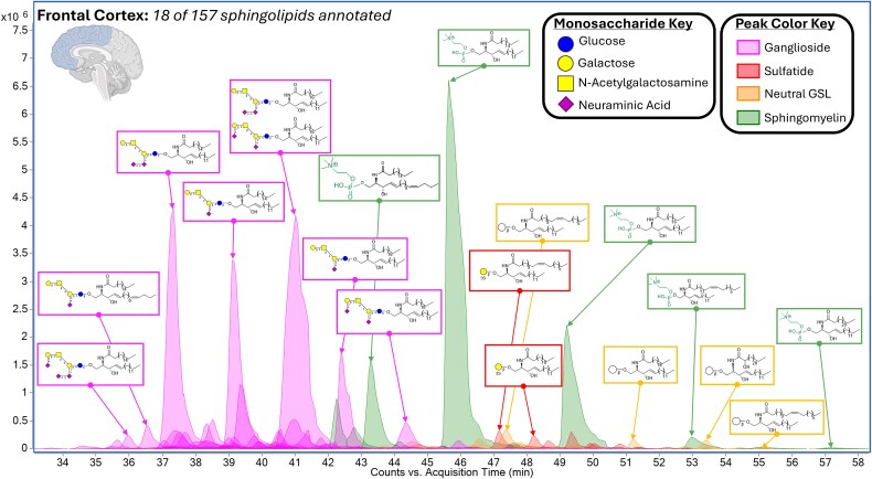
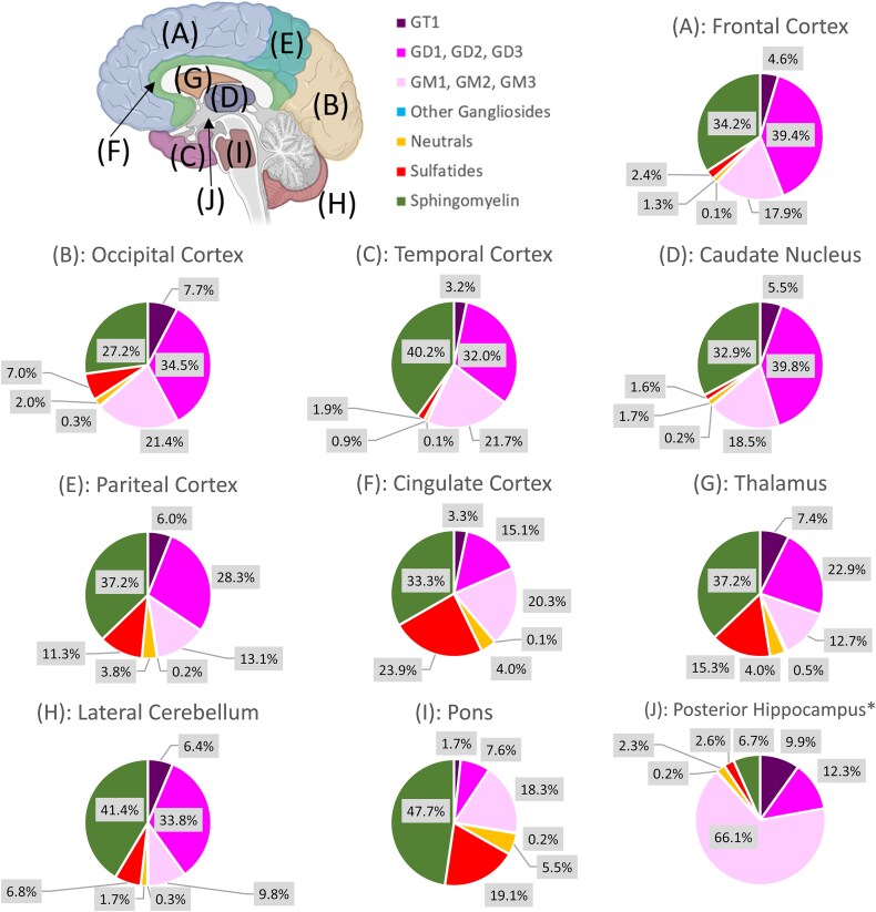
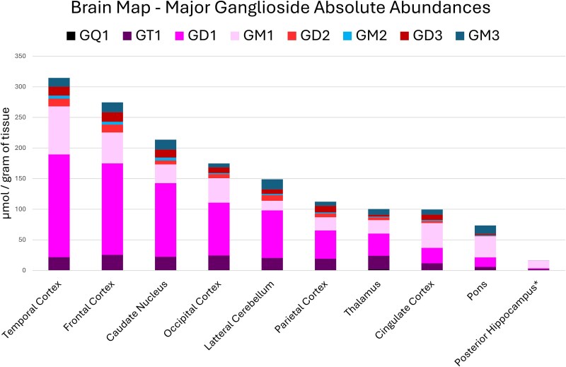
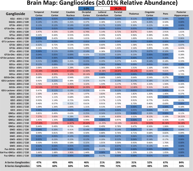
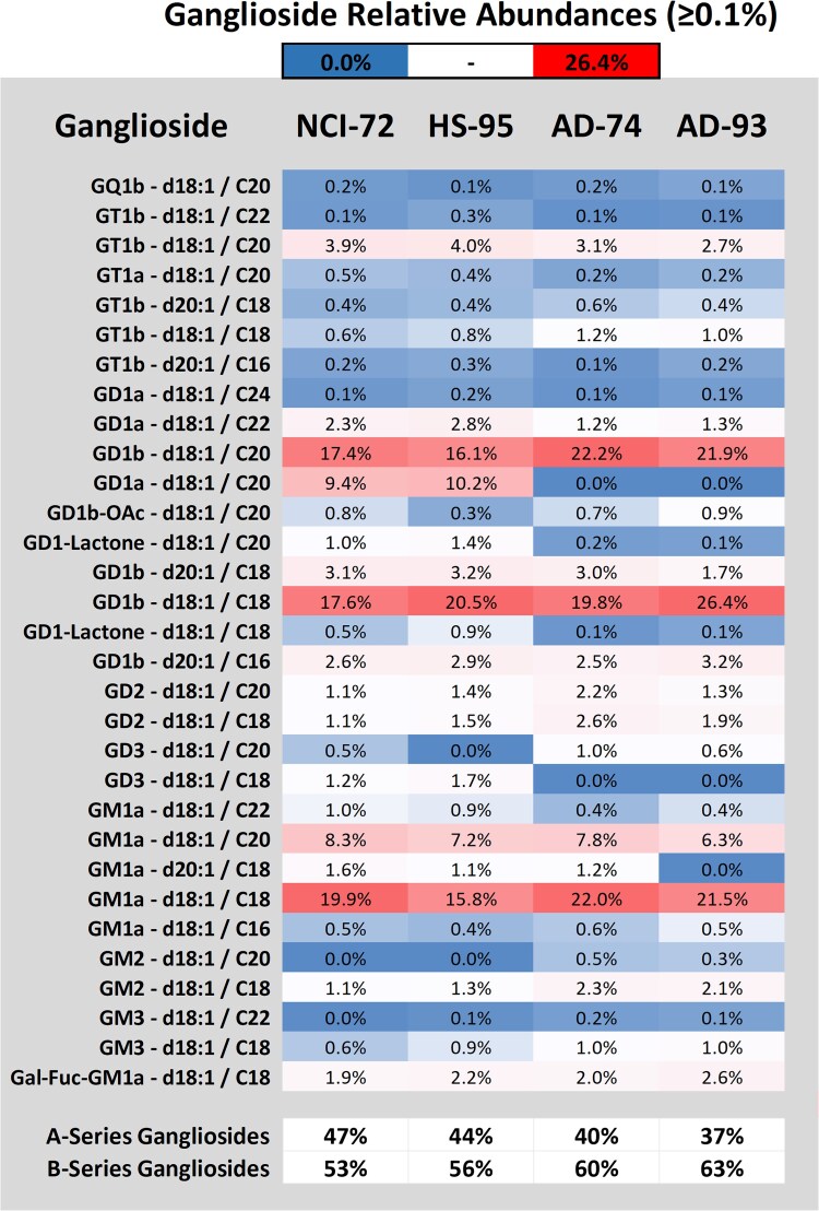
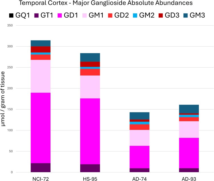
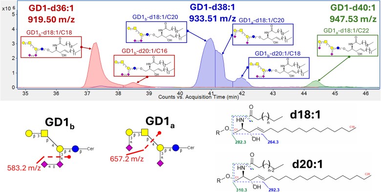
Glycosphingolipids are a unique class of bioactive lipids responsible for lateral membrane organization and signaling found in high abundance in the central nervous system. Using nanoflow MEA Chip Q/ToF mass spectrometry, we profiled the intact glycosphingolipids of the elderly human brain in a region-specific manner. By chromatographic separation of glycan and ceramide isomers, we determined gangliosides to be the highest source of heterogeneity between regions with the expression of a- and b-series glycan structures. Investigation of these trends showed that specific glycan structures were, in part, determined by the structure of their lipid backbone. This study provides insight into the dynamic process of membrane remodeling in the brain during aging.

Keywords: MEA Chip; Nanoflow HPLC-Q; ToF; brain map; glycosphingolipids.


Conflict of interest statement

None declared.

Figures

Fig. 1
Fig. 1
Example chromatogram of human brain tissue by nanoHPLC-MEAchip-Q/ToF analysis. This chromatographic separation, annotating 18 of 157 identified sphingolipids, was used to profile the frontal cortex of a 72-year-old male with no sign of cognitive impairment. Inset structures were assigned by matching biologically relevant molecular structures to accurate mass precursor ions, isotopic ratios, structurally relevant product ions from collision-induced dissociation, and elution patterns.
Fig. 2
Fig. 2
Region specific sphingolipid profile of the major headgroups in ten functional brain regions from relative abundance values obtained from a 72-year-old male with no sign of cognitive impairment. Each headgroup category includes all the observed ceramide structures associated with the depicted headgroups.
Fig. 3
Fig. 3
Absolute quantitation of the ganglioside profile in ten functional brain regions from relative abundance values obtained from a 72-year-old male with no sign of cognitive impairment. Each representative ganglioside included both the a- and b-series glycan structures for the major ceramide structures d18:1/C16, C18, C20, and C22 when observed in the depicted region.
Fig. 4
Fig. 4
Relative abundance heatmap of all identified intact gangliosides (≥0.01%) with summation of overall a- and b-series structures for ten functional brain regions from a 72-year-old male with no sign of cognitive impairment.
Fig. 5
Fig. 5
Relative abundances of gangliosides with ≥0.1% observed in the temporal cortex for comparison of age matched controls NCI-72 & HS-95 to autopsy confirmed Alzheimer disease subjects AD-74 & AD-93. Summation of overall a- and b-series structures included all depicted ceramide structures.
Fig. 6
Fig. 6
Absolute quantitation of the major ganglioside species observed in the temporal cortex for comparison of age matched controls NCI-72 & HS-95 to autopsy confirmed Alzheimer disease subjects AD-74 & AD-93. Each representative ganglioside included both a- and b-series glycan structures for the major ceramide structures d18:1/C16, C18, C20, and C22.
Fig. 7
Fig. 7
Example ganglioside elution profile and identifying MS2 product ions produced by nanoHPLC-MEAchip-Q/ToF analysis. Depicted isomeric structures include glycan a- & b-series isomers (GD1a & GD1b) with ceramide isomers (d18:1/CN & d20:1/CN).

Similar articles

References

    1. Ariga T, McDonald MP, Robert KY. 2008. Role of ganglioside metabolism in the pathogenesis of Alzheimer’s disease - a review. J Lipid Res. 49:1157–1175. 10.1194/jlr.R800007-JLR200. - DOI - PMC - PubMed
    1. Arumugam S et al. 2021. Ceramide structure dictates glycosphingolipid Nanodomain assembly and function. Nat Commun. 12:3675. - PMC - PubMed
    1. Bhaduri A, Neumann EK, Kriegstein AR, Sweedler JV. 2021. Identification of lipid heterogeneity and diversity in the developing human brain. JACS Au. 1:2261–2270. 10.1021/jacsau.1c00393. - DOI - PMC - PubMed
    1. Blumenreich S et al. 2022. Elevation of gangliosides in four brain regions from Parkinson’s disease patients with a GBA mutation. Npj Parkinson’s Dis. 8:99. - PMC - PubMed
    1. Boggs JM, Gao W, Hirahara Y. 2008. Myelin glycosphingolipids, Galactosylceramide and Sulfatide, participate in carbohydrate–carbohydrate interactions between apposed membranes and may form Glycosynapses between oligodendrocyte and/or myelin membranes. Biochim Biophys Acta (BBA). 1780:445–455. - PubMed