Functional analysis of promoter element 2 within the viral polymerase gene of an emerging paramyxovirus, Sosuga virus
- PMID: 40207914
- PMCID: PMC12054172
- DOI: 10.1128/spectrum.00534-25
Functional analysis of promoter element 2 within the viral polymerase gene of an emerging paramyxovirus, Sosuga virus
Abstract
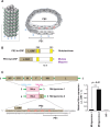
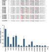
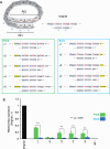
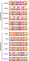
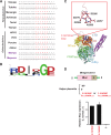
Paramyxovirus genomes carry bipartite promoters at the 3' ends of both their genome and antigenome, thereby initiating RNA synthesis, which requires the viral polymerase to recognize two elements: the primary promoter element 1 (PE1) and the secondary promoter element 2 (PE2). We have previously shown that the antigenomic PE2 (agPE2) in many viruses in the Rubulavirinae subfamily is located within the coding region of the viral RNA polymerase L gene. Sosuga virus (SOSV), belonging to the Rubulavirinae subfamily, is highly pathogenic to humans, thus necessitating high-level containment facilities for infectious virus research. The use of a minigenome system permits studies of viral RNA synthesis at lower biosafety levels. Because minigenomes of negative-strand RNA viruses generally comprise only the untranslated regions, agPE2 within the L coding region-such as those found in Rubulavirinae like SOSV-is typically omitted. However, generating an SOSV minigenome that retains agPE2 led to a pronounced increase in activity, enabling a detailed examination of the role of agPE2 in SOSV replication. In many Rubulavirinae, the agPE2 not only acts as a promoter but also encodes part of the L protein, resulting in a distinct motif at the C-terminus of the L protein. We have further shown that this motif is preserved even in Rubulavirinae that no longer contain the agPE2 within the L gene.IMPORTANCEParamyxoviruses are classified into three major subfamilies: Orthoparamyxovirinae, Avulavirinae, and Rubulavirinae. All paramyxovirus genomes and antigenomes possess bipartite promoters, comprising two elements: promoter element 1 (PE1) at the 3' end and promoter element 2 (PE2) located internally. We previously revealed that, in many Rubulavirinae, the antigenomic PE2 lies within the coding region of the viral RNA polymerase L gene. In this study, we used Sosuga virus, a member of the Rubulavirinae subfamily, to elucidate the role of antigenomic PE2 in viral replication. Because the PE2 region encodes part of the L protein, its presence leads to a distinctive motif at the C-terminus of L protein. Notably, this motif is conserved in all Rubulavirinae, including those that do not harbor the antigenomic PE2 within their L gene, indicating its importance in viral propagation.
Keywords: RNA polymerases; negative-strand RNA virus; paramyxovirus; promoters; viral replication.
Conflict of interest statement
Yusuke Matsumoto receives compensation from Denka Co., Ltd. The other authors declare no competing interests.
Figures

Similar articles
-
Phylogenetic analysis of the promoter element 2 of paramyxo- and filoviruses.Microbiol Spectr. 2024 May 2;12(5):e0041724. doi: 10.1128/spectrum.00417-24. Epub 2024 Apr 12. Microbiol Spectr. 2024. PMID: 38606982 Free PMC article.
-
A functional antigenomic promoter for the paramyxovirus simian virus 5 requires proper spacing between an essential internal segment and the 3' terminus.J Virol. 1998 Jan;72(1):10-9. doi: 10.1128/JVI.72.1.10-19.1998. J Virol. 1998. PMID: 9420195 Free PMC article.
-
A Minigenome Study of Hazara Nairovirus Genomic Promoters.J Virol. 2019 Mar 5;93(6):e02118-18. doi: 10.1128/JVI.02118-18. Print 2019 Mar 15. J Virol. 2019. PMID: 30626667 Free PMC article.
-
The latest advancements in Sosuga virus (SOSV) research.Front Microbiol. 2024 Nov 1;15:1486792. doi: 10.3389/fmicb.2024.1486792. eCollection 2024. Front Microbiol. 2024. PMID: 39552644 Free PMC article. Review.
-
Paramyxovirus RNA synthesis, mRNA editing, and genome hexamer phase: A review.Virology. 2016 Nov;498:94-98. doi: 10.1016/j.virol.2016.08.018. Epub 2016 Aug 25. Virology. 2016. PMID: 27567257 Review.
References
LinkOut - more resources
Full Text Sources

