. 2025 May 20;99(5):e0227524.
doi: 10.1128/jvi.02275-24. Epub 2025 Apr 10.

Cell binding, uptake, and infection of influenza A virus using recombinant antibody-based receptors

Affiliations

Cell binding, uptake, and infection of influenza A virus using recombinant antibody-based receptors

Oluwafemi F Adu et al. J Virol. .

Abstract

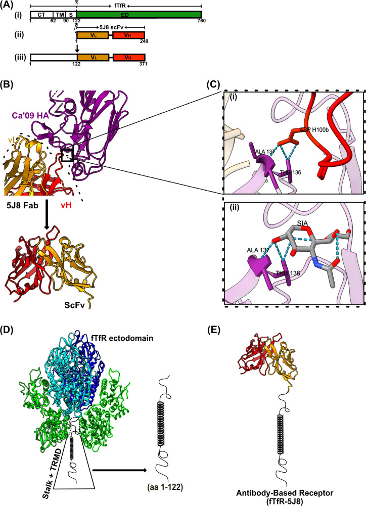
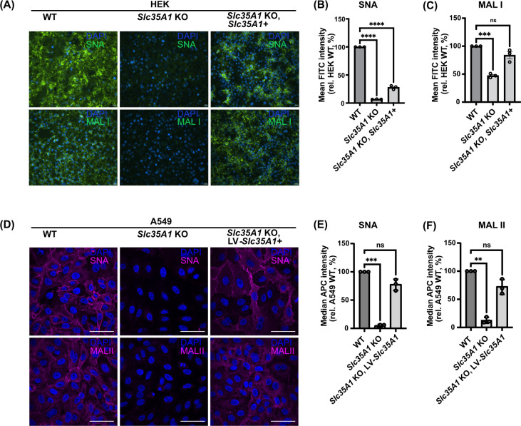
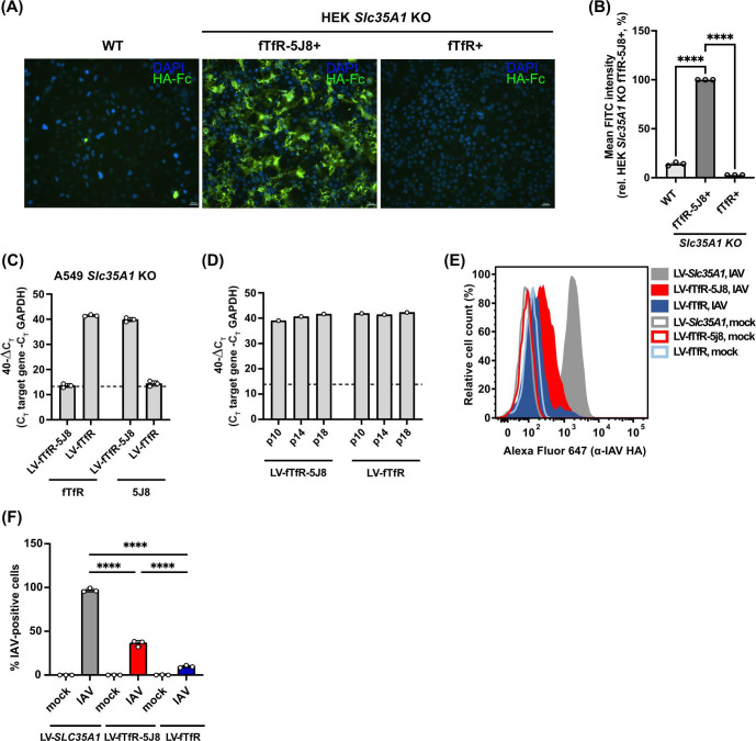
Human and avian influenza A viruses bind to sialic acid (Sia) receptors on cells as their primary receptors, and this results in endocytic uptake of the virus. While the role of Sia on glycoproteins and/or glycolipids for virus entry is crucial, the roles of the carrier proteins are still not well understood. Furthermore, it is still unclear how receptor binding leads to infection, including whether the receptor plays a structural or other roles beyond being a simple tether. To enable the investigation of the receptor binding and cell entry processes in a more controlled manner, we have designed a protein receptor for pandemic H1 influenza A viruses. The engineered receptor possesses the binding domains of an anti-HA antibody prepared as a single-chain variable fragment (scFv) fused with the stalk, transmembrane, and cytoplasmic sequences of the feline transferrin receptor type-1 (fTfR). When expressed in cells that lack efficient display of Sia due to a knockout of the Slc35A1 gene, which encodes for the solute carrier family 35 transporter (SLC35A1), the anti-H1 receptor was displayed on the cell surface, bound virus, or hemagglutinin proteins, and the virus was efficiently endocytosed into the cells. Infection occurred at similar levels to those seen after reintroducing Sia expression, and lower affinity receptor mutants displayed enhanced infections. Treatment with clathrin-mediated endocytosis (CME) inhibitors significantly reduced viral entry, indicating that virus rescue by the antibody-based receptor follows a similar internalization route as Sia-expressing cells.IMPORTANCEInfluenza A viruses primarily circulate among avian reservoir hosts but can also jump species, causing outbreaks in mammals, including humans. A key interaction of the viruses is with host cell sialic acids, which vary in chemical form, in their linkages within the oligosaccharide, and in their display on various surface glycoproteins or glycolipids with differing properties. Here, we report a new method for examining the processes of receptor binding and uptake into cells during influenza A virus infection, by use of an engineered HA-binding membrane glycoprotein, where antibody variable domains are used to bind the virus, and the transferrin receptor uptake structures mediate efficient entry. This will allow us to test and manipulate the processes of cell binding, entry, and infection.

Keywords: endocytosis; experimental tools; host-cell interactions; orthomyxovirus; receptor binding.


Conflict of interest statement

The authors declare no conflict of interest.

Figures

Fig 1
Fig 1
Design of antibody-based IAV receptor. (A) Cartoon showing the construction and cloning of the fTfR-5J8 chimera. (i, ii) Heavy and light variable chains of MAb 5J8 were cloned as scFv constructs in-frame onto residues 1–122 of the fTfR (iii) construct. (B) Crystal structure of Fab 5J8: A/Ca/07/2009-HA1 (PDB: 4M5Z). Purple (HA), gold (Fab vL), and red (Fab vH). (C) Magnified H-bond interactions of HCDR3 (red) Asp 100 interactions with (i) Ala 137 and Thr 136 HA residues. (ii) Identical H-bond interactions of Sia with Ala 137 and Thr 136 HA residues (PDB:3UBE) (36). (D) Cartoon of the TfR structure showing the ectodomain with transmembrane (TRMD) and stalk domains (aa 1–122). (E) Cartoon of antibody-based receptor monomer (scFv from Fab 5J8) fused to the stalk and transmembrane and cytoplasmic domains of the fTfR). Colors in the fTfR are similar to the different domains as defined for the human TfR; green (bottom half), protease-like domain; green (top half), apical domain; cyan and blue, helical domain (37).
Fig 2
Fig 2
Validating Sia expression in HEK293 and A549 WT and Slc35A1 KO conditions. (A) Representative unmodified fluorescent microscopy images of fixed cells stained with DAPI (blue) and fluorescein-labeled SNA or MAL I (green). Scale bar represents 20 µm. Relative fluorescence level of (B) SNA and (C) MAL I stained live cells quantified by flow cytometry with more than 10,000 cells (events) analyzed for each stained condition in n = 3 biological experiments. Mean fluorescence intensities (MFIs) were normalized relative to HEK WT Sia expression (B, C). Background (unstained) subtracted MFI values were analyzed using PRISM software. Error bars show mean ± standard mean error. Statistics were calculated using two-way analysis of variance with Tukey’s multiple comparisons test. P < 0.05; ****P < 0.0001, ***P < 0.001. (D) Unmodified (WT) and Slc35A1 KO A549 cells transduced with an Slc35A1 encoding lentivirus (LV-Slc35A1) were fixed and stained with biotinylated SNA or MAL II (magenta) and DAPI (blue). The Alexa Fluor 647-streptavidin signal intensities were assessed via microscopy. Images are representative of n = 3 independent replicates. Scale bar represents 50 µm. (E, F) Cells from (D) were incubated with biotinylated SNA or MAL II and the APC-streptavidin in live cells analyzed via flow cytometry. Following subtraction of the median APC signal of corresponding background samples, the median APC signal was normalized to that of WT cells. Data are means ± standard error of mean/standard deviation from n = 3 independent experiments. Statistical significance was inferred by two-tailed one-sample t-test with a theoretical mean of 100. ***P < 0.001, **P < 0.01.
Fig 3
Fig 3
Binding analysis of HA-probe or live virus to antibody-based receptor-expressing cells. (A) Representative unmodified fluorescent microscopic images of fixed cells incubated with HA-hFc probe and detected with fluorescein-labeled anti-human Fc and DAPI (blue). Scale bar represents 20 µm. (B) Relative fluorescent intensities of live HA-hFc-stained cells were obtained via flow cytometry, with more than 10,000 cells (events) analyzed for each stained condition from n = 3 biological experiments. Fluorescent intensities were normalized relative to fTfR-5J8-expressing cells. Error bars show mean ± standard mean error. Statistics were calculated using two-way analysis of variance (ANOVA) with Tukey’s multiple comparisons test. P < 0.05; ****P < 0.0001. (C) The fTfR and fTfR-5J8 expression was analyzed in A549 Slc35A1 KO cells transduced with lentiviruses encoding fTfR (LV-fTfR) or fTfR-5J8 (LV-fTfR-5J8) via RT-qPCR. Primers targeting GAPDH, the extracellular domain of fTfR and 5J8, were utilized. Data are 40-∆CT values from n = 3 independent experiments. A CT of 40 was assigned to samples where no amplification was detected. (D) The receptor expression stability in cells from (C) was analyzed via RT-qPCR by evaluating RNA collected at passage numbers 10, 14, and 18 (p10, p14, and p18, respectively). (E) Cells from (C) and A549 Slc35A1 KO cells stably expressing SLC35A1 (LV-Slc35A1) were inoculated with A/Netherlands/602/09 at an MOI of 25 for 1.5 h on ice. Cells were incubated with an anti-HA antibody, and the signal from bound virus was quantified via flow cytometry. A representative histogram from n = 3 independent experiments is shown. (F) Quantification of the percentage of IAV positive cells from (E). The gating strategy was established using the mock-infected sample. Data are means ± standard deviation, and statistical significance was inferred by one-way ANOVA with Sidak’s multiple comparisons test. ****P < 0.0001.
Fig 4
Fig 4
Antibody-based receptor rescues IAV infection in the absence of Sia. (A) Representative fluorescence microscopy images of fixed Ca’09-infected cells. Receptors (fTfR-5J8 and full-length fTfR) were transiently expressed on HEK Slc35A1 KO for 24 h before viral infection (MOI = 0.2) for 8 h. Cells were fixed and stained with anti-NP antibody (green) and DAPI (blue). (B, C) Infection data for Ca’09 or PR8 infected cells, respectively. Four different fields of view were imaged for each condition, and percent infection was determined. Data were normalized to HEK WT infected cells. (D) Infection curve for the Neth/09-Renilla at MOI = 3 in A549 LV-fTfR, LV-fTfR-5J8, and LV-Slc35A1 cells. (E) AUC plot from D, where AUC values of LV-fTfR-5J8 and LV-fTfR are shown relative to LV-Slc35A1. ND, no infection detected; NS, not significant. All experiments were performed in three independent replicates (n = 3). Error bars show mean ± standard mean error. Statistics were calculated using two-way analysis of variance with Tukey’s multiple comparisons test. *P < 0.05; **P < 0.01, ****P < 0.0001.
Fig 5
Fig 5
Receptor mutants binding and infection analysis. (A) Fab 5J8 and A/Ca/07/2009 HA complex (PDB: 4M5Z) showing interacting residues between antibody complementarity-determining region (CDRs) and the HA antigen. Intermolecular contacts were visualized using Chimera X (53) (green dashed lines). Red (HCDR3 loop), gold (LCDR3), and purple (receptor binding site [RBS] pocket, HA antigen). (B) Cartoon showing the fTfR-5J8 construct (WT) and the Flag tag version (WT [Flag+]). Mutagenesis was carried out using the WT (Flag+) construct. (C) Receptor: HA-hFc binding analysis data. Fixed HEK Slc35A1 KO cells transiently expressing receptors were incubated with an anti-Flag antibody and the HA-hFc probe. Fluorescent signals were detected using flow cytometry on the FITC (anti-Flag) and APC (HA-hFc) channels. Binding strength to the HA-hFc probe was quantified as the relative mean fluorescence intensity (MFI) of APC in Flag+ cells, normalized to the mean FITC signal. The Flag+ population was gated using cells expressing only fTfR or original WT receptor. (D) Selected receptor mutants transiently expressed in HEK Slc35A1 KO cells were infected with the Ca’09 virus at an MOI of 0.5 for 8 h. Fixed cells were permeabilized and stained with an anti-NP antibody to detect infected (NP+) cells via flow cytometry on the FITC channel. Mean FITC intensity was expressed relative to the WT (Flag+) control. Final MFI values were calculated by subtracting the signal intensity of mock-infected cells. All experiments were performed in three independent replicates (n = 3). Error bars show mean ± standard mean error. Statistics were calculated using two-way analysis of variance with Tukey’s multiple comparisons test. *P < 0.05; **P < 0.01; ***P < 0.001; ****P < 0.0001. Only significant values are indicated in the graphs.
Fig 6
Fig 6
IAV entry mediated by the antibody-based receptor is dynamin- and clathrin-dependent. A549 Slc35A1 KO cells stably expressing SLC35A1 (LV-Slc35A1) or fTfR-5J8 (LV-5J8-TfR) were pre-treated with dynasore (40 µM–20 µM), pitstop 2 (15 µM–10 µM), NH4Cl (25 mM), or DMSO (0.15%) for 30 min at 37°C and then infected with the indicated viruses at an MOI of 3 in their presence. (A) Cell viability at 24 h post-inhibitor treatment relative to the DMSO control. (B) Neth/09-Renilla infection curves. (C) AUC values from (B) until 9 h post-infection were normalized to the DMSO-treated sample within each cell line. (D) AUC values for VSV-GFP and RSV-GFP infection curves. AUC values for VSV-GFP or RSV-GFP infections were calculated until 11 or 27 h post-infection, respectively, and were normalized to the corresponding DMSO-treated samples. (E–G) A549 LV-Slc35A1 and LV-5J8-fTfR cells were pre-treated with bafilomycin (10 nM) or DMSO (0.1%) for 2 h at 37°C and inoculated with Neth/09-Renilla as in (A). (E) Cell viability at 3 h post-inhibitor treatment relative to the DMSO control. (F, G) Neth/09-Renilla infection curves (F) and the corresponding total AUC values until 9 h post-infection (G). (A–F) Data are means ± standard deviation from n = 3 independent experiments. Statistical significance was determined by two-tailed one-sample t-test with a theoretical mean of 100 (C, D) or by two-tailed unpaired t-test. *P < 0.05, **P < 0.01; ns, not significant. The dashed line indicates 80% cell viability (A, E), the luminescence from the mock-infected sample (B, F) or the AUC for the mock-infected sample (G).
Fig 7
Fig 7
Summary of the new receptor prepared here and its potential for analysis of the IAV entry pathways. HEK and A549 Slc35A1 KO cells were prepared, allowing us to examine the rescue of IAV uptake, internalization, and infection in the absence of efficient Sia surface expression by using the alternative receptor. Schematics of the Sia biosynthetic pathway are shown (summarized in reference 68), indicating the accumulation of CMP-Sia in the cytosol due to the gene deletion of the antiporter SLC35A1. UDP-GlcNAC, uridine diphosphate N-acetylglucosamine; ManNAC-6-P, N-acetyl-mannosamine 6-phosphate; GNE, UDP-GlcNAc 2-epimerase/ManNAc-6-kinase.

Similar articles

Cited by

References

    1. Karakus U, Pohl MO, Stertz S. 2020. Breaking the convention: sialoglycan variants, coreceptors, and alternative receptors for influenza A virus entry. J Virol 94:e01357. doi:10.1128/JVI.01357-19 - DOI - PMC - PubMed
    1. de Vries E, Du W, Guo H, de Haan CAM. 2020. Influenza A virus hemagglutinin–neuraminidase–receptor balance: preserving virus motility. Trends Microbiol 28:57–67. doi:10.1016/j.tim.2019.08.010 - DOI - PMC - PubMed
    1. Luo M. 2012. Influenza virus entry. Adv Exp Med Biol 726:201–221. doi:10.1007/978-1-4614-0980-9_9 - DOI - PMC - PubMed
    1. Chu VC, Whittaker GR. 2004. Influenza virus entry and infection require host cell N-linked glycoprotein. Proc Natl Acad Sci USA 101:18153–18158. doi:10.1073/pnas.0405172102 - DOI - PMC - PubMed
    1. Chien YAA, Alford BK, Wasik BR, Weichert WS, Parrish CR, Daniel S. 2023. Single particle analysis of H3N2 influenza entry differentiates the impact of the sialic acids (Neu5Ac and Neu5Gc) on virus binding and membrane fusion. J Virol 97:e0146322. doi:10.1128/jvi.01463-22 - DOI - PMC - PubMed

MeSH terms

Substances

LinkOut - more resources