. 2025 May 13;93(5):e0057424.
doi: 10.1128/iai.00574-24. Epub 2025 Apr 10.

A Salmonella enterica serovar Typhimurium genome-wide CRISPRi screen reveals a role for type 1 fimbriae in evasion of antibody-mediated agglutination

Affiliations

A Salmonella enterica serovar Typhimurium genome-wide CRISPRi screen reveals a role for type 1 fimbriae in evasion of antibody-mediated agglutination

Samantha K Lindberg et al. Infect Immun. .

Abstract

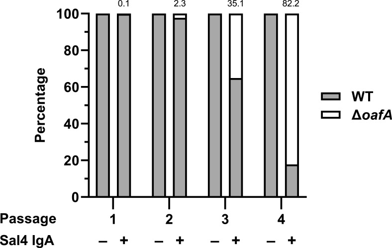
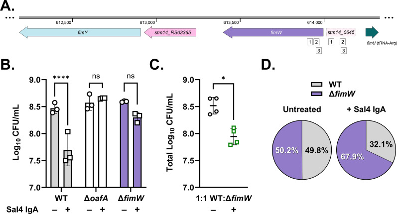
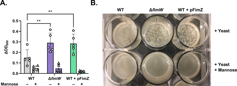
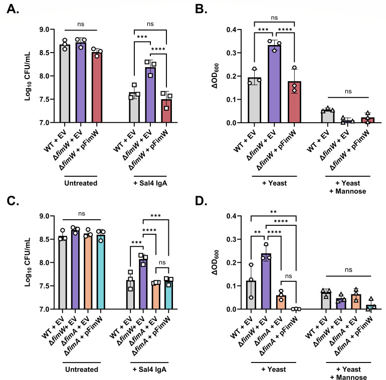
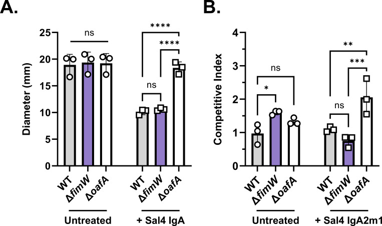
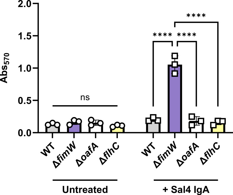
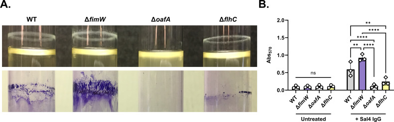
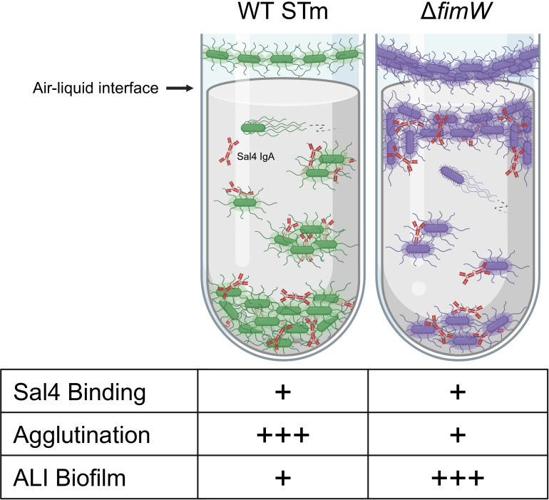
The O5-specific monoclonal IgA antibody, Sal4, mediates the conversion of Salmonella enterica serovar Typhimurium (STm) from virulent, free-swimming cells to non-motile, multicellular biofilm-like aggregates within a matter of hours. We hypothesize that the rapid transition from an invasive to a non-invasive state is an adaptation of STm to Sal4 IgA exposure. In this report, we performed a genome-wide CRISPR interference (CRISPRi) screen to identify STm genes that influence multicellular aggregate formation in response to Sal4 IgA treatment. From a customized library of >36,000 spacers, ~1% (373) were enriched at the top of the culture supernatant after two consecutive rounds of Sal4 IgA treatment. The enriched spacers mapped to a diversity of targets, including genes involved in O-antigen modification, cyclic-di-GMP metabolism, outer membrane biosynthesis/signaling, and invasion/virulence, with the most frequently targeted gene being fimW, which encodes a negative regulator of type 1 fimbriae (T1F) expression. Generation of a STm ΔfimW strain confirmed that the loss of FimW activity results in a hyperfimbriated phenotype and evasion of Sal4 IgA-mediated agglutination in solution. Closer examination of the fimW mutant revealed its propensity to form biofilms at the air-liquid interface in response to Sal4 exposure, suggesting that T1F "primes" STm to transition from a planktonic to a sessile state, possibly by facilitating bacterial attachment to abiotic surfaces. These findings shed light on the mechanism by which IgA antibodies influence STm virulence in the intestinal environment.

Keywords: Salmonella; adhesion; antibody; immunity; mucosal.


Conflict of interest statement

The authors declare no conflict of interest.

Figures

Fig 1
Fig 1
Repeated Sal4 IgA treatment enriches for O5- STm in a predominantly O5+ population. Mid-log phase cultures of WT (SL174; lacZ+) and ΔoafA (SL180; lacZ-) STm were washed in PBS, combined at a ratio of approximately 6,000:1, and then left untreated or treated with 15 µg/mL of Sal4 IgA. At 5 h post-treatment, 100 µL of the culture from the top of the supernatant was collected and plated on LB agar containing X-gal to determine the relative composition of each strain. This portion of the culture was passaged, and the assay procedure was repeated the following day for a total of four rounds of treatment. Data represent the percentage of each strain (with the value for ΔoafA listed above the bar for the Sal4-treated groups) averaged from two biological replicates each with two technical replicates.
Fig 2
Fig 2
A STm fimW mutant evades Sal4-mediated agglutination in vitro. (A) Gene organization of fimW and neighboring genetic features, including the positive fimbrial regulator gene fimY, the putative phosphodiesterase-encoding gene stm14_RS03365, the upstream region of fimW (previously annotated as stm14_0645), and the tRNA-Arg-encoding gene fimU. White boxes represent enriched spacers (32 nucleotides) identified in the screen analysis that target complementary sequences within fimW and stm14_0645. For panels B and C, clearance of bacterial cells from the top of the culture supernatant of homogeneous and mixed cultures of the indicated strains was quantified by plating colony-forming units (CFUs) following 2 h of treatment with 15 μg/mL Sal4 IgA. (B) Data represent three biological replicates with error bars representing the standard deviation of the mean. Statistical significance was determined by ordinary two-way analysis of variance (ANOVA) with Šídák’s multiple comparison test. Asterisks (****) indicate P < 0.0001 and ns = not significant. (C) Data was obtained from four biological replicates with error bars representing the standard deviation of the mean. Statistical significance was determined by paired t-test (* indicates P < 0.05). (D) Percent composition of WT (SL174; lacZ+) and ΔfimW (SL164; lacZ-) recovered from the air–liquid interface as determined by blue–white screening of LB + X-gal plates. Values represent the average percentage of each strain from four biological replicates.
Fig 3
Fig 3
The fimW mutant efficiently agglutinates yeast in a mannose-sensitive manner. (A) Cultures of STm WT (SL174), ΔfimW (SL164), and WT + pFimZ (SL218; induced with 0.2% L-arabinose) were incubated statically for 48 h at 37 °C prior to centrifugation and resuspension in fresh LB. Cultures were mixed with yeast (final concentration: 10 mg/mL) either in the absence (hexagons) or presence of 3% (w/v) D-mannose (triangles) in a 12-well plate, as detailed in the Materials and Methods. The turbidity of the wells at 600 nm (OD600) was measured via spectrophotometry immediately after the addition of yeast. Data were obtained from five biological replicates with error bars representing the standard deviation of the mean. Statistical significance was determined by two-way ANOVA followed by Tukey’s post-hoc multiple comparisons test. Asterisks (**) indicate P < 0.01 and ns = not significant. (B) Representative image showing the extent of yeast agglutination when mixed with each STm strain in the absence and presence of mannose in the 12-well plate format.
Fig 4
Fig 4
Overexpression of T1F reduces STm susceptibility to Sal4-mediated agglutination in the snow globe assay. (A) Recovered CFU/mL of STm WT + pBAD24-EV (empty vector; EV), ΔfimW + EV, and ΔfimW + pBAD24-fimW (pFimW) cultures in the snow globe assay. (B) Quantification of mannose-sensitive yeast agglutination of the STm WT + EV, ΔfimW + EV, and ΔfimW + pFimW strains. (C) Recovered CFU/mL of STm WT + EV, ΔfimW + EV, ΔfimA + EV, and ΔfimA + pFimW cultures in the snow globe assay. (D) Quantification of mannose-sensitive yeast agglutination of WT + EV, ΔfimW + EV, ΔfimA + EV, and ΔfimA + pFimW strains. For panels A and C, the indicated strains were grown to mid-log phase in the presence of 0.02% arabinose, washed in PBS, and either left untreated (circles) or treated with 15 μg/mL of Sal4 IgA (squares). After 2 h of treatment, the top of the supernatant was collected and plated on LB agar to measure CFU. For panels B and D, the indicated strains were incubated statically for 48 h in LB containing 0.02% arabinose at 37°C prior to centrifugation and resuspension in LB. Cultures were mixed with 10mg/mL yeast in the presence (triangles) and absence (hexagons) of 3% mannose in a 12-well plate, and the optical density of the wells at 600nm (OD600) was measured via spectrophotometry. The strains used are SL257, SL253, SL255, SL289, and SL291. For all panels, data were obtained from three biological replicates with error bars representing the standard deviation of the mean. Statistical significance was determined by two-way ANOVA followed by Tukey’s post hoc multiple comparisons test. Asterisks (**, ***, ****) indicate P < 0.01, P < 0.001, and P < 0.0001, respectively, and ns = not significant.
Fig 5
Fig 5
The fimW mutant is susceptible to additional Sal4-induced effects. (A) Plates of 0.3% LB agar with and without 5.0 μg/mL Sal4 IgA were stab inoculated with 1.0 μL of overnight cultures of WT (SL174), ΔfimW (SL164), and ΔoafA (SL180) and then incubated at 37°C for 4.5 h. Plates were imaged, and the diameter (mm) of bacterial migration was measured using Fiji. Data represent three biological experiments each averaged from three technical replicates. Statistical significance was determined by two-way ANOVA followed by Tukey’s post-hoc multiple comparisons test. Asterisks (****) indicate P < 0.0001 and ns = not significant. (B) WT (SL174; lacZ+) was mixed 1:1 with WT (SL239; lacZ-), ΔfimW (SL164; lacZ-), and ΔoafA (SL180; lacZ-) and incubated for 15 min with 15µg/mL of purified recombinant Sal4 IgA2m1 before addition to HeLa cell monolayers in 96-well microtiter plates. Plates were centrifuged at 1,000 xg for 10 min (rotating the plate after 5 min) to promote bacteria–cell contact. After 1 h of incubation at 37 °C, monolayers were treated with 100 µg/mL gentamicin and incubated again for 1 h to eliminate extracellular bacteria. Cells were washed and lysed with 1% Triton X-100 diluted in Ca2+ and Mg2+-free PBS, and the resulting suspension was plated on LB containing X-gal to enable blue-white screening of CFUs. The competitive index [(% strain A output/% strain B output)/(% strain A input/% strain B input)] was calculated for each treatment group. Data represent three biological experiments, each averaged from three technical replicates. Statistical significance was determined by two-way ANOVA followed by Tukey’s post-hoc multiple comparisons test. Asterisks (*, **, ***) indicate P < 0.05, P < 0.01, and P < 0.001, respectively, and ns = not significant.
Fig 6
Fig 6
Overexpression of T1F enhances Sal4 IgA-mediated biofilm production on polystyrene. Mid-log phase cultures of WT (SL174), ΔfimW (SL164), ΔoafA (SL180), and ΔflhC (SL202) were washed with LB, standardized to an OD600 value of 1.0, and transferred to a 12-well polystyrene tissue culture plate. Cultures were left untreated (circles) or treated with 15 μg/mL Sal4 IgA (squares) at 23°C with shaking (200 rpm) for 1 h. The culture media was aspirated, and the biofilms were heat-fixed at 60°C for 1 h. Biofilms were stained with 0.1% crystal violet and washed with distilled water for 5 min, then the crystal violet stain was solubilized with 30% acetic acid for 5 min. Absorbance of crystal violet was quantified at Abs570. Data represent three biological experiments, and error bars represent standard deviation of the mean. Statistical significance was determined by two-way ANOVA followed by Tukey’s post-hoc multiple comparisons test. Asterisks (****) indicate P < 0.0001 and ns = not significant.
Fig 7
Fig 7
Crystal violet staining of agglutination-evading mutants following Sal4 IgG treatment. Mid-log phase cultures of WT (SL174), ΔfimW (SL164), ΔoafA (SL180), and ΔflhC (SL202) were washed with LB, standardized to an OD600 value of 1.0, and transferred to 16 × 125 mm borosilicate glass tubes. Cultures were mixed 1:1 with LB with (diamonds) and without (circles) 30 μg/mL Sal4 IgG and then incubated at 23°C with shaking (200 rpm) for 1 h (for a final concentration of 15 μg/mL Sal4). The culture media was aspirated, and the tubes were heat-fixed at 60°C for 1 h. Biofilms were stained with 0.1% crystal violet and washed with distilled water for 5 min each, then the stain was solubilized with 30% acetic acid for 5 min. Dissolved crystal violet was transferred to a 96-well plate prior to measurement of absorbance at 570 nm (Abs570). (A) Representative images of the indicated strains 1 h p.t. (top) and with remaining crystal violet stain after the final wash step (bottom). (B) Data represent three biological experiments, and error bars represent standard deviation of the mean. Statistical significance was determined by two-way ANOVA followed by Tukey’s post-hoc multiple comparisons test. Asterisks (**, ****) indicate P < 0.01 and P < 0.0001, respectively. ns = not significant.
Fig 8
Fig 8
Summary model of Sal4-induced impacts on WT and ΔfimW STm strains in vitro. The monoclonal IgA Sal4 binds to the O5 antigen expressed by both WT (green; left) and ΔfimW (purple; right) STm and induces a form of outer membrane stress that signals the bacteria to transition from a planktonic to sessile state, either proximal to an abiotic surface or in suspension. The majority of the WT population agglutinates and forms dense aggregates at the bottom of the culture tube. A small subpopulation of WT STm remains at the top of the supernatant near the air–liquid interface (ALI) where the bacteria can adhere to the surface. Hyperexpression of type 1 fimbriae on the bacterial surface, occurring through inhibition of fimW, enables STm to evade Sal4-mediated agglutination and enhances cell adhesion at the air–liquid interface. Free-floating aggregates and surface-adhered microcolonies both bound by Sal4 IgA continue to produce EPS, resulting in the formation of biofilms. Schematic created in BioRender.

Similar articles

References

    1. Troeger C, Forouzanfar M, Rao PC, Khalil I, Brown A, Reiner RC, Fullman N, Thompson RL, Abajobir A, Ahmed M, et al. . 2017. Estimates of global, regional, and national morbidity, mortality, and aetiologies of diarrhoeal diseases: a systematic analysis for the global burden of disease study 2015. Lancet Infect Dis 17:909–948. doi:10.1016/S1473-3099(17)30276-1 - DOI - PMC - PubMed
    1. Demissie GD, Yeshaw Y, Aleminew W, Akalu Y. 2021. Diarrhea and associated factors among under five children in sub-Saharan Africa: evidence from demographic and health surveys of 34 sub-Saharan countries. PLoS ONE 16:e0257522. doi:10.1371/journal.pone.0257522 - DOI - PMC - PubMed
    1. Mather AE, Reid SWJ, Maskell DJ, Parkhill J, Fookes MC, Harris SR, Brown DJ, Coia JE, Mulvey MR, Gilmour MW, Petrovska L, de Pinna E, Kuroda M, Akiba M, Izumiya H, Connor TR, Suchard MA, Lemey P, Mellor DJ, Haydon DT, Thomson NR. 2013. Distinguishable epidemics of multidrug-resistant Salmonella Typhimurium DT104 in different hosts. Science 341:1514–1517. doi:10.1126/science.1240578 - DOI - PMC - PubMed
    1. Feasey NA, Masesa C, Jassi C, Faragher EB, Mallewa J, Mallewa M, MacLennan CA, Msefula C, Heyderman RS, Gordon MA. 2015. Three epidemics of invasive multidrug-resistant Salmonella bloodstream infection in Blantyre, Malawi, 1998-2014. Clin Infect Dis 61 Suppl 4:S363–71. doi:10.1093/cid/civ691 - DOI - PMC - PubMed
    1. Okoro CK, Barquist L, Connor TR, Harris SR, Clare S, Stevens MP, Arends MJ, Hale C, Kane L, Pickard DJ, Hill J, Harcourt K, Parkhill J, Dougan G, Kingsley RA. 2015. Signatures of adaptation in human invasive Salmonella Typhimurium ST313 populations from sub-Saharan Africa. PLoS Negl Trop Dis 9:e0003611. doi:10.1371/journal.pntd.0003611 - DOI - PMC - PubMed

Publication types

MeSH terms

LinkOut - more resources