. 2025 Apr 10;10(10):e189330.
doi: 10.1172/jci.insight.189330. eCollection 2025 May 22.

The PERK/ATF4 pathway is required for metabolic reprogramming and progressive lung fibrosis

Affiliations

The PERK/ATF4 pathway is required for metabolic reprogramming and progressive lung fibrosis

Jyotsana Pandey et al. JCI Insight. .

Abstract

Asbestosis is a prototypical type of fibrosis that is progressive and does not resolve. ER stress is increased in multiple cell types that contribute to fibrosis; however, the mechanism(s) by which ER stress in lung macrophages contributes to fibrosis is poorly understood. Here, we show that ER stress resulted in protein kinase RNA-like ER kinase (PERK; Eif2ak3) activation in humans with asbestosis. Similar results were seen in asbestos-injured mice. Mice harboring a conditional deletion of Eif2ak3 were protected from fibrosis. Lung macrophages from asbestosis individuals had evidence of metabolic reprogramming to fatty acid oxidation (FAO). Eif2ak3fl/fl mice had increased oxygen consumption rate (OCR), whereas OCR in Eif2ak3-/- Lyz2-cre mice was reduced to control levels. PERK increased activating transcription factor 4 (Atf4) expression, and ATF4 bound to the Ppargc1a promoter to increase its expression. GSK2656157, a PERK-specific inhibitor, reduced FAO, Ppargc1a, and Aft4 in lung macrophages and reversed established fibrosis in mice. These observations suggest that PERK is a therapeutic target to reverse established fibrosis.

Keywords: Fatty acid oxidation; Fibrosis; Immunology; Macrophages; Pulmonology.


Figures

Figure 1
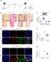
Figure 1. Asbestos activates PERK in lung macrophages.
(A) Lung macrophages from normal and asbestosis humans were obtained by bronchoalveolar lavage (BAL) and subjected to immunoblot analysis (n = 4). Densitometry of (B) phosphorylated (p-) PERK, (C) p-eIF2α, and (D) p-IRE1α in humans. (E) WT mice were exposed to man-made vitreous fiber (MMVF) or asbestos (100 μg intratracheally; i.t.). BAL was performed on day 21, and lung macrophages were subjected to immunoblot analysis (n = 3). Densitometry of (F) p-PERK, (G) p-eIF2α, and (H) p-IRE1α from exposed mice. (I) Macrophages were exposed to vehicle (Con), asbestos (Asb), tunicamycin (TUN), thapsigargin (TH), or 4PBA (PBA) and subjected to immunoblot analysis. Densitometry of (J) p-PERK and (K) p-eIF2α (n = 3). (L) Macrophages were exposed to vehicle or asbestos and stained for p-PERK. The staining was imaged by confocal microscopy, scale bars at 10 μm and 40×. (M) Quantification of mean fluorescence intensity (n = 3). Data shown as mean ± SEM. Two-tailed Student’s t test in BD, FH, and M. One-way ANOVA with Tukey’s post hoc comparison in J and K. *P ≤ 0.05, **P ≤ 0.01, and ****P ≤ 0.0001.
Figure 2
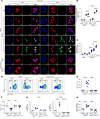
Figure 2. PERK is required for asbestos-induced lung fibrosis.
(A) Schematic representation for animal study: Eif2ak3fl/fl and Eif2ak3–/– Lyz2-cre littermates were exposed to MMVF or asbestos. Lung macrophages and lung tissues were isolated at 21 days and subjected to histology, hydroxyproline, and confocal imaging. OCR, oxygen consumption rate. (B) Masson’s trichome staining, scale bars at 100 μm and 10×. (C) Hydroxyproline assay (n = 5–7). Inset, immunoblot analysis of PERK in BAL cells from Eif2ak3fl/fl and Eif2ak3–/– Lyz2-cre mice. (D) Lung tissue sections from MMVF- or asbestos-injured mice were stained with p-PERK and F4/80. The staining was imaged by confocal microscopy, scale bars at 10 μm and 40×. (E) Quantification of mean fluorescence intensity (n = 6). (F) Lung tissue sections were stained with Col1a and α-SMA and imaged by confocal microscopy, scale bars at 10 μm and 40×. (G) Quantification of mean fluorescence intensity (n = 3). Data shown as mean ± SEM. One-way ANOVA with Tukey’s post hoc comparison in C. Two-tailed Student’s t test in E and G. ***P ≤ 0.001, ****P ≤ 0.0001. (See also Supplemental Figure 1.)
Figure 3
Figure 3. PERK activation increases in a time-dependent manner in BAL cells.
(A) Lung macrophages were obtained by BAL from Eif2ak3fl/fl and Eif2ak3–/– Lyz2-cre littermates from MMVF- or asbestos-injured mice. RAMs and MDMs were FACS-sorted on days 0, 5, 10, 15, and 21 and stained for p-PERK and F4/80. The staining was imaged by confocal microscopy, scale bars at 10 μm and 40×. (B) Quantification of mean fluorescence intensity (n = 7). (C) Hydroxyproline assay in lung tissues at indicated time points (n = 3–6). Eif2ak3fl/fl and Eif2ak3–/– Cx3cr1creER mice were administered tamoxifen and exposed to MMVF or asbestos. BAL was performed 21 days after exposure. (D) Representative flow plots with percentages and number of (E) MDMs (n = 6) and (F) RAMs (n = 4–6). FSC, forward scatter. (G) Eif2ak3 expression in FACS-sorted BAL cells (n = 3). (H) Hydroxyproline analysis in lung tissues (n = 6). Data shown as mean ± SEM. One-way ANOVA with Tukey’s post hoc comparison. *P ≤ 0.05, **P ≤ 0.01, ***P ≤ 0.001, and ****P ≤ 0.0001. (See also Supplemental Figure 2.)
Figure 4
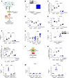
Figure 4. Asbestos-induced PERK activation mediates metabolic reprogramming.
Lung macrophages were obtained by BAL from normal and asbestosis humans. (A) Acetyl-CoA concentration measured by fluorometry in humans (n = 5). (B) TCA metabolites measured by mass spectrometry in humans and shown as fold-change (n = 4–5). (C) Ratio of NAD+/NADH in humans measured by fluorometry (n = 4). (D) Immunoblot analysis of CPT1A and (E) densitometry of CPT1A in BAL cell mitochondrial fractions from humans (n = 4). WT mice were exposed to MMVF or asbestos (100 μg i.t.). VDAC, voltage-dependent anion channel. (F) Lung macrophages from exposed mice were subjected to mass spectrometry. l-carnitine was normalized to total ion chromatography (TIC) (n = 3–4). (G) Hydroxyproline assay in lung tissues at the designated time points (n = 5). Cpt1a mRNA expression in (H) BAL cells isolated at day 21 from exposed Eif2ak3fl/fl and Eif2ak3–/– Lyz2-cre mice (n = 4) and (I) FACS-sorted RAMs and MDMs isolated from BAL at day 21 from Eif2ak3fl/fl and Eif2ak3–/– Cx3cr1creER mice (n = 3). (J) OCR kinetics in BAL cells isolated at day 21 from exposed Eif2ak3fl/fl and Eif2ak3–/– Lyz2-cre mice (n = 3). Oligo, oligomycin; FCCP, carbonyl cyanide p-trifluoromethoxyphenylhydrazone; Rot/Anti A, rotenone/antimycin A; min-μg, minute/μg protein. Data shown as mean ± SEM. Two-tailed Student’s t test in A, C, and E. One-way ANOVA with Tukey’s post hoc comparison in FI. *P ≤ 0.05, **P ≤ 0.01, ***P ≤ 0.001, and ****P ≤ 0.0001. (See also Supplemental Figure 3.)
Figure 5
Figure 5. PERK activates PGC-1α by increasing ATF4 in lung macrophages.
(A) Schematic representation of PERK pathway activation and downstream signaling molecules. CHOP, C/EBP homologous protein. (B) Lung macrophages from normal and asbestosis humans were obtained by BAL. Densitometry of immunoblot. Inset, immunoblot analysis of PGC-1α (n = 3). (C) Macrophages were cotransfected with renilla luciferase plasmid, pGL3-Ppargc1a luciferase promoter, and empty, PERKWT, or PERKDN and exposed to asbestos (24 hours). Ppargc1a promoter activity was determined by measuring firefly and renilla luciferase (n = 3). (D) ATF3 (n = 3), (E) ATF4 (n = 3), and (F) PPARGC1A mRNA expression (n = 3) in transfected macrophages exposed to asbestos. (G) Macrophages were exposed to control or asbestos and subjected to ChIP with antibodies against ATF3 or ATF4 followed by real-time PCR to determine Ppargc1a promoter binding (n = 3–9). (H) Lung macrophages were obtained from normal and asbestosis humans by BAL. Densitometry of immunoblot. Inset, immunoblot analysis of ATF4 (n = 4). (I) Macrophages were cotransfected with pGL3-Ppargc1a luciferase promoter combined with scramble or ATF4 siRNA, and empty or PERKWT, and exposed to asbestos. Ppargc1a promoter activity (n = 3–4). Inset, immunoblot analysis for ATF4. (J) Schematic illustration of ATF4 binding site on Ppargc1a promoter in the cAMP response element (CRE) domain and mutation sites on specific residues. (K) Ppargc1a promoter luciferase activity in macrophages transfected with empty, PERKWT, or pGL3-Ppargc1a luciferase mutant and exposed to asbestos (n = 3). (L) Ppargc1a mRNA expression in BAL isolated at day 21 from exposed Eif2ak3fl/fl and Eif2ak3–/– Lyz2-cre mice (n = 4). (M) Ppargc1a mRNA expression in FACS-sorted RAMs and MDMs isolated at day 21 from exposed Eif2ak3fl/fl and Eif2ak3–/– Cx3cr1creER mice (n = 3). Data shown as mean ± SEM. Two-tailed Student’s t test in B and H. One-way ANOVA with Tukey’s post hoc comparison in CG, I, and KM. *P ≤ 0.05, **P ≤ 0.01, ***P ≤ 0.001, and ****P ≤ 0.0001. (See also Supplemental Figure 4.)
Figure 6
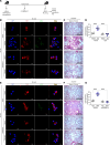
Figure 6. Pharmacological inhibition of PERK reverses established lung fibrosis.
(A) Schematic of animal study. Thirteen days after exposure, GSK2656157 (GSK, 30 mg/kg i.p.) was administered daily to mice. (B) Lung macrophages were obtained by BAL from MMVF- or asbestos-exposed mice and subjected to staining for p-eIF2α and F4/80. The staining was imaged by confocal microscopy, scale bars at 10 μm or 40×. (C) Masson’s trichrome staining in representative micrographs from n = 4–5 mice per condition, scale bar at 100 μm and 10×. (D) Hydroxyproline assay in lung tissues (n = 4–5). (E) Lung macrophages were obtained by BAL from control or bleomycin-exposed mice and subjected to staining for p-eIF2α and F4/80, scale bars at 10 μm or 40×. (F) Masson’s trichrome staining in representative micrographs from (n = 4–5) mice per condition, scale bar at 100 μm and 10×. (G) Hydroxyproline assay in lung tissues (n = 4–5). Data shown as mean ± SEM. One-way ANOVA with Tukey’s post hoc comparison. ****P < 0.0001. (See also Supplemental Figure 5.)
Figure 7
Figure 7. Pharmacological inhibition of PERK abrogates FAO in mice with established fibrosis.
Thirteen days after exposure, GSK (30 mg/kg i.p.) was administered daily to mice. (A) FAO in BAL macrophages from asbestos- or MMVF-exposed mice was measured by OCR with the addition of BSA or BSA:palmitate (PMT) using Seahorse XF96 bioanalyzer (Agilent Technologies) (n = 4–5). min-μg, minute/μg protein. Total RNA was isolated from lung macrophages and subjected to real-time PCR for (B) Ppargc1a (n = 3) and (C) Atf4 mRNA expression (n = 3). (D) FAO in macrophages from control or bleomycin-exposed mice was measured by OCR with the addition of BSA or PMT using Seahorse XF96 bioanalyzer (n = 3–5). Total RNA was isolated from lung macrophages and subjected to real-time PCR for (E) Ppargc1a (n = 3) and (F) Atf4 mRNA expression (n = 3). Data shown as mean ± SEM. One-way ANOVA with Tukey’s post hoc comparison in B and C. Two-tailed Student’s t test in E and F. **P ≤ 0.01, ****P ≤ 0.0001. (See also Supplemental Figure 6.)
Figure 8
Figure 8. PERK regulates macrophage pro-fibrotic gene expression.
Lung macrophages from normal and asbestosis humans were obtained by BAL. mRNA expression of (A) TGFB1 (n = 5), (B) IL10 (n = 5–7), (C) ARG1 (n = 6), and (D) MRC1 (n = 6) in humans. (E) Tgfb1 (n = 4), (F) Il10 (n = 4), and (G) Pdgfb (n = 4) mRNA expression in BAL isolated at day 21 from exposed Eif2ak3fl/fl and Eif2ak3–/– Lyz2-cre mice. (H) Tgfb1 (n = 3) and (I) Pdgfb (n = 3) mRNA expression in FACS-sorted BAL cells isolated at day 21 from exposed Eif2ak3fl/fl and Eif2ak3–/– Cx3cr1creER mice. (J) Active TGF-β1 (n = 4–5) and (K) PDGF-BB (n = 4–5) in BAL fluid harvested at day 21 from WT mice administered GSK 13 days after exposure to MMVF or asbestos. Data shown as mean ± SEM. Two-tailed Student’s t test in AD. One-way ANOVA with Tukey’s post hoc comparison in EK. *P ≤ 0.05, ***P ≤ 0.001, and ****P ≤ 0.0001. (See also Supplemental Figure 7.)

References

    1. Attfield MD, et al. Changing patterns of pneumoconiosis mortality--United States, 1968–2000. MMWR Morb Mortal Wkly Rep. 2004;53(28):627–632. - PubMed
    1. King TE., Jr A phase 3 trial of pirfenidone in patients with idiopathic pulmonary fibrosis. N Engl J Med. 2014;370(22):2083–2092. doi: 10.1056/NEJMoa1402582. - DOI - PubMed
    1. Richeldi L, et al. Efficacy and safety of nintedanib in idiopathic pulmonary fibrosis. N Engl J Med. 2014;370(22):2071–2082. doi: 10.1056/NEJMoa1402584. - DOI - PubMed
    1. Harraz MM, et al. SOD1 mutations disrupt redox-sensitive Rac regulation of NADPH oxidase in a familial ALS model. J Clin Invest. 2008;118(2):659–670. - PMC - PubMed
    1. He C, et al. Accelerated development of pulmonary fibrosis via Cu,Zn-superoxide dismutase-induced alternative activation of macrophages. J Biol Chem. 2013;288(28):20745–20757. doi: 10.1074/jbc.M112.410720. - DOI - PMC - PubMed

MeSH terms